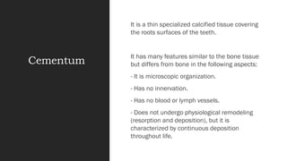
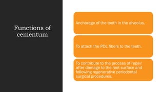
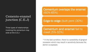
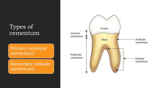
Coronal and radicular pulp
Apical foramen
Accessory canal
Functions of dental pulp
Components of dental pulp
Functions of pulpal extracellular matrix
Organization of cells in the pulp
The principle cells of the pulp
The pathways of collagen synthesis
Matrix and ground substances
Vasculature and lymphatic supply
Innervation of Dentin- pulp complex
Disorders of the dental pulp
Advances in pulp vitality testing
Cementum also commonly known as root cementum , is a highly mineralized tissue covering the entire root surface.
Cementum is also often referred to as a bone-like tissue. Cementum contains two types of fibers, mainly extrinsic (Sharpey's) fibers and intrinsic fibers. Fibroblasts and cementoblasts are the fiber secreting cells.
Dentinogenesis is the formation of dentin, a substance that forms the majority of teeth. Dentinogenesis is performed by odontoblasts, which are a special type of biological cell on the outer wall of dental pulps, and it begins at the late bell stage of a tooth development. The different stages of dentin formation after differentiation of the cell result in different types of dentin: mantle dentin, primary dentin, secondary dentin, and tertiary dentin.
Upload By : Ahmed Ali Abbas
Babylon University College of Dentistry
download this file from Website on google theoptimalsmile.wix.com/dentistry
Oral histology
This Slide, gives a Brief introduction to the Anatomy of the tooth specifically the outer shell, the enamel, including the structures, development and abnormalities.
Created by Dr. Mohsen S. Mohamed
For Ozident.com
It is a presentation in detail about the strongest structure of the oral cavity "ENAMEL". It is a simple topic but people find it difficult to learn about it. I hope my presentation is a simple method to learn about it. I would like to thank my professors for assign me this project and i learn't a lot from it and still learning my basics daily.
Indian Dental Academy: will be one of the most relevant and exciting training center with best faculty and flexible training programs for dental professionals who wish to advance in their dental practice,Offers certified courses in Dental implants,Orthodontics,Endodontics,Cosmetic Dentistry, Prosthetic Dentistry, Periodontics and General Dentistry.
this ppt includes information regarding a dental tissue know as the cementum.
It is a hard tissue covering the roots of the teeth.
this ppt includes, the composition, formation, types, cells and functions the cementum plays in maintaining the integrity of the tooth .
Cementum is the calcified, avascular mesenchymal tissue that forms the outer covering of the anatomic root.
it describes the development of cementum and its various types.
cementum is avascular connective tissue
it attaches tooth to alveolar bone via pdl in this seminar i explained about charecterestics of cementum &anamolies and some pathological conditions associated with it
Cementum /certified fixed orthodontic courses by Indian dental academy Indian dental academy
Welcome to Indian Dental Academy
The Indian Dental Academy is the Leader in continuing dental education , training dentists in all aspects of dentistry and offering a wide range of dental certified courses in different formats.
Indian dental academy has a unique training program & curriculum that provides students with exceptional clinical skills and enabling them to return to their office with high level confidence and start treating patients
State of the art comprehensive training-Faculty of world wide repute &Very affordable.
Upload By : Ahmed Ali Abbas
Babylon University College of Dentistry
download this file from Website on google theoptimalsmile.wix.com/dentistry
Oral histology
Cementum
Periodontal Ligament
Alveolar bone
Gingiva facing the tooth
Outline
- Anterior & Posterior triangle of the neck, and sub-divided triangles of them.
- Origin, Insertion, Innervation and Function of all muscle of Anterior & Posterior triangle of the neck.
Seminar about diagnostic methods that used in endodontic which include:
i. Case History.
ii. Clinical Examination.
iii. Radiographs.
iv. Pulp Vitality Tests.
Macroeconomics- Movie Location
This will be used as part of your Personal Professional Portfolio once graded.
Objective:
Prepare a presentation or a paper using research, basic comparative analysis, data organization and application of economic information. You will make an informed assessment of an economic climate outside of the United States to accomplish an entertainment industry objective.
Palestine last event orientationfvgnh .pptxRaedMohamed3
An EFL lesson about the current events in Palestine. It is intended to be for intermediate students who wish to increase their listening skills through a short lesson in power point.
2024.06.01 Introducing a competency framework for languag learning materials ...Sandy Millin
http://sandymillin.wordpress.com/iateflwebinar2024
Published classroom materials form the basis of syllabuses, drive teacher professional development, and have a potentially huge influence on learners, teachers and education systems. All teachers also create their own materials, whether a few sentences on a blackboard, a highly-structured fully-realised online course, or anything in between. Despite this, the knowledge and skills needed to create effective language learning materials are rarely part of teacher training, and are mostly learnt by trial and error.
Knowledge and skills frameworks, generally called competency frameworks, for ELT teachers, trainers and managers have existed for a few years now. However, until I created one for my MA dissertation, there wasn’t one drawing together what we need to know and do to be able to effectively produce language learning materials.
This webinar will introduce you to my framework, highlighting the key competencies I identified from my research. It will also show how anybody involved in language teaching (any language, not just English!), teacher training, managing schools or developing language learning materials can benefit from using the framework.
The French Revolution, which began in 1789, was a period of radical social and political upheaval in France. It marked the decline of absolute monarchies, the rise of secular and democratic republics, and the eventual rise of Napoleon Bonaparte. This revolutionary period is crucial in understanding the transition from feudalism to modernity in Europe.
For more information, visit-www.vavaclasses.com
Introduction to AI for Nonprofits with Tapp NetworkTechSoup
Dive into the world of AI! Experts Jon Hill and Tareq Monaur will guide you through AI's role in enhancing nonprofit websites and basic marketing strategies, making it easy to understand and apply.
Honest Reviews of Tim Han LMA Course Program.pptxtimhan337
Personal development courses are widely available today, with each one promising life-changing outcomes. Tim Han’s Life Mastery Achievers (LMA) Course has drawn a lot of interest. In addition to offering my frank assessment of Success Insider’s LMA Course, this piece examines the course’s effects via a variety of Tim Han LMA course reviews and Success Insider comments.
Read| The latest issue of The Challenger is here! We are thrilled to announce that our school paper has qualified for the NATIONAL SCHOOLS PRESS CONFERENCE (NSPC) 2024. Thank you for your unwavering support and trust. Dive into the stories that made us stand out!
Biological screening of herbal drugs: Introduction and Need for
Phyto-Pharmacological Screening, New Strategies for evaluating
Natural Products, In vitro evaluation techniques for Antioxidants, Antimicrobial and Anticancer drugs. In vivo evaluation techniques
for Anti-inflammatory, Antiulcer, Anticancer, Wound healing, Antidiabetic, Hepatoprotective, Cardio protective, Diuretics and
Antifertility, Toxicity studies as per OECD guidelines
2. Cementum
It is a thin specialized calcified tissue covering
the roots surfaces of the teeth.
It has many features similar to the bone tissue
but differs from bone in the following aspects:
- It is microscopic organization.
- Has no innervation.
- Has no blood or lymph vessels.
- Does not undergo physiological remodeling
(resorption and deposition), but it is
characterized by continuous deposition
throughout life.
3. Functions of
cementum
Anchorage of the tooth in the alveolus.
To attach the PDL fibers to the teeth.
To contribute to the process of repair
after damage to the root surface and
following regenerative periodontal
surgical procedures.
4. Cemento-enamel
junction (C.E.J)
* In the last condition, there is a possibility of gingival
recession which may result in sensitivity because the
dentin is exposed.
Three types of relationships
involving the cementum may
exist at the C.E.J:-
Cementum overlaps the enamel
(60%-65%)
Edge-to edge (butt joint (30%)
Cementum and enamel fail to
meet (5%-10%)
7. Types of cementum:
There are two types of cementum:
1. Primary (acellular cementum):-
Is the first to be formed in conjunction with root formation and tooth eruption , it does not contain cells
and sharpey's fibers make up most of its structure . Generally it covers the cervical third of the root
2. Secondary (cellular cementum):-
Which is formed after tooth eruption and in response to functional demands, therefore it grows faster
and over a thin layer of acellular cementum at the apical third of the root and furcations of multirooted
teeth . This type of cementum contains cells (cementocytes), but sharpey's fibers occupy a smaller
portion of this type of cementum. Cellular cementum is less calcified than the acellular type.
8. Both acellular cementum
and cellular cementum
are arranged in lamellae
separated by incremental
lines parallel to the long
axis of the root. These
lines represent “rest
periods” in cementum
formation and they are
more mineralized than
the adjacent cementum
9. Structures of
cementum
Fibrous elements (collagen fibers)
which is composed of type I (90%)
and type III (about 5%) collagens.
Cellular elements
Calcified interfibrillar matrix
Cementum consist of:
10. 1. Fibrous elements:
There are two types:
a. Extrinsic fibers (sharpey's fibers): which are the embedded portion of the principal fibers of the
PDL and are formed by the fibroblast cells.
Sharpey's fibers make up most of the structure of acellular cementum and they are inserted at
right angles to the root surface and penetrate deep into the cementum.
b. Intrinsic fibers: These fibers are produced by cementoblast cells and are oriented more or less
parallel to the long axis of the root and form a cross banding arrangement with sharpey's fibers
12. 2. Cellular elements:
The cells associated with cementum are few and generally resides within the PDL:
a. cementoblast cells: responsible for the formation of both cellular and acellular cementum.
b. cementocyte cells: are found only in cellular cementum , they are located within spaces
(lacunae) that communicate with each other through canaliculi for transportation of nutrients
through the cementum and contribute to the maintenance of the vitality of this tissue.
c. fibroblast cells: these cells belong to the PDL where they are responsible for synthesis of
principal fibers but since these fibers become embedded in cementum, fibroblasts indirectly
participate in the formation of cementum.
d. cementoclast cells: these cells are responsible for extensive root resorption that lead to
primary teeth exfoliation.
13. Permenant teeth do not undergo physiologic resorption but localized cemental resorption
may occur which appears as concavities in the root surface and may be caused by local or
systemic causes.
- Local conditions include, trauma from occlusion, orthodontic movement, cyst and occur
on mesial surfaces in association with mesial drift.
- Among systemic conditions are calcium deficiency and hypothyroidism.
Reversal line: The newly formed cementum is demarcated from the root by a deeply
staining irregular line which delineates the border of the previous resorption.
Trauma from occlusion: Forces that exceed the adaptive capacity of the periodontium and
produce injury.
16. 3. Interfibrillar matrix:
These are proteoglycans, glycoproteins and phosphoproteins formed by cementoblast cells.
Proteoglycans are most likely to play a role in regulating cell-cell and cell-matrix interactions both during
normal development and during the regeneration of cementum.
17. Mineralization
of cementum
Occurs by the deposition of hydroxyapatite
crystals:
- first within the collagen fibers.
- later upon the fiber surface.
- finally in the interfibrillar matrix.
Cellular cementum is less calcified than
acellular cementum.
Cementum mineralizalion is less than that of
the bone, enamel and dentin.
18. Permeability of
cementum
In very young animals, acellular cementum
and cellular cementum are very permeable
and permit the diffusion of dyes from the
pulp and external root surface.
The canaliculi in cellular cementum is some
areas are contagious with the dentinal
tubule.
The permeability of cementum diminishes
with age.
19. Exposure of
cementum to
the oral
environment
Cementum becomes exposed to the oral
environment in cases of gingival recession
and as a result of the loss of attachment in
pocket formation.
The cementum is sufficiently permeable to
be penetrated in these cases by organic
substances, organic ions and bacteria.
Bacterial invasion of the cementum occurs
frequently in individuals with periodontal
disease, and cementum caries can develop.
20. Development
of cementum
Both cellular and acellular cementum are produced by
cementoblast cells.
Cementoid is first formed which is a non-calcified tissue
containing collagen fibrils distributed in matrix.
Cementum is characterized by continuous deposition and
increase in thickness throughout ife. A thin layer of
cementum noted on recently erupted tooth will tend to
increase thickness with age.
Cementum formation is most rapid in the apical regions to
compensates for tooth eruption and attrition.
The thickness of cementum is more pronounced in the
apical third and in the furcation areas than the cervical
portion.
Cementum is thicker in distal than in mesial surfaces
because of functional stimulation from mesial drift over
time.
21. Hypercementosis
Refers to a prominant thickening of the
cementum.
It is largely an age-related phenomenon.
It may be locailzed to one tooth e.g. tooth
without antagonists or with periapical lesion.
Sometimes affect the entire dentition that
may occur in patients with paget's disease.
It could pose a problem if an affected tooth
requires extraction.
22. Cemental aplasia or hypoplasia
Refers to an absence or paucity of cellular cementum
23. Ankylosis
Fusion of the cementum and alveolar bone with
obliteration of the PDL.
It results in resorption of the cementum and its gradual
replacement by bone tissue and it may develop after
chronic periapical inflammation and occlusal trauma.
Clinically, ankylosed teeth lack the physiologic mobility of
normal teeth as well as proprioception is lost because
pressure receptors in the PDL. are deleted or do not
function correctly.
Furthermore, the physiologic drifting and eruption of
teeth can no longer occur.
* When implants are placed in the jaw, healing results in
bone that is formed in direct apposition to the implant
without intervening CT, this maybe interpreted as a form
of ankyloses.