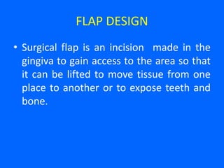
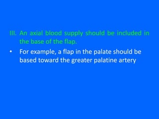
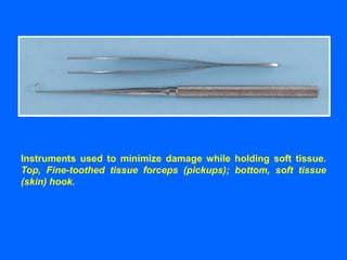
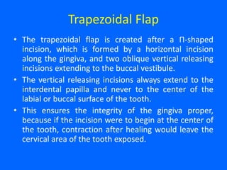
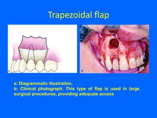
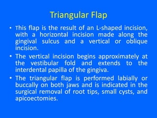
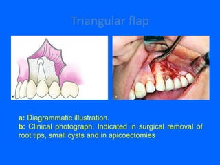
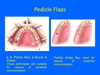
The basic necessities required for oral surgery
includes adequate visibility that depends
upon adequate access, adequate light, and a
surgical field free of excess blood and other
fluids.
Adequate access not only requires the
patient's ability to open the mouth
widely, but it also may require surgically
created exposure.
Retraction of tissues away from the
operative field provides much of the
necessary access.
Improved access is also gained by the
creation of surgical flaps.
Access to the site of operation is gained
by cutting the skin or mucous membrane
and by dissecting through this incision to
lay back a flap.
A few basic principles are important to
remember when performing incisions.
1. A sharp blade of the proper size should
be used.
• A sharp blade allows incisions to be made cleanly,
without unnecessary damage caused by repeated
strokes.
• The rate at which a blade dulls depends on the
resistance of tissues through which the blade cuts.
• Bone and ligamental tissues dull blades more
rapidly than does buccal mucosa.
• Therefore the surgeon should change blades
whenever the knife does not seem to be incising
easily.
2. A firm, continuous stroke should be
used when incising.
• Repeated, tentative strokes increase both
the amount of damaged tissue within a
wound and the amount of bleeding,
thereby impairing wound healing.
• Long, continuous strokes are preferred to
short, interrupted ones.
3. The surgeon should carefully avoid
cutting vital structures when incising.
• No patient's microanatomy is exactly the same. Therefore to
avoid unintentionally cutting large vessels or nerves, the
surgeon must incise only deeply enough to define the next
layer.
• Vessels can be more easily controlled before they are
completely divided, and important nerves can usually be
freed from adjacent tissue and retracted away from the area
to be incised.
• In addition, when using a scalpel the surgeon's focus must
remain on the blade to avoid accidentally cutting structures
such as the lips when introducing and removing the blade to
and from the mouth.
4. incisions through epithelial surfaces
that the surgeon plans to
reapproximate should be made with
the blade held perpendicular to the
epithelial surface.
• This angle produces squared wound
edges that are both easier to reorient
properly during suturing and less
susceptible to necrosis of the wound
edges as a result of ischemia.
5. incisions in the oral cavity should be
properly placed.
• It is more desirable to incise through attached gingiva and
over healthy bone than through unattached gingiva and over
unhealthy or missing bone.
• Properly placed incisions allow the wound margins to be
sutured over intact, healthy bone that is at least a few
millimeters away from the damaged bone, thereby providing
support for the healing wound.
• Incisions placed near the teeth for extractions should be
made in the gingival sulcus, unless the clinician feels it is
necessary to excise the marginal gingiva or to leave the
marginal gingiva untouched.
FLAP DESIGN
• Surgical flap is an incision made in the
gingiva to gain access to the area so that
it can be lifted to move tissue from one
place to another or to expose teeth and
bone.
PRINCIPLES OF FLAP DESIGN
I. The apex (tip) of a flap should never be
wider than the base, unless a major artery is
present in the base.
• Flaps should have sides that either run
parallel to each other or, preferably,
converge moving from the base to the apex
of the flap.
II. The length of the flap should be no
more than twice the width of the base.
III. An axial blood supply should be included in
the base of the flap.
• For example, a flap in the palate should be
based toward the greater palatine artery
IV. The base of flaps should not be excessively
twisted, stretched, or grasped with anything
that might damage vessels, because these
maneuvers can compromise the blood
supply feeding and draining the flap.
A, Principles of flap
design. In general, flap
base dimension (x) must
not be less than height
dimension (y), and
preferably flap should
have x = 2y.
B, When releasing,
incision is used to reflect
a two-sided flap; incision
should be designed to
maximize flap blood
supply by leaving wide
base. Design on left is
correct; design on right is
incorrect.
C, When "buttonhole"
occurs near free edge of
flap, blood supply to flap
tissue on side of hole
away from flap base is
compromised.
V. Flap margin dehiscence (separation) is
prevented by approximating the edges
of the flap over healthy bone, by gently
handling the flap's edges, and by not
placing the flap under tension.
• Dehiscence exposes underlying bone,
producing pain, bone loss, and
increased scarring.
VI. It is preferable to create a flap at the
onset of surgery that is large enough for
the surgeon to avoid either tearing it or
interrupting surgery to enlarge it.
Three types of properly designed oral soft tissue flaps.
A, Horizontal and single vertical incisions used to create two-sided flap.
B, Horizontal and two vertical incisions used to create three-sided flap.
C, Single horizontal incision used to create single-sided (envelope) flap.
VII.The flap itself must be larger than the
bone deficit so that the flap margins,
when sutured, are resting on intact,
healthy bone and not over missing or
unhealthy bone, thus preventing flap
dehiscence and tearing.
TISSUE HANDLING
• The difference between an acceptable and an excellent
surgical outcome often rests on how the surgeon handles the
tissues.
• The use of proper incision and flap design techniques plays a
role; however, tissue also must be handled carefully.
• Excessive pulling or crushing, extremes of temperature,
desiccation, or the use of unphysiologic chemicals easily
damages tissue.
• Therefore, the surgeon should use care whenever touching
tissue. When tissue forceps are used, they should not be
pinched together too tightly; rather, they should be used
delicately to hold tissue.
• When possible, toothed forceps or tissue hooks should be
used to hold tissue.
Instruments used to minimize damage while holding soft tissue.
Top, Fine-toothed tissue forceps (pickups); bottom, soft tissue
(skin) hook.
• In addition, tissues should not be over
aggressively retracted to gain greater surgical
access.
• This includes not pulling excessively to retract
the cheeks or the tongue during surgery.
• When bone is cut, copious amounts of
irrigation should be used to decrease the
amount of bone damage from frictional heat.
• Soft tissue should also be protected from
frictional heat or direct trauma from drilling
equipment.
• Tissue should not be allowed to desiccate; open
wounds should be frequently moistened or
covered with a damp sponge if the surgeon is not
working on them for a while.
• Finally, only physiologic substances should come
in contact with living tissue. For example, tissue
forceps used to place a specimen into formalin
during a biopsy procedure should not be returned
to the wound until any contaminating formalin is
thoroughly removed.
• The surgeon who handles tissue gently and
physiologically is rewarded with grateful patients
whose wounds heal with less frequent
complications.
Types of Flaps
Trapezoidal Flap
• The trapezoidal flap is created after a Π-shaped
incision, which is formed by a horizontal incision
along the gingiva, and two oblique vertical releasing
incisions extending to the buccal vestibule.
• The vertical releasing incisions always extend to the
interdental papilla and never to the center of the
labial or buccal surface of the tooth.
• This ensures the integrity of the gingiva proper,
because if the incision were to begin at the center of
the tooth, contraction after healing would leave the
cervical area of the tooth exposed.
• A satisfactory surgical field is ensured when
the incision extends at least one or two teeth
on either side of the area of bone removal.
• The fact that the base of the resulting flap is
broader than its free gingival margin ensures
the necessary adequate blood supply for the
healing process.
• The trapezoidal flap is suitable for extensive
surgical procedures, especially when the
triangular flap would not provide adequate
access.
Trapezoidal flap
a: Diagrammatic illustration.
b: Clinical photograph. This type of flap is used in large
surgical procedures, providing adequate access
• Advantages. Provides excellent access,
allows surgery to be performed on more
than one or two teeth, produces no
tension in the tissues, allows easy
reapproximation of the flap to its original
position and hastens the healing process.
• Disadvantages. Produces a defect in the
attached gingiva (recession of gingiva).
Triangular Flap
• This flap is the result of an L-shaped incision,
with a horizontal incision made along the
gingival sulcus and a vertical or oblique
incision.
• The vertical incision begins approximately at
the vestibular fold and extends to the
interdental papilla of the gingiva.
• The triangular flap is performed labially or
buccally on both jaws and is indicated in the
surgical removal of root tips, small cysts, and
apicoectomies.
Triangular flap
a: Diagrammatic illustration.
b: Clinical photograph. Indicated in surgical removal of
root tips, small cysts and in apicoectomies
• Advantages: Ensures an adequate blood
supply, satisfactory visualization, very good
stability and reapproximation; it is easily
modified with a small releasing incision, or an
additional vertical incision, or even
lengthening of the horizontal incision.
• Disadvantages: Limited access to long roots,
tension is created when the flap is held with a
retractor, and it causes a defect in the
attached gingiva.
Envelope Flap
• This type of flap is the result of an extended horizontal
incision along the cervical lines of the teeth.
• The incision is made in the gingival sulcus and extends
along four or five teeth. The tissue connected to the
cervical lines of these teeth and the interdental papillae
is thus freed.
• The envelope flap is used for surgery of incisors,
premolars and molars, on the labial or buccal and palatal
or lingual surface, and is usually indicated when the
surgical procedure involves the cervical lines of the teeth
labially (or buccally) and palatally (or lingually),
apicoectomy (palatal root), removal of impacted teeth,
cysts, etc.
Envelope Flap
a: Diagrammatic illustration (buccal). b: Clinical
photograph (palatal).
• Advantages: Avoidance of vertical incision and
easy reapproximation to original position.
• Disadvantages: Difficult reflection (mainly
palatally), great tension with a risk of the ends
tearing, limited visualization in apicoectomies,
limited access, possibility of injury of palatal
vessels and nerves, defect of attached gingiva.
Semilunar Flap
• This flap is the result of a curved incision, which
begins just beneath the vestibular fold and has a
bow-shaped course with the convex part towards
the attached gingiva.
• The lowest point of the incision must be at least
0.5 cm from the gingival margin, so that the
blood supply is not compromised. Each end of
the incision must extend at least one tooth over
on each side of the area of bone removal.
• The semilunar flap is used in apicoectomies and
removal of small cysts and root tips.
Semilunar Flap
a: Diagrammatic illustration. b: Clinical photograph. It is used in
apicoectomies and removal of small cysts and root tips
• Advantages: Small incision and easy
reflection, no recession of gingiva around the
prosthetic restoration, no intervention at the
periodontium, easier oral hygiene compared
to other types of flaps.
• Disadvantages: Possibility of the incision
being performed right over the bone lesion
due to miscalculation, scarring mainly in the
anterior area, difficulty of reapproximation
and suturing due to absence of specific
reference points, limited access and
visualization, tendency to tear.
Flap Resulting from Y-shaped Incision
• An incision is made along the midline of
the palate, as well as two anterolateral
incisions, which are anterior to the
canines.
• This type of flap is indicated in surgical
procedures involving the removal of
small exostoses.
Flap Resulting from shaped Incision
• This type of flap is used in larger exostoses,
and is basically an extension of the Y-shaped
incision.
• The difference is that two more posterolateral
incisions are made, which are necessary for
adequate access to the surgical field.
• This flap is designed such that major branches
of the greater palatine artery are not severed.
a Y-shaped and b -shaped incisions.
Pedicle Flaps
• The three main types of pedicle flaps used for
closure of an oroantral communication are:
buccal, palatal, and bridge flaps.
Pedicle Flaps
a, b. Pedicle flaps. a Buccal. b
Palatal.
These techniques are suitable
for closure of oroantral
communication
Pedicle bridge flap, used for
closure of oroantral
communication

5 incisions

  • 2.
    The basic necessitiesrequired for oral surgery includes adequate visibility that depends upon adequate access, adequate light, and a surgical field free of excess blood and other fluids.
  • 3.
    Adequate access notonly requires the patient's ability to open the mouth widely, but it also may require surgically created exposure. Retraction of tissues away from the operative field provides much of the necessary access. Improved access is also gained by the creation of surgical flaps.
  • 4.
    Access to thesite of operation is gained by cutting the skin or mucous membrane and by dissecting through this incision to lay back a flap. A few basic principles are important to remember when performing incisions.
  • 5.
    1. A sharpblade of the proper size should be used. • A sharp blade allows incisions to be made cleanly, without unnecessary damage caused by repeated strokes. • The rate at which a blade dulls depends on the resistance of tissues through which the blade cuts. • Bone and ligamental tissues dull blades more rapidly than does buccal mucosa. • Therefore the surgeon should change blades whenever the knife does not seem to be incising easily.
  • 6.
    2. A firm,continuous stroke should be used when incising. • Repeated, tentative strokes increase both the amount of damaged tissue within a wound and the amount of bleeding, thereby impairing wound healing. • Long, continuous strokes are preferred to short, interrupted ones.
  • 7.
    3. The surgeonshould carefully avoid cutting vital structures when incising. • No patient's microanatomy is exactly the same. Therefore to avoid unintentionally cutting large vessels or nerves, the surgeon must incise only deeply enough to define the next layer. • Vessels can be more easily controlled before they are completely divided, and important nerves can usually be freed from adjacent tissue and retracted away from the area to be incised. • In addition, when using a scalpel the surgeon's focus must remain on the blade to avoid accidentally cutting structures such as the lips when introducing and removing the blade to and from the mouth.
  • 8.
    4. incisions throughepithelial surfaces that the surgeon plans to reapproximate should be made with the blade held perpendicular to the epithelial surface. • This angle produces squared wound edges that are both easier to reorient properly during suturing and less susceptible to necrosis of the wound edges as a result of ischemia.
  • 9.
    5. incisions inthe oral cavity should be properly placed. • It is more desirable to incise through attached gingiva and over healthy bone than through unattached gingiva and over unhealthy or missing bone. • Properly placed incisions allow the wound margins to be sutured over intact, healthy bone that is at least a few millimeters away from the damaged bone, thereby providing support for the healing wound. • Incisions placed near the teeth for extractions should be made in the gingival sulcus, unless the clinician feels it is necessary to excise the marginal gingiva or to leave the marginal gingiva untouched.
  • 12.
    FLAP DESIGN • Surgicalflap is an incision made in the gingiva to gain access to the area so that it can be lifted to move tissue from one place to another or to expose teeth and bone.
  • 13.
    PRINCIPLES OF FLAPDESIGN I. The apex (tip) of a flap should never be wider than the base, unless a major artery is present in the base. • Flaps should have sides that either run parallel to each other or, preferably, converge moving from the base to the apex of the flap.
  • 14.
    II. The lengthof the flap should be no more than twice the width of the base.
  • 15.
    III. An axialblood supply should be included in the base of the flap. • For example, a flap in the palate should be based toward the greater palatine artery
  • 16.
    IV. The baseof flaps should not be excessively twisted, stretched, or grasped with anything that might damage vessels, because these maneuvers can compromise the blood supply feeding and draining the flap.
  • 17.
    A, Principles offlap design. In general, flap base dimension (x) must not be less than height dimension (y), and preferably flap should have x = 2y. B, When releasing, incision is used to reflect a two-sided flap; incision should be designed to maximize flap blood supply by leaving wide base. Design on left is correct; design on right is incorrect. C, When "buttonhole" occurs near free edge of flap, blood supply to flap tissue on side of hole away from flap base is compromised.
  • 18.
    V. Flap margindehiscence (separation) is prevented by approximating the edges of the flap over healthy bone, by gently handling the flap's edges, and by not placing the flap under tension. • Dehiscence exposes underlying bone, producing pain, bone loss, and increased scarring.
  • 19.
    VI. It ispreferable to create a flap at the onset of surgery that is large enough for the surgeon to avoid either tearing it or interrupting surgery to enlarge it.
  • 20.
    Three types ofproperly designed oral soft tissue flaps. A, Horizontal and single vertical incisions used to create two-sided flap. B, Horizontal and two vertical incisions used to create three-sided flap. C, Single horizontal incision used to create single-sided (envelope) flap.
  • 21.
    VII.The flap itselfmust be larger than the bone deficit so that the flap margins, when sutured, are resting on intact, healthy bone and not over missing or unhealthy bone, thus preventing flap dehiscence and tearing.
  • 22.
  • 23.
    • The differencebetween an acceptable and an excellent surgical outcome often rests on how the surgeon handles the tissues. • The use of proper incision and flap design techniques plays a role; however, tissue also must be handled carefully. • Excessive pulling or crushing, extremes of temperature, desiccation, or the use of unphysiologic chemicals easily damages tissue. • Therefore, the surgeon should use care whenever touching tissue. When tissue forceps are used, they should not be pinched together too tightly; rather, they should be used delicately to hold tissue. • When possible, toothed forceps or tissue hooks should be used to hold tissue.
  • 24.
    Instruments used tominimize damage while holding soft tissue. Top, Fine-toothed tissue forceps (pickups); bottom, soft tissue (skin) hook.
  • 25.
    • In addition,tissues should not be over aggressively retracted to gain greater surgical access. • This includes not pulling excessively to retract the cheeks or the tongue during surgery. • When bone is cut, copious amounts of irrigation should be used to decrease the amount of bone damage from frictional heat. • Soft tissue should also be protected from frictional heat or direct trauma from drilling equipment.
  • 26.
    • Tissue shouldnot be allowed to desiccate; open wounds should be frequently moistened or covered with a damp sponge if the surgeon is not working on them for a while. • Finally, only physiologic substances should come in contact with living tissue. For example, tissue forceps used to place a specimen into formalin during a biopsy procedure should not be returned to the wound until any contaminating formalin is thoroughly removed. • The surgeon who handles tissue gently and physiologically is rewarded with grateful patients whose wounds heal with less frequent complications.
  • 27.
  • 28.
    Trapezoidal Flap • Thetrapezoidal flap is created after a Π-shaped incision, which is formed by a horizontal incision along the gingiva, and two oblique vertical releasing incisions extending to the buccal vestibule. • The vertical releasing incisions always extend to the interdental papilla and never to the center of the labial or buccal surface of the tooth. • This ensures the integrity of the gingiva proper, because if the incision were to begin at the center of the tooth, contraction after healing would leave the cervical area of the tooth exposed.
  • 29.
    • A satisfactorysurgical field is ensured when the incision extends at least one or two teeth on either side of the area of bone removal. • The fact that the base of the resulting flap is broader than its free gingival margin ensures the necessary adequate blood supply for the healing process. • The trapezoidal flap is suitable for extensive surgical procedures, especially when the triangular flap would not provide adequate access.
  • 30.
    Trapezoidal flap a: Diagrammaticillustration. b: Clinical photograph. This type of flap is used in large surgical procedures, providing adequate access
  • 31.
    • Advantages. Providesexcellent access, allows surgery to be performed on more than one or two teeth, produces no tension in the tissues, allows easy reapproximation of the flap to its original position and hastens the healing process. • Disadvantages. Produces a defect in the attached gingiva (recession of gingiva).
  • 32.
    Triangular Flap • Thisflap is the result of an L-shaped incision, with a horizontal incision made along the gingival sulcus and a vertical or oblique incision. • The vertical incision begins approximately at the vestibular fold and extends to the interdental papilla of the gingiva. • The triangular flap is performed labially or buccally on both jaws and is indicated in the surgical removal of root tips, small cysts, and apicoectomies.
  • 33.
    Triangular flap a: Diagrammaticillustration. b: Clinical photograph. Indicated in surgical removal of root tips, small cysts and in apicoectomies
  • 34.
    • Advantages: Ensuresan adequate blood supply, satisfactory visualization, very good stability and reapproximation; it is easily modified with a small releasing incision, or an additional vertical incision, or even lengthening of the horizontal incision. • Disadvantages: Limited access to long roots, tension is created when the flap is held with a retractor, and it causes a defect in the attached gingiva.
  • 35.
    Envelope Flap • Thistype of flap is the result of an extended horizontal incision along the cervical lines of the teeth. • The incision is made in the gingival sulcus and extends along four or five teeth. The tissue connected to the cervical lines of these teeth and the interdental papillae is thus freed. • The envelope flap is used for surgery of incisors, premolars and molars, on the labial or buccal and palatal or lingual surface, and is usually indicated when the surgical procedure involves the cervical lines of the teeth labially (or buccally) and palatally (or lingually), apicoectomy (palatal root), removal of impacted teeth, cysts, etc.
  • 36.
    Envelope Flap a: Diagrammaticillustration (buccal). b: Clinical photograph (palatal).
  • 37.
    • Advantages: Avoidanceof vertical incision and easy reapproximation to original position. • Disadvantages: Difficult reflection (mainly palatally), great tension with a risk of the ends tearing, limited visualization in apicoectomies, limited access, possibility of injury of palatal vessels and nerves, defect of attached gingiva.
  • 38.
    Semilunar Flap • Thisflap is the result of a curved incision, which begins just beneath the vestibular fold and has a bow-shaped course with the convex part towards the attached gingiva. • The lowest point of the incision must be at least 0.5 cm from the gingival margin, so that the blood supply is not compromised. Each end of the incision must extend at least one tooth over on each side of the area of bone removal. • The semilunar flap is used in apicoectomies and removal of small cysts and root tips.
  • 39.
    Semilunar Flap a: Diagrammaticillustration. b: Clinical photograph. It is used in apicoectomies and removal of small cysts and root tips
  • 40.
    • Advantages: Smallincision and easy reflection, no recession of gingiva around the prosthetic restoration, no intervention at the periodontium, easier oral hygiene compared to other types of flaps. • Disadvantages: Possibility of the incision being performed right over the bone lesion due to miscalculation, scarring mainly in the anterior area, difficulty of reapproximation and suturing due to absence of specific reference points, limited access and visualization, tendency to tear.
  • 41.
    Flap Resulting fromY-shaped Incision • An incision is made along the midline of the palate, as well as two anterolateral incisions, which are anterior to the canines. • This type of flap is indicated in surgical procedures involving the removal of small exostoses.
  • 42.
    Flap Resulting fromshaped Incision • This type of flap is used in larger exostoses, and is basically an extension of the Y-shaped incision. • The difference is that two more posterolateral incisions are made, which are necessary for adequate access to the surgical field. • This flap is designed such that major branches of the greater palatine artery are not severed.
  • 43.
    a Y-shaped andb -shaped incisions.
  • 44.
    Pedicle Flaps • Thethree main types of pedicle flaps used for closure of an oroantral communication are: buccal, palatal, and bridge flaps.
  • 45.
    Pedicle Flaps a, b.Pedicle flaps. a Buccal. b Palatal. These techniques are suitable for closure of oroantral communication Pedicle bridge flap, used for closure of oroantral communication