Downloaded 519 times


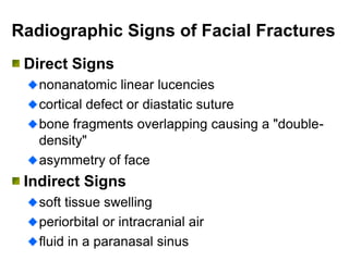


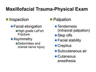


This document discusses various types of facial trauma and fractures. It provides details on examining and evaluating patients with facial trauma, including important physical exam findings. It also reviews specific fractures like orbital fractures, LeFort fractures, mandibular fractures, and nasal bone fractures. Radiographic signs of facial fractures and emergency management of airway and hemorrhage are also covered.


