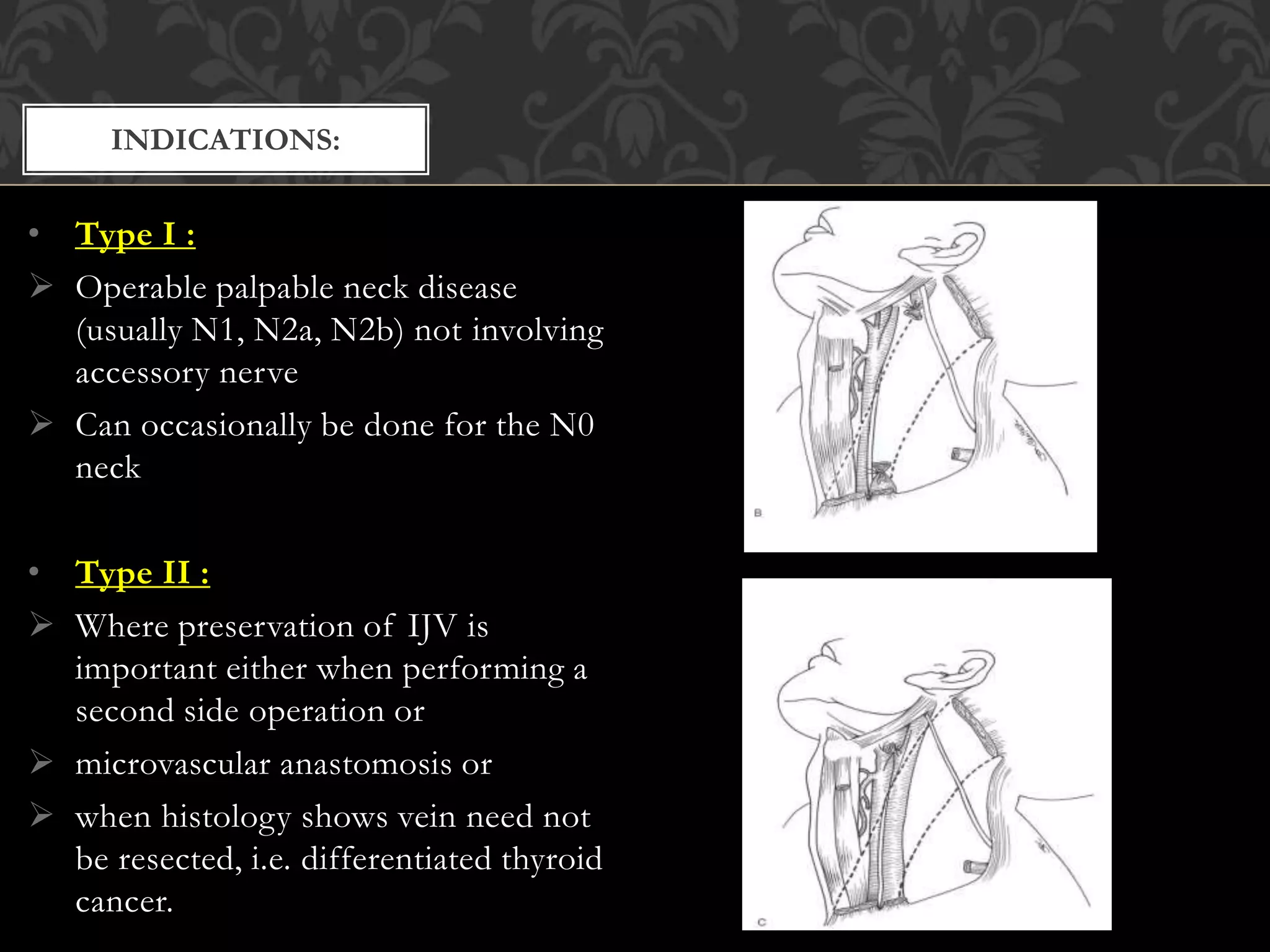
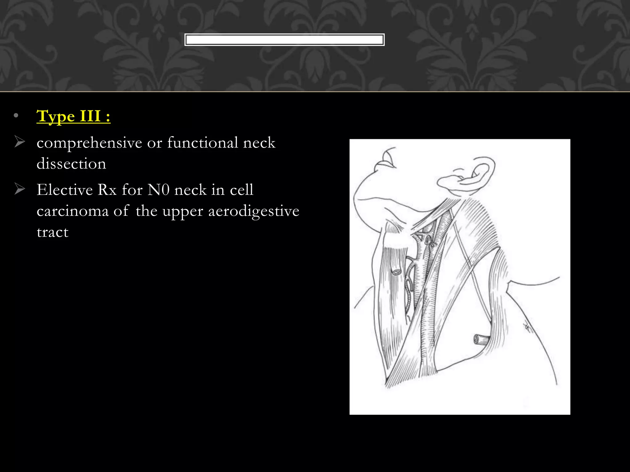
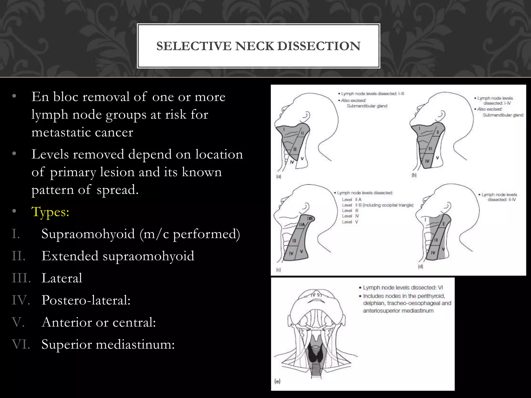
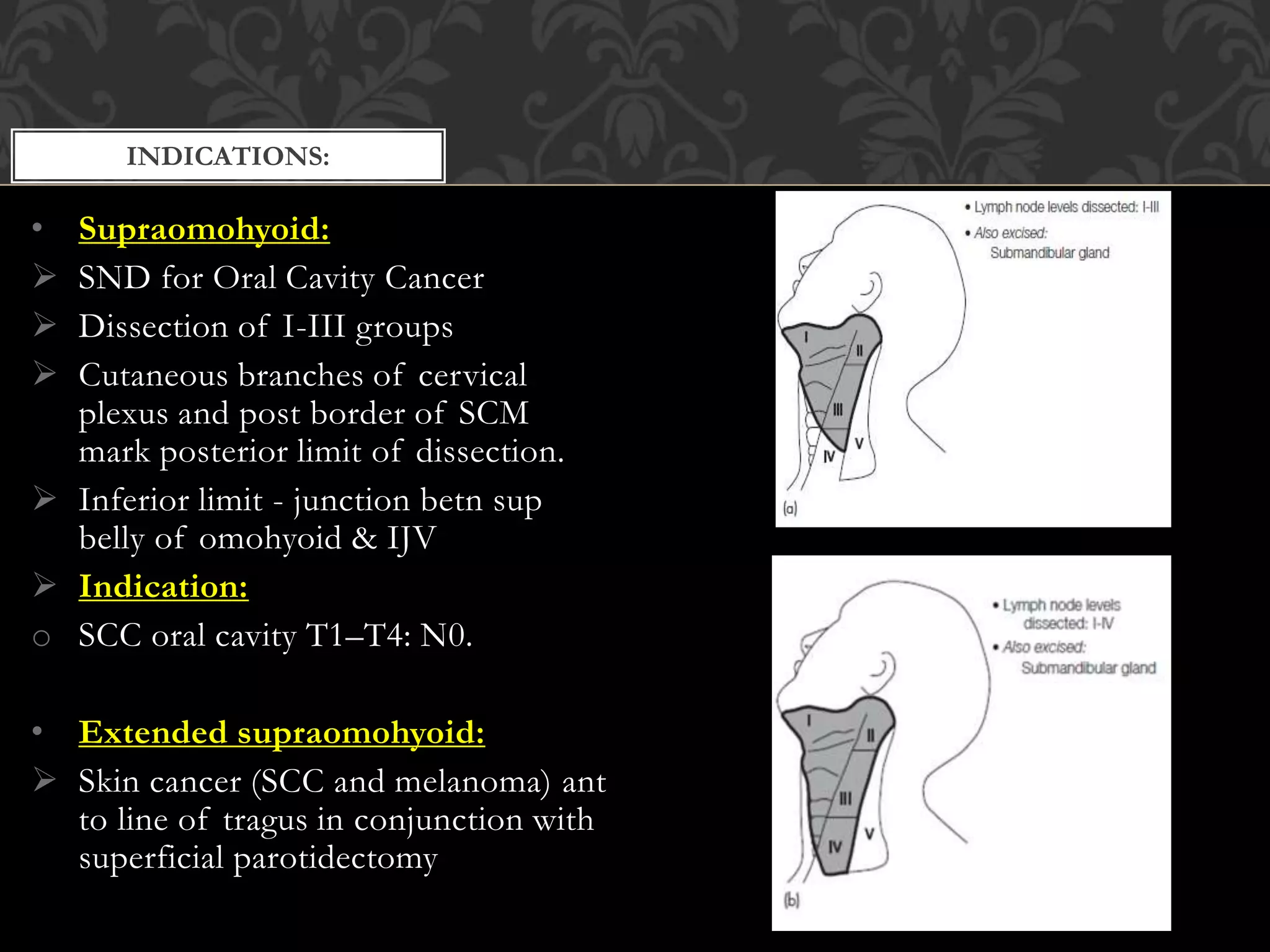
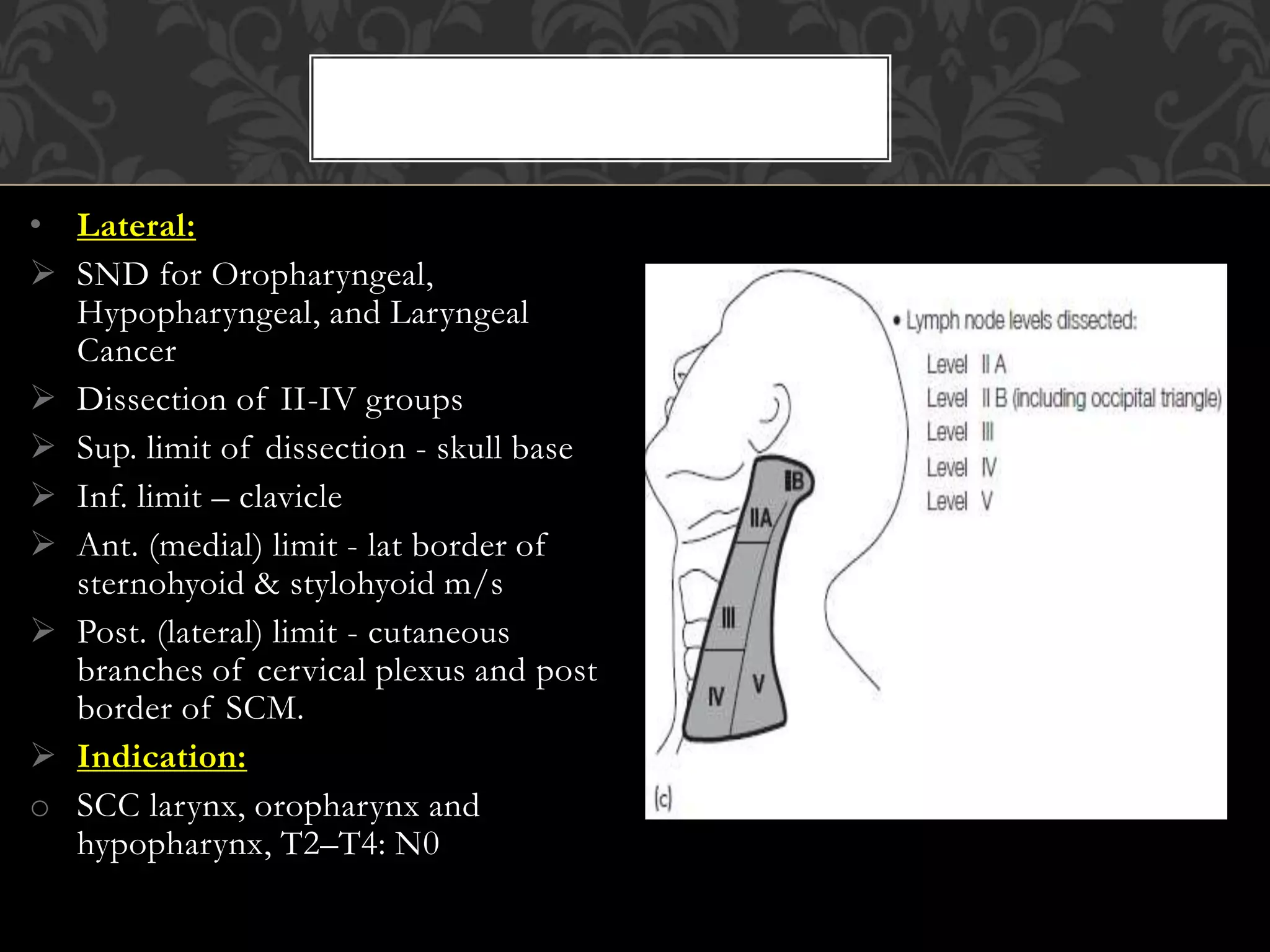
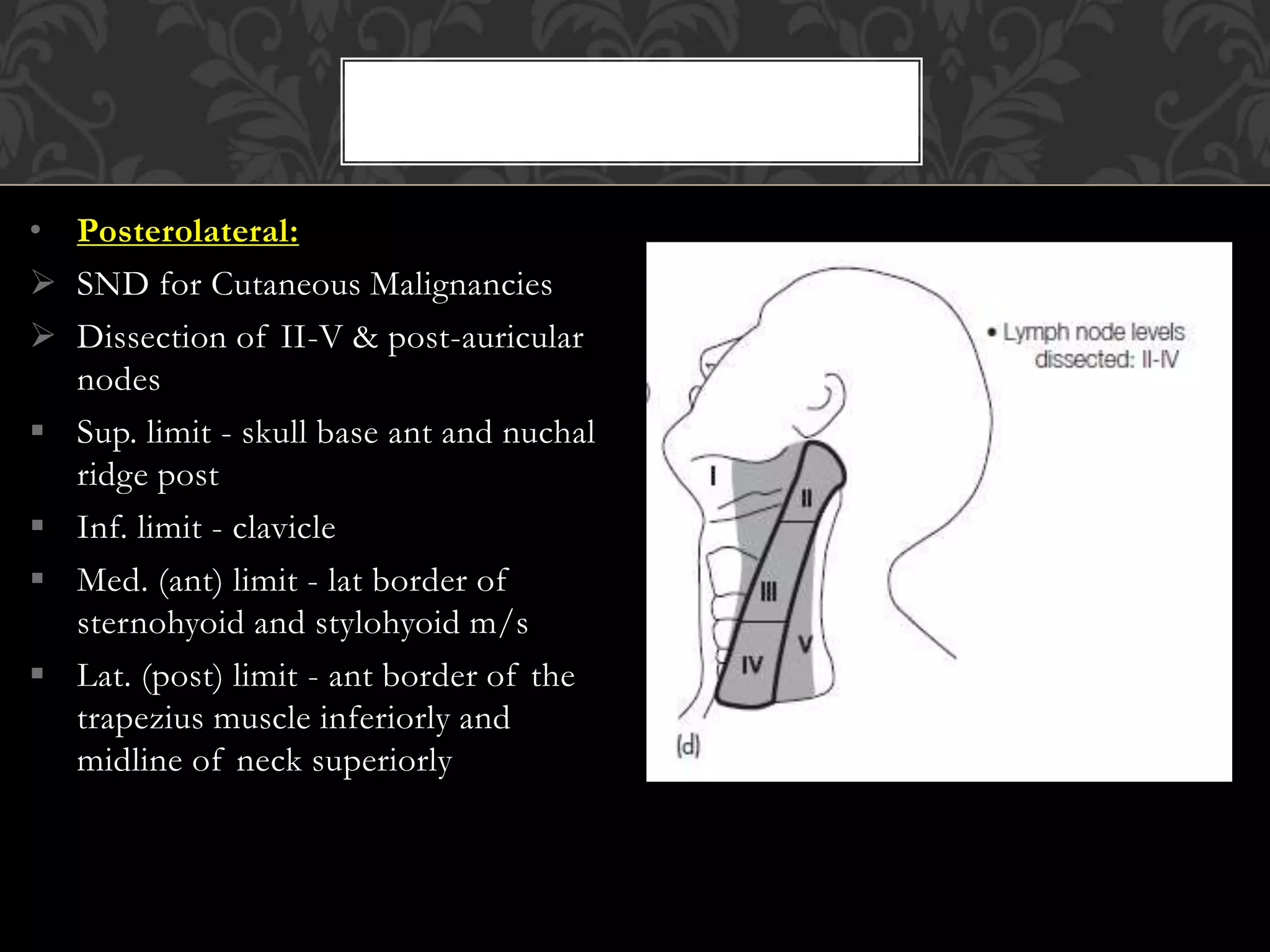
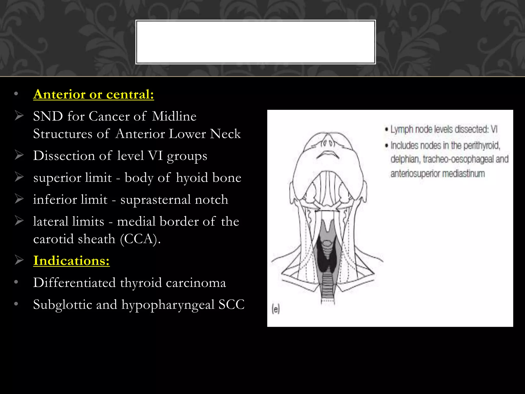
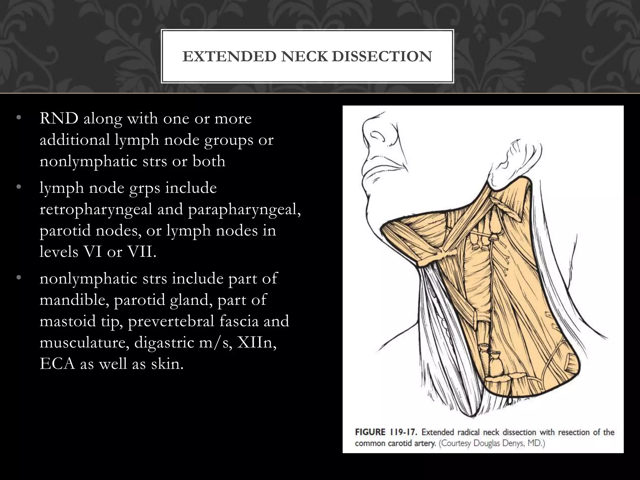
This document provides information about neck dissection procedures. It discusses the history and evolution of neck dissection, defines the different types of neck dissections including radical, modified radical and selective neck dissections, and describes the key steps in performing a modified radical neck dissection. Complications of neck dissection such as hemorrhage, wound infection, and carotid artery rupture are also summarized.
Maxillectomy and craniofacial resection Mamoon Ameen
all maxillectomy types in detail and maxillofacial resection ,indications ,contraindications ,preoperative asssessment and detail techniques and rehabilitations
This powerpoint describes the types of maxillectomy & operative steps for total maxillectomy. It also enumerates various flaps used for reconstruction of maxillectomy defect.
New Directions in Targeted Therapeutic Approaches for Older Adults With Mantl...i3 Health
i3 Health is pleased to make the speaker slides from this activity available for use as a non-accredited self-study or teaching resource.
This slide deck presented by Dr. Kami Maddocks, Professor-Clinical in the Division of Hematology and
Associate Division Director for Ambulatory Operations
The Ohio State University Comprehensive Cancer Center, will provide insight into new directions in targeted therapeutic approaches for older adults with mantle cell lymphoma.
STATEMENT OF NEED
Mantle cell lymphoma (MCL) is a rare, aggressive B-cell non-Hodgkin lymphoma (NHL) accounting for 5% to 7% of all lymphomas. Its prognosis ranges from indolent disease that does not require treatment for years to very aggressive disease, which is associated with poor survival (Silkenstedt et al, 2021). Typically, MCL is diagnosed at advanced stage and in older patients who cannot tolerate intensive therapy (NCCN, 2022). Although recent advances have slightly increased remission rates, recurrence and relapse remain very common, leading to a median overall survival between 3 and 6 years (LLS, 2021). Though there are several effective options, progress is still needed towards establishing an accepted frontline approach for MCL (Castellino et al, 2022). Treatment selection and management of MCL are complicated by the heterogeneity of prognosis, advanced age and comorbidities of patients, and lack of an established standard approach for treatment, making it vital that clinicians be familiar with the latest research and advances in this area. In this activity chaired by Michael Wang, MD, Professor in the Department of Lymphoma & Myeloma at MD Anderson Cancer Center, expert faculty will discuss prognostic factors informing treatment, the promising results of recent trials in new therapeutic approaches, and the implications of treatment resistance in therapeutic selection for MCL.
Target Audience
Hematology/oncology fellows, attending faculty, and other health care professionals involved in the treatment of patients with mantle cell lymphoma (MCL).
Learning Objectives
1.) Identify clinical and biological prognostic factors that can guide treatment decision making for older adults with MCL
2.) Evaluate emerging data on targeted therapeutic approaches for treatment-naive and relapsed/refractory MCL and their applicability to older adults
3.) Assess mechanisms of resistance to targeted therapies for MCL and their implications for treatment selection
Ethanol (CH3CH2OH), or beverage alcohol, is a two-carbon alcohol
that is rapidly distributed in the body and brain. Ethanol alters many
neurochemical systems and has rewarding and addictive properties. It
is the oldest recreational drug and likely contributes to more morbidity,
mortality, and public health costs than all illicit drugs combined. The
5th edition of the Diagnostic and Statistical Manual of Mental Disorders
(DSM-5) integrates alcohol abuse and alcohol dependence into a single
disorder called alcohol use disorder (AUD), with mild, moderate,
and severe subclassifications (American Psychiatric Association, 2013).
In the DSM-5, all types of substance abuse and dependence have been
combined into a single substance use disorder (SUD) on a continuum
from mild to severe. A diagnosis of AUD requires that at least two of
the 11 DSM-5 behaviors be present within a 12-month period (mild
AUD: 2–3 criteria; moderate AUD: 4–5 criteria; severe AUD: 6–11 criteria).
The four main behavioral effects of AUD are impaired control over
drinking, negative social consequences, risky use, and altered physiological
effects (tolerance, withdrawal). This chapter presents an overview
of the prevalence and harmful consequences of AUD in the U.S.,
the systemic nature of the disease, neurocircuitry and stages of AUD,
comorbidities, fetal alcohol spectrum disorders, genetic risk factors, and
pharmacotherapies for AUD.
Title: Sense of Taste
Presenter: Dr. Faiza, Assistant Professor of Physiology
Qualifications:
MBBS (Best Graduate, AIMC Lahore)
FCPS Physiology
ICMT, CHPE, DHPE (STMU)
MPH (GC University, Faisalabad)
MBA (Virtual University of Pakistan)
Learning Objectives:
Describe the structure and function of taste buds.
Describe the relationship between the taste threshold and taste index of common substances.
Explain the chemical basis and signal transduction of taste perception for each type of primary taste sensation.
Recognize different abnormalities of taste perception and their causes.
Key Topics:
Significance of Taste Sensation:
Differentiation between pleasant and harmful food
Influence on behavior
Selection of food based on metabolic needs
Receptors of Taste:
Taste buds on the tongue
Influence of sense of smell, texture of food, and pain stimulation (e.g., by pepper)
Primary and Secondary Taste Sensations:
Primary taste sensations: Sweet, Sour, Salty, Bitter, Umami
Chemical basis and signal transduction mechanisms for each taste
Taste Threshold and Index:
Taste threshold values for Sweet (sucrose), Salty (NaCl), Sour (HCl), and Bitter (Quinine)
Taste index relationship: Inversely proportional to taste threshold
Taste Blindness:
Inability to taste certain substances, particularly thiourea compounds
Example: Phenylthiocarbamide
Structure and Function of Taste Buds:
Composition: Epithelial cells, Sustentacular/Supporting cells, Taste cells, Basal cells
Features: Taste pores, Taste hairs/microvilli, and Taste nerve fibers
Location of Taste Buds:
Found in papillae of the tongue (Fungiform, Circumvallate, Foliate)
Also present on the palate, tonsillar pillars, epiglottis, and proximal esophagus
Mechanism of Taste Stimulation:
Interaction of taste substances with receptors on microvilli
Signal transduction pathways for Umami, Sweet, Bitter, Sour, and Salty tastes
Taste Sensitivity and Adaptation:
Decrease in sensitivity with age
Rapid adaptation of taste sensation
Role of Saliva in Taste:
Dissolution of tastants to reach receptors
Washing away the stimulus
Taste Preferences and Aversions:
Mechanisms behind taste preference and aversion
Influence of receptors and neural pathways
Impact of Sensory Nerve Damage:
Degeneration of taste buds if the sensory nerve fiber is cut
Abnormalities of Taste Detection:
Conditions: Ageusia, Hypogeusia, Dysgeusia (parageusia)
Causes: Nerve damage, neurological disorders, infections, poor oral hygiene, adverse drug effects, deficiencies, aging, tobacco use, altered neurotransmitter levels
Neurotransmitters and Taste Threshold:
Effects of serotonin (5-HT) and norepinephrine (NE) on taste sensitivity
Supertasters:
25% of the population with heightened sensitivity to taste, especially bitterness
Increased number of fungiform papillae
Pulmonary Thromboembolism - etilogy, types, medical- Surgical and nursing man...VarunMahajani
Disruption of blood supply to lung alveoli due to blockage of one or more pulmonary blood vessels is called as Pulmonary thromboembolism. In this presentation we will discuss its causes, types and its management in depth.
Couples presenting to the infertility clinic- Do they really have infertility...Sujoy Dasgupta
Dr Sujoy Dasgupta presented the study on "Couples presenting to the infertility clinic- Do they really have infertility? – The unexplored stories of non-consummation" in the 13th Congress of the Asia Pacific Initiative on Reproduction (ASPIRE 2024) at Manila on 24 May, 2024.
Acute scrotum is a general term referring to an emergency condition affecting the contents or the wall of the scrotum.
There are a number of conditions that present acutely, predominantly with pain and/or swelling
A careful and detailed history and examination, and in some cases, investigations allow differentiation between these diagnoses. A prompt diagnosis is essential as the patient may require urgent surgical intervention
Testicular torsion refers to twisting of the spermatic cord, causing ischaemia of the testicle.
Testicular torsion results from inadequate fixation of the testis to the tunica vaginalis producing ischemia from reduced arterial inflow and venous outflow obstruction.
The prevalence of testicular torsion in adult patients hospitalized with acute scrotal pain is approximately 25 to 50 percent
Recomendações da OMS sobre cuidados maternos e neonatais para uma experiência pós-natal positiva.
Em consonância com os ODS – Objetivos do Desenvolvimento Sustentável e a Estratégia Global para a Saúde das Mulheres, Crianças e Adolescentes, e aplicando uma abordagem baseada nos direitos humanos, os esforços de cuidados pós-natais devem expandir-se para além da cobertura e da simples sobrevivência, de modo a incluir cuidados de qualidade.
Estas diretrizes visam melhorar a qualidade dos cuidados pós-natais essenciais e de rotina prestados às mulheres e aos recém-nascidos, com o objetivo final de melhorar a saúde e o bem-estar materno e neonatal.
Uma “experiência pós-natal positiva” é um resultado importante para todas as mulheres que dão à luz e para os seus recém-nascidos, estabelecendo as bases para a melhoria da saúde e do bem-estar a curto e longo prazo. Uma experiência pós-natal positiva é definida como aquela em que as mulheres, pessoas que gestam, os recém-nascidos, os casais, os pais, os cuidadores e as famílias recebem informação consistente, garantia e apoio de profissionais de saúde motivados; e onde um sistema de saúde flexível e com recursos reconheça as necessidades das mulheres e dos bebês e respeite o seu contexto cultural.
Estas diretrizes consolidadas apresentam algumas recomendações novas e já bem fundamentadas sobre cuidados pós-natais de rotina para mulheres e neonatos que recebem cuidados no pós-parto em unidades de saúde ou na comunidade, independentemente dos recursos disponíveis.
É fornecido um conjunto abrangente de recomendações para cuidados durante o período puerperal, com ênfase nos cuidados essenciais que todas as mulheres e recém-nascidos devem receber, e com a devida atenção à qualidade dos cuidados; isto é, a entrega e a experiência do cuidado recebido. Estas diretrizes atualizam e ampliam as recomendações da OMS de 2014 sobre cuidados pós-natais da mãe e do recém-nascido e complementam as atuais diretrizes da OMS sobre a gestão de complicações pós-natais.
O estabelecimento da amamentação e o manejo das principais intercorrências é contemplada.
Recomendamos muito.
Vamos discutir essas recomendações no nosso curso de pós-graduação em Aleitamento no Instituto Ciclos.
Esta publicação só está disponível em inglês até o momento.
Prof. Marcus Renato de Carvalho
www.agostodourado.com
These lecture slides, by Dr Sidra Arshad, offer a quick overview of physiological basis of a normal electrocardiogram.
Learning objectives:
1. Define an electrocardiogram (ECG) and electrocardiography
2. Describe how dipoles generated by the heart produce the waveforms of the ECG
3. Describe the components of a normal electrocardiogram of a typical bipolar leads (limb II)
4. Differentiate between intervals and segments
5. Enlist some common indications for obtaining an ECG
Study Resources:
1. Chapter 11, Guyton and Hall Textbook of Medical Physiology, 14th edition
2. Chapter 9, Human Physiology - From Cells to Systems, Lauralee Sherwood, 9th edition
3. Chapter 29, Ganong’s Review of Medical Physiology, 26th edition
4. Electrocardiogram, StatPearls - https://www.ncbi.nlm.nih.gov/books/NBK549803/
5. ECG in Medical Practice by ABM Abdullah, 4th edition
6. ECG Basics, http://www.nataliescasebook.com/tag/e-c-g-basics
2. • Systematic removal of lymph nodes, along with their surrounding
fibrofatty tissue, from various compartments of neck
• Aim : to remove neck lymph nodes into which cancer cells may have
migrated
• Metastases may originate from tumours of oral cavity, tongue,
nasopharynx, oropharynx, hypopharynx, and larynx, as well as the thyroid,
parotid and posterior scalp.
INTRODUCTION:
3. • Therapeutic neck dissection is used when metastatic cervical
lymphadenopathy is clinically evident.
• Elective neck dissection is used to remove lymph node groups in pts
who have clinically node-negative disease and who have increased risk of
harboring occult disease in neck
• Salvage neck dissection is done when metastatic disease is clinically
evident in the neck after previous treatment
4. • 1888 - Jawdynski described en bloc resection with resection of carotid,
internal jugular vein and sternocleidomastoid muscle.
• 1906 - George W. Crile of the Cleveland Clinic describes radical neck
dissection.
• 1957 - Hayes Martin describes routine use of radical neck dissection for
control of neck metastases.
• 1967 - Oscar Suarez and E. Bocca describe a more conservative operation
which preserves SAN, IJV and SCM.
• Last 3 decades - Further operations have been described to selectively
remove the involved regional lymph groups.
HISTORY OF NECK DISSECTION:
6. • Suggested by Suen and Goepfert (1997)
• Biologic significance for lymphatic drainage depending on site of tumor
IMPORTANCE OF SUBDIVISIONS:
7. “N” classification – AJC (1997)
NX: Regional lymph nodes cannot be
assessed
N0: No regional lymph node
metastasis
N1: Metastasis in single ipsilateral
lymph node, 3 cm or less in greatest
dimension
N2a: single ipsilateral lymph node >3
cm but <6 cm in greatest dimension
N2b: multiple ipsilateral lymph nodes,
none >6 cm
N2c: bilateral or contralateral nodes,
<6 cm
N3: lymph node >6 cm
STAGING OF HEAD AND NECK
CANCER
8. • Thyroid and nasopharynx have different staging based on tumor behavior
and prognosis
• Staging in nasopharyngeal cancer
N1: unilateral
N2: bilateral (Both are above supraclavicular fossa & < 6 cm)
N3: > 6 cm or in supraclavicular fossa
• Staging in thyroid cancer
N1a: ipsilateral
N1b: midline / bilateral / contralateral
9.
10. Principles of Classification
• RND: standard basic procedure for
cervical lymphadenectomy, all other
represent one or more modifications
• MRND: When modification of RND
involves preservation of one or more
non-lymphatic strs.
• SND: When modification involves
one or more lymph node groups that
are routinely removed in RND.
• Extended RND: When modification
involves removal of additional lymph
node groups or non-lymphatic
structures relative to RND.
CLASSIFICATION OF NECK
DISSECTIONS
11. • Removal of all ipsilateral cervical
lymph node groups that extend from
Body of mandible superiorly to
Clavicle inferiorly and from
Contralateral anterior belly of
digastric & lat border of strap
muscles anteriorly to
Ant border of trapezius posteriorly
RADICAL NECK DISSECTION
12. • extensive lymph node metastases
with extension beyond capsule of
node or nodes that involves SAN
and IJV.
• Untreatable primary tumor
• Unfit form major surgery
• Distant metastasis
• Significant b/l neck diseases
INDICATIONS: CONTRA-INDICATIONS:
13. • En bloc removal of lymph node–
bearing tissue from one side of the
neck (I-V)
• Unlike RND, it preserves SAN, IJV,
and/or SCM
• TYPES:
I. Type I preserves SAN
II. Type II preserves SAN & IJV
III. Type III preserves SAN, IJV &
SCM
MODIFIED RADICAL NECK
DISSECTION:
14. • Type I :
Operable palpable neck disease
(usually N1, N2a, N2b) not involving
accessory nerve
Can occasionally be done for the N0
neck
• Type II :
Where preservation of IJV is
important either when performing a
second side operation or
microvascular anastomosis or
when histology shows vein need not
be resected, i.e. differentiated thyroid
cancer.
INDICATIONS:
15. • Type III :
comprehensive or functional neck
dissection
Elective Rx for N0 neck in cell
carcinoma of the upper aerodigestive
tract
16. • Reduce postsurgical shoulder pain and shoulder dysfunction
• Improve cosmetic outcome
• Reduce likelihood of bilateral IJV resection
BENEFIT OF MRND:
17. • En bloc removal of one or more
lymph node groups at risk for
metastatic cancer
• Levels removed depend on location
of primary lesion and its known
pattern of spread.
• Types:
I. Supraomohyoid (m/c performed)
II. Extended supraomohyoid
III. Lateral
IV. Postero-lateral:
V. Anterior or central:
VI. Superior mediastinum:
SELECTIVE NECK DISSECTION
18. • Supraomohyoid:
SND for Oral Cavity Cancer
Dissection of I-III groups
Cutaneous branches of cervical
plexus and post border of SCM
mark posterior limit of dissection.
Inferior limit - junction betn sup
belly of omohyoid & IJV
Indication:
o SCC oral cavity T1–T4: N0.
• Extended supraomohyoid:
Skin cancer (SCC and melanoma) ant
to line of tragus in conjunction with
superficial parotidectomy
INDICATIONS:
19. • Lateral:
SND for Oropharyngeal,
Hypopharyngeal, and Laryngeal
Cancer
Dissection of II-IV groups
Sup. limit of dissection - skull base
Inf. limit – clavicle
Ant. (medial) limit - lat border of
sternohyoid & stylohyoid m/s
Post. (lateral) limit - cutaneous
branches of cervical plexus and post
border of SCM.
Indication:
o SCC larynx, oropharynx and
hypopharynx, T2–T4: N0
20. • Posterolateral:
SND for Cutaneous Malignancies
Dissection of II-V & post-auricular
nodes
Sup. limit - skull base ant and nuchal
ridge post
Inf. limit - clavicle
Med. (ant) limit - lat border of
sternohyoid and stylohyoid m/s
Lat. (post) limit - ant border of the
trapezius muscle inferiorly and
midline of neck superiorly
21. • Anterior or central:
SND for Cancer of Midline
Structures of Anterior Lower Neck
Dissection of level VI groups
superior limit - body of hyoid bone
inferior limit - suprasternal notch
lateral limits - medial border of the
carotid sheath (CCA).
Indications:
• Differentiated thyroid carcinoma
• Subglottic and hypopharyngeal SCC
22. • Sup. Mediastinum:
Differentiated and medullary thyroid
carcinoma
Subglottic laryngeal and
hypopharyngeal SCC
Cervical oesophageal carcinoma
23. • RND along with one or more
additional lymph node groups or
nonlymphatic strs or both
• lymph node grps include
retropharyngeal and parapharyngeal,
parotid nodes, or lymph nodes in
levels VI or VII.
• nonlymphatic strs include part of
mandible, parotid gland, part of
mastoid tip, prevertebral fascia and
musculature, digastric m/s, XIIn,
ECA as well as skin.
EXTENDED NECK DISSECTION
24. • compartmental removal of lymph
nodes limited to one or two
contiguous neck levels
• INDICATION:
removal of lymph node disease as/w
supraglottic cancer
residual disease following
chemoradiation that is confined to a
single level
SUPERSELECTIVE NECK
DISSECTION
25. • lymphoscintigraphy and sentinel
lymph node biopsy (SLNB)
• powerful adjunct to surgical
treatment
• minimally invasive, can accurately
stage clinically occult neck
LYMPHOSCINTIGRAPHY-DIRECTED
NECK DISSECTION
26. • Position:
Supine
Roll placed beneath shoulders to
optimally extend neck.
Skin is prepped and draped to allow
full exposure of both sides of neck
with clear visualization of
surrounding landmarks
TECHNIQUE:
27. • Optimal exposure of all lymph node
levels to be dissected (I -V)
• Preserve as much blood supply as
possible
• Flaps raised should be broadly
based, sup or inf
• Should avoid any trifurcations,
particularly those that overlie carotid
sheath
• Incisions that fit these criteria
Hockey stick
Boomerang
McFee incision
Apron incision (b/l ND)
INCISION:
28. Y type (or Crile)
Schobinger incision
Modified Schobinger incision
horizontal-T (Hetter) incision
Utility incision
DIFFERENT INCISIONS:
29. • Raised in subplatysmal plane
• Major corners of consternation:
Lower end of internal jugular vein.
Junction of lateral border of clavicle
with lower edge of trapezius.
Upper end of internal jugular vein.
Submandibular triangle.
• Minor corners of consternation:
Retropharyngeal nodes.
Parapharyngeal nodes.
Chaissaignac’s triangle.
RAISING THE FLAP:
30. • Step 1:
incision is made through skin, subcutaneous fat, and platysma muscle
superior flap is elevated
submandibular gland fascia is then incised
Resection of fat and lymph nodes from submental triangle (Level Ia)
submental triangle is resected inferiorly to hyoid bone with electro-cautery.
Deep plane of dissection is mylohyoid muscles
OPERATIVE STEPS FOR MRND:
31.
32.
33. • Step 2:
addresses Level Ib
submandibular gland capsule is
dissected from gland in a superior
direction in a subcapsular plane
Resection of fat and lymph nodes
tucked anteriorly and deeply between
ant belly of digastric & mylohyoid
m/s
34. Facial artery and vein are identified by blunt dissection with a fine haemostat
Facial lymph nodes; if present, are dissected
Divided and tied close to submandibular gland so as not to injure marginal
mandibular nerve
This frees up gland superiorly, which can then be reflected away from
mandible
addresses the lingual nerve, submandibular duct, and XIIn
35.
36.
37. • Step 3:
fascia along lateral aspect of
digastric divided
EJV divided
post belly of digastric exposed along
its entire length
Identification of XIIn deep to veins
that cross nerve
Sternomastoid branch of occipital
artery that tethers XIIn identified
Dividing this artery releases XIIn
Then courses vertically and leads
directly to ant border of IJV
38.
39.
40.
41. • Step 4:
fatty tissue in Level II dissected
XIn which may course lateral,
medial or very rarely through IJV
identified
transverse process of C1 vertebra
can be palpated immediately post
to XIn and IJV
42. • Step 5:
directed at anterior neck
anteriorly based subplatysmal flap
raised
exposing omohyoid and SCM
muscle inferiorly down the clavicle
anterior jugular vein left in elevated
flap
Omohyoid divided and levels II, III
cleared
43. • Step 6:
Posteriorly-based flap elevated
Platysma is often absent posteriorly
hence flap may be very thin
Dissection continues until ant
border of trapezius is reached
44. • Step 7:
dissecting out XIn and mobilizing Level IIb
XIn is identified by dissecting at post border of SCM, approx 1-2cm post to
point where greater auricular nerve curves around m/s
Once XIn exposed and freed from IJV, it is exposed distally to where it
disappears behind trapezius m/s
Freed completely and branches sectioned to SCM
45.
46.
47. • Step 8:
dissection of Level IIb and
transposition of the XIn
SCM is divided below mastoid.
exposing fat at top of Level IIb
dissection is carried deeper until
deep muscles of neck that run in a
posteroinferior direction appear
dissection is then directed postero-
inferiorly, where greater occipital
nerve (C1) is divided
Level IIb and IIa are then dissected
off
XIn is now trans-located posteriorly
48.
49.
50. • Step 9:
clavicular and sternal heads of
SCM divided
not to dissect immediately lateral to
IJV, as right lymphatic duct (right
neck) or thoracic duct (left neck)
may be injured; chyle leak
EJV is divided and ligated and
omohyoid divided
Supraclavicular fat exposed.
Brachial plexus, phrenic nerve &
transverse cervical vessels identified
51.
52.
53.
54. • Step 10:
freeing inferolateral part of Level V
Identifying and dividing
supraclavicular nerves
Incision of fatty vascular pedicle
containing transverse cervical artery
and vein
isolation and division of transverse
cervical artery and its proximity to
XIn.
55.
56. • Step 11:
anterograde dissection of Levels II
-V with scalpel
dissection proceeds over a broad
front until entire cervical plexus has
been exposed
cervical plexus nerves are each
divided, taking care not to injure
phrenic nerve
This brings carotid sheath into view
carotid sheath is incised along full
course of vagus nerve, and neck
dissection specimen is stripped off
the IJV
57.
58.
59.
60. • Step 12:
final step is to:
strip neck dissection specimen off
infrahyoid strap muscles
to identify and preserve superior
thyroid vascular pedicle, and
to deliver neck dissection specimen
• Closure:
61. • HAEMORRHAGE:
perioperative or postoperative
Damage to IJV at its upper or lower end before it has been ligated
Secondary haemorrhage may occur as a result of carotid artery rupture
• WOUND INFECTION:
four most important factors
1. Contamination of surgical field.
2. Contamination of surgical field as
operation involves in-continuity RND and primary excision
3. Postoperative haematoma which then becomes infected.
4. Flap necrosis and wound breakdown.
COMPLICATIONS
62. • CAROTID ARTERY RUPTURE:
Following necrosis of arterial wall d/to infection
preoperative radiotherapy is implicated in most series
• CHYLOUS FISTULA:
More usually, a leak of fluid occurs when lower end of jugular vein is
being dissected
Mild leak, i.e. < 100mL/day: conservative management
Major leak: re-explore wound to identify source of leak and oversew
it
63. • PNEUMOTHORAX
disease lower neck, apical pleura may be damaged during dissection
• NERVE INJURIES:
standard radical neck dissection the nerves which are deliberately divided are:
accessory nerve;
branches of the cervical plexus.
descendens hypoglossi
Other nerves that may be damaged by accident include:
facial nerve or its mandibular or cervical division;
hypoglossal and lingual nerves;
vagus, symphathetic trunk, phrenic nerve or brachial plexus.