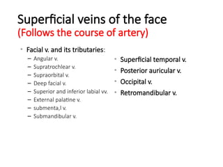
Superficial veins ofthe face
(Follows the course of artery)
• Facial v. and its tributaries:
– Angular v.
– Supratrochlear v.
– Supraorbital v.
– Deep facial v.
– Superior and inferior labial vv.
– External palatine v.
– submenta,l v.
– Submandibular v.
• Superficial temporal v.
• Posterior auricular v.
• Occipital v.
• Retromandibular v.
13.
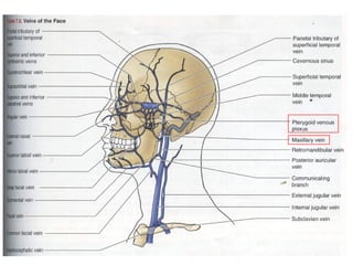
Deep veins ofthe face
• Maxillary v.
• Pterygoid venous plexus
• In the infratemporal fossa
• Between the temporal and lateral pterygoid muscles
15.
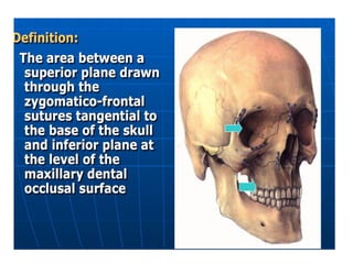
Definition
• Maxillofacial trauma@ Facial trauma
• Defined as any physical trauma to the face including
• Soft tissue injuries (burns, lacerations and bruises)
• Fractures of the facial bones (nasal fractures, fractures
of the jaw)
• Trauma such as eye injuries
16.
Aetiology
• Assaults (#1in developed country)
• MVA – 70% d/t speeding (#1 in developing country)
• Falls
• Sport injury
• Industrial injuries
• Animal bites
• Burns
• War injuries
Primary Survey
• A- airway with cervical spine protection
• B - breathing
• C - circulation with haemorrhage control
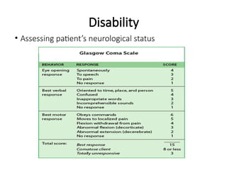
• D - disability or neurological status
• E - exposure and environment
19.
Secure the airway
Look,listen and feel for any signs indicating obstruction:
1) respiratory distress
2) decreased conscious level
3) noisy breathing/stridor
20.
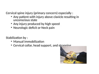
Cervical spine injury(primary concern) especially :
• Any patient with injury above clavicle resulting in
unconscious state
• Any injury produced by high speed
• Neurologic deficit or Neck pain
Stabilization by :
• Manual immobilization
• Cervical collar, head support, and strapping
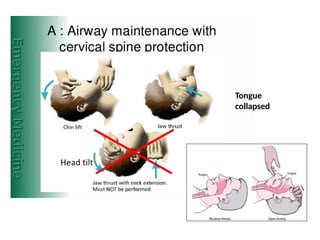
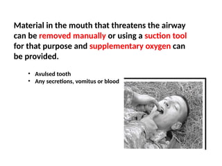
Material in themouth that threatens the airway
can be removed manually or using a suction tool
for that purpose and supplementary oxygen can
be provided.
• Avulsed tooth
• Any secretions, vomitus or blood
23.
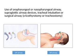
Use of oropharyngealor nasopharyngeal airway,
supraglottic airway devices, tracheal intubation or
surgical airway (cricothyrotomy or tracheostomy)
OP airway NP airway
24.
Breathing
• Only assessbreathing after airway is secured
• Examined the chest:
1. Inspection: adequate and symmetrical excursion,
bruising, open wound, tachypnea
2. palpation: position of trachea
3. percussion: hyper-resonance/ dullness
4. auscultation: abnormal/ absent breath sound
25.
Circulation with Haemorrhage
Control
•Assess by looking for external bleeding
• Visible signs of shock:
1. pallor
2. prolonged capillary refill
3. clammy and cool skin
4. tachycardia
5. diminished/ absent pulse pressure
26.
• External bleedingis controlled by direct pressure
• Two cannulae sited for administration of fluid and
blood
• Blood sample can be drawn for baseline diagnostic
tests and transfusion cross-matching
• Crystalloid intravenous fluid can be given to maintain
the cardiac output
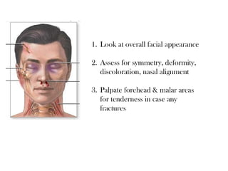
1. Look atoverall facial appearance
2. Assess for symmetry, deformity,
discoloration, nasal alignment
3. Palpate forehead & malar areas
for tenderness in case any
fractures
Ears
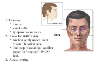
1. Examine
• Pinnae
•canal walls
• tympanic membranes
2. Look for Battle’s sign
• Suction gently under direct
vision if blood in canal
• Put drop of canal fluid on filter
paper for “ring sign” CSF
leak
3. Assess hearing
33.
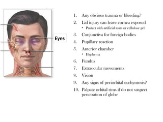
1. Any obvioustrauma or bleeding?
2. Lid injury can leave cornea exposed
• Protect with artificial tears or cellulose gel
3. Conjunctiva for foreign bodies
4. Pupillary reaction
5. Anterior chamber
• Hyphema
6. Fundus
7. Extraocular movements
8. Vision
9. Any signs of periorbital ecchymosis?
10. Palpate orbital rims if do not suspect
penetration of globe
Eyes
34.
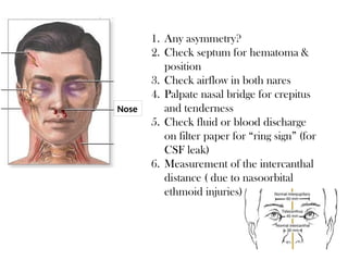
1. Any asymmetry?
2.Check septum for hematoma &
position
3. Check airflow in both nares
4. Palpate nasal bridge for crepitus
and tenderness
5. Check fluid or blood discharge
on filter paper for “ring sign” (for
CSF leak)
6. Measurement of the intercanthal
distance ( due to nasoorbital
ethmoid injuries)
Nose
35.
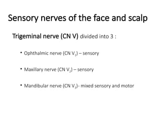
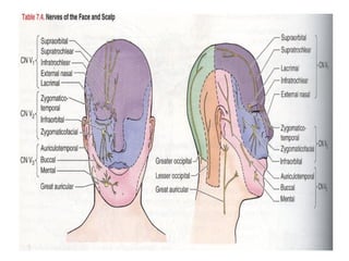
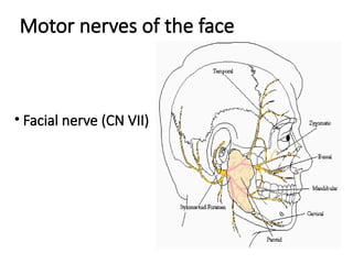
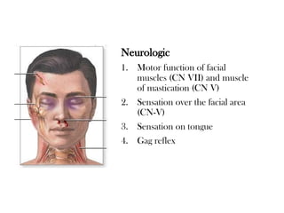
Neurologic
1. Motor functionof facial
muscles (CN VII) and muscle
of mastication (CN V)
2. Sensation over the facial area
(CN-V)
3. Sensation on tongue
4. Gag reflex
36.
History approach
History canbe obtained from the patient, orwitnesses/ accompanying
family members :
• How did the accident occur?
• When did the accident occur?
• What are the specifics of the injury:
(Type of the object contacted, direction of the contact?)
• Was there any loss of consciousness?
• What symptoms experienced by the patient:
(pain/ altered sensation/ visual changes/ malocclusion)
• Complete review of medical conditions, medication / allergies
37.
Physical examination
• Theface and cranium carefully inspected – any evidence of
trauma : laceration, abrasion, contusions, area of
edema/hematoma/ possible contour defect/ ecchymosis
* periorbital ecchymosis indicateorbital rim or zygomatic
complex fractures
* Battle’s sign ( bruises behind ear)suggest basilar skull fracture
* ecchymosis in the floor of mouth indicate anterior mandibular
fracture
http://www.sciencedirect.com/science/article/pii/0030422056900251
http://emedicine.medscape.com/article/434875-overview
38.
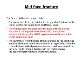
Mid face fracture
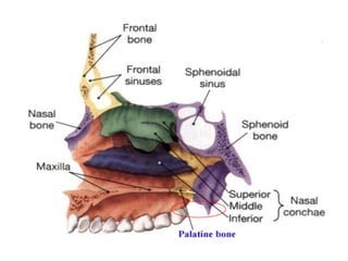
Theface is divided into equal thirds :
• The upper face, from the hairline to the glabella. Fractures in this
region involve the frontal bone and frontal sinus.
• The midface, from the glabella to the base of the columella.
Fractures is this region involve the maxilla, nasal bones,
nasoethmoidal complex (NOE), zygomaticomaxillary complex
(ZMC), and orbital floor.
• The lower face, from the base of the columella to the soft tissue
menton. The lower third is subdivided in an upper third from the
columella base to the lip commissure and two lower thirds from
the lower lip to menton. Fractures in this region involve
the dentoalveolar segments and the mandible.
41.
* Area ofstrength
Vertical and horizontal pillars
Muscular attachment
* Area of weakness
Sutures
Lining tissues and air-filled cavities
42.
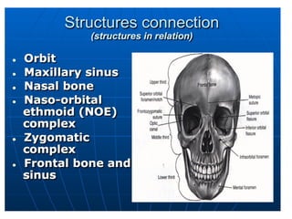
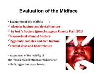
Evaluation of theMidface
• Evaluation of the midface :
Alveolar fracture and dental fracture
Le Fort ‘s fracture ((french surgeon Rane Le Fort 1901)
Naso-orbital ethmoid fracture
Zygomatic complex and arch fracture
Frontal sinus and bone fracture
• Assessment of the mobility of
the maxilla isolated structure/combination
with the zygoma or nasal bones.
43.
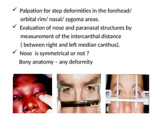
Palpation forstep deformities in the forehead/
orbital rim/ nasal/ zygoma areas.
Evaluation of nose and paranasal structures by
measurement of the intercanthal distance
( between right and left median canthus).
Nose is symmetrical or not ?
Bony anatomy – any deformity
44.
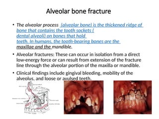
Alveolar bone fracture
•The alveolar process (alveolar bone) is the thickened ridge of
bone that contains the tooth sockets (
dental alveoli) on bones that hold
teeth. In humans, the tooth-bearing bones are the
maxillae and the mandible.
• Alveolar fractures: These can occur in isolation from a direct
low-energy force or can result from extension of the fracture
line through the alveolar portion of the maxilla or mandible.
• Clinical findings include gingival bleeding, mobility of the
alveolus, and loose or avulsed teeth.
45.
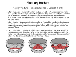
Maxillary fracture
Maxillary fractures:These are classified as Le Fort I, II, or III
• LeFort I fracture is a horizontal maxillary fracture across the inferior aspect of the maxilla
and separates the alveolar process containing the maxillary teeth and hard palate from the
rest of the maxilla. The fracture extends through the lower third of the septum and
includes the medial and lateral maxillary sinus walls extending into the palatine bones and
pterygoid plates.
• LeFort II fracture is a pyramidal fracture starting at the nasal bone and extending through
the ethmoid and lacrimal bones; downward through the zygomaticomaxillary suture;
continuing posteriorly and laterally through the maxilla, below the zygoma; and into the
pterygoid plates.
• LeFort III fracture or craniofacial disjunction is a separation of all of the facial bones from
the cranial base with simultaneous fracture of the zygoma, maxilla, and nasal bones. The
fracture line extends posterolaterally through ethmoid bones, orbits, and pterygomaxillary
suture into the sphenopalatine fossa. See the image below :
46.
Presentation of themaxillary fractures :
• Potential findings of LeFort I fracture include facial edema and
mobility of the hard palate and maxillary alveolus and teeth.
• Clinical presentations of LeFort II fractures include facial edema,
telecanthus, subconjunctival hemorrhage, mobility of the maxilla
at the nasofrontal suture, epistaxis, and possible CSF rhinorrhea.
• Characteristic findings of LeFort III fractures include massive
edema with facial rounding, elongation, and flattening. An
anterior open bite may be present due to posterior and inferior
displacement of the midfacial skeleton. Movement of all facial
bones in relation to the cranial base with manipulation of the
teeth and hard palate, epistaxis, and CSF rhinorrhea may also be
found upon physical examination.
• http://emedicine.medscape.com/article/434875-overview#a1
47.
Nasoethmoidal fractures (NOE):
•As the incidence of high-speed, high-force
accidents has increased over the decades, so too
has the number of such fractures. Due to the
degree of force and the vectors involved, NOE
fractures rarely occur as isolated events. Associated
injures often include central nervous system injury,
cribriform plate fracture, cerebrospinal fluid
rhinorrhea, and fractures of the frontal bone,
orbital floor, and middle third of the face, as well as
injury to the lacrimal system.
48.
Zygomaticomaxillary complex fractures(ZMC):
• These fractures result from direct trauma. Fracture
lines extend through zygomaticotemporal,
zygomaticofrontal, and zygomaticomaxillary sutures
and the articulation with the greater wing of the
sphenoid bone. The majority of these fractures
result from trauma inflicted in altercations followed
by MVA.
49.
Frontal bone fractures
•Usually result from a high velocity blunt trauma to
the forehead (e.g. MVA). The anterior and/or
posterior table of the frontal sinus may be involved.
More than one-third of patients with frontal sinus
fractures are likely to have concomitant intracranial
injury.
50.
Mandibular fractures
• Thesecan occur in multiple locations secondary to
the U-shape of the jaw and the weak condylar
neck. Fractures occur secondary to direct or
indirect facial injury, including motor vehicle
accidents, falls, sports, and assaults with blunt
weapons or guns. Close to half of all patients with
maxillofacial injuries have concomitant mandibular
fractures.
51.
Diagnosis
History & Evalutionof Midface ( PE)
Investigation :
• Laboratory studies
Laboratory studies should be ordered based on the patient's
medical history, current condition and planned surgical
procedure.
• Imaging studies
CT scan is the gold standard imaging technique to diagnose
maxillofacial fractures. The sensitivity and negative predictive
values of a routine nonenhanced head CT scan for fracture
surveillance was found to be 100%
However, when the patient sustains a low impact injury to the mandible, a panoramic x-ray
should be the initial screening imaging technique.
52.
Indications for treatment
Physicalsigns of a fracture of the maxilla.
Evidence of a fractured maxilla on imaging.
Disruption of the occlusion of the teeth.
Displacement of the maxilla.
Post traumatic facial deformity.
Fractured or displaced teeth.
Cerebrospinal fluid leak.
Abnormal eye movement or restriction of eye movement.
Occlusion of the nasolacrimal duct.
Sensory or motor nerve deficit.
Other evidence of loss of function
53.
Treatment
• General medicaltherapy: Administer oxygen and isotonic
crystalloid fluids. Administer packed red blood cells if
indicated. Check the tetanus status of the patient and
administer indicated.
• Antibiotics: Administer antibiotics for open fractures until the
fractures are repaired and the soft tissue wounds are closed.
• Pain management: Use oral medications for minor injuries and
parenteral medications if the patient cannot take oral
medications (ie, nothing by mouth [NPO]). For anti-
inflammatory control, use ibuprofen, naproxen, or ketorolac
(Toradol). For central control, use narcotics (eg, codeine,
oxycodone, hydrocodone, meperidine, morphine).
54.
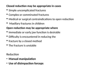
Closed reduction maybe appropriate in cases
Simple uncomplicated fractures
Complex or comminuted fractures
Medical or surgical contraindications to open reduction
Maxillary fractures in children
Open reduction may be appropriate where
Immediate or early jaw function is desirable
Difficulty is encountered in reducing the
fracture by a closed method
The fracture is unstable
Reduction
• Manual manipulation
• Use of disimpaction forceps
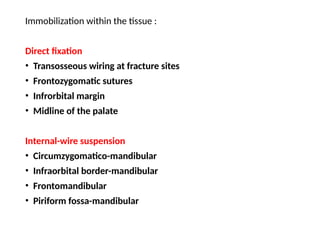
Immobilization within thetissue :
Direct fixation
• Transosseous wiring at fracture sites
• Frontozygomatic sutures
• Infrorbital margin
• Midline of the palate
Internal-wire suspension
• Circumzygomatico-mandibular
• Infraorbital border-mandibular
• Frontomandibular
• Piriform fossa-mandibular
57.
Support via themaxillary sinus by filling materials
• Ribbon gauze
• Balloon
• Folly catheter
• Polyethylene material
http://emedicine.medscape.com/article/434875-overview
Editor's Notes
#8 Facial nerve runs anteriorly > engulfed b parotid gland > forms parotid plexus > 5 branches arises from the parotid plexus
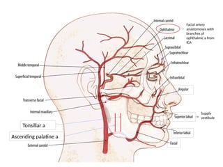
#11 It gives a branch, the transverse facial artery, which courses through the face parallel to the parotid duct.
#14 Vein divided into superficial (which follows the course of artery) and deep vein with is boxed in red.
Pterygoid plexus is located in the infratemporal fossa (between temporal bone and lateral pterygoid muscle)
The veins drains into internal and external jugular vein
#23 Tracheal intubation (inserting a tube into the airway to assist breathing) may be difficult or impossible due to swelling.
Nasal intubation may be contraindicated in the presence of facial trauma because if there is an undiscovered fracture at the base of the skull, the tube could be forced through it and into the brain.
If facial injuries prevent oraotracheal or nasotracheal intubation, a surgical airway can be placed to provide an adequate airway.
Although cricothyrotomy and tracheostomy can secure an airway when other methods fail, they are used only as a last resort because of potential complications and the difficulty of the procedures.
#33 Periorbital ecchymosis : raccoon’s eye (base of skul #)

![Treatment
• General medical therapy: Administer oxygen and isotonic
crystalloid fluids. Administer packed red blood cells if
indicated. Check the tetanus status of the patient and
administer indicated.
• Antibiotics: Administer antibiotics for open fractures until the
fractures are repaired and the soft tissue wounds are closed.
• Pain management: Use oral medications for minor injuries and
parenteral medications if the patient cannot take oral
medications (ie, nothing by mouth [NPO]). For anti-
inflammatory control, use ibuprofen, naproxen, or ketorolac
(Toradol). For central control, use narcotics (eg, codeine,
oxycodone, hydrocodone, meperidine, morphine).](https://image.slidesharecdn.com/maxillfacialtraumaclassificationandmanagement-250522042345-11cda69d/85/maxillfacial-trauma-classification-and-management-pptx-53-320.jpg)