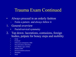
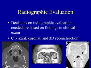
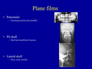
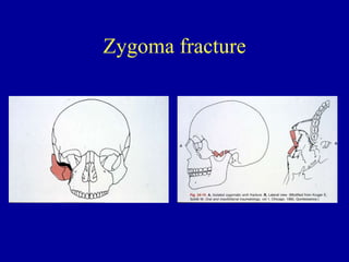
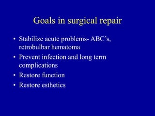
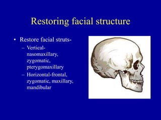
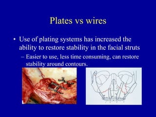
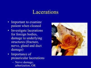
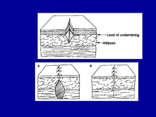
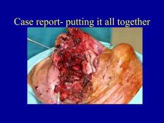
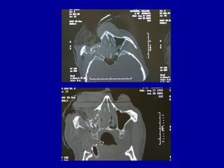
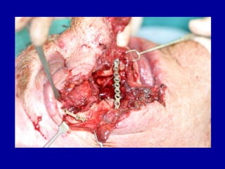
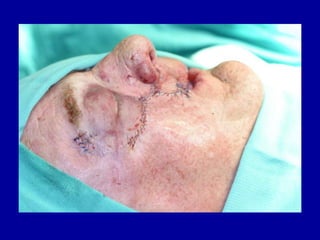
SlideShare a Scribd company logo
Major Midface Trauma
Steven Edlund DDS
Lecturer Dept of Oral and
Maxillofacial Surgery
Goals
• learn the basics a maxillofacial trauma
exam
• understand how to identify common
fractures and their complications
• basics of treatment fractures
• laceration management and complications
Specific Objectives (test
material)
• understand the importance of always performing an exam in the same sequence
• know how to examine cranial nerves
• know what an aferrent pupilary defect is
• know how to identify a Lefort I,II, or III fracture, a zygoma and zygomatic arch fracture,
orbital floor fracture, frontal sinus fracture, and naso-orbital-ethmoidal (NOE) fracture
from a physical exam
• understand the role of radiology (particularly CT) in the evaluation and management of
midface trauma
• define Lefort I, II, and III fractures and understand how they differ from Lefort
osteotomies (may be helpful to look up the osteotomies from other lectures)
• know the areas of fracture in zygoma fractures
• know what NOE fractures are, and know what nasal fractures are and timing of
treatment
• understand the basics of managing facial fractures (when surgery is appropriate, surgical
approaches, goals of treatment)
• understand the importance of evaluation the facial nerve in pre-auricular lacerations
• know the order of treatment in laceration repair
Trauma Exam
• Know your ABC’s
– A- airway
– B- breathing
– C- circulation
– D- differential diagnosis
• Patient stability is first and foremost goal
Trauma Exam Continued
• Always proceed in an orderly fashion
– Form a pattern and always follow it
1. General overview
• Facial/cervical symmetry
2. Top down- lacerations, contusions, foreign
bodies, palpate for boney steps and mobility
• Scalp
• Forehead
• Orbits/eyes-entrapment, APD
• Nose-deviation, rhinorrhea
• Ears-Battles sign, otorhea
• Midface/ZMC
• Intraoral-dental occlusion
• Mandible
• Neck
Radiographic Evaluation
• Decisions on radiographic evaluation
needed are based on findings in clinical
exam
• CT- axial, coronal, and 3D reconstruction
Plane films
• Panoramic
– Screening maxilla and mandible
• PA skull
– Skull and mandibular fractures
• Lateral skull
– Nose, sinus, maxilla
Plane films continued
• Waters view
– Maxilla, maxillary sinus
• Submental vertex
– Zygomztic arch
CT vs plane films
• CT has become the standard of care where
available, for evaluation of midface skeletal
trauma
• Both have strengths and weaknesses, but
plane films are rarely ordered today
Fractures
• Basic classification
– Greenstick- seen in children; more like a bend than a
clean break
– Simple- clean break along a single line with minimal
disruption of soft tissues; can be displaced or non-
displaced.
– Compound- broken bone that is displaced through the
integument
– Cominuted- multiple little pieces; looks like its
shatteren
Lefort Fractures
• Defined by Renee LeFort in early 1900’s
• Dropped skulls and viewed midface fracture
patterns
• Basic patterns were found based on the direction
of the blow to the face
• These fractures can occur in combination
• Can often be detected with bimanual palpation and
manipulation
LeFort I
• Separates the maxilla and pterygoid plates from the
skull, in a transverse direction, at the level of the
lateral aspect of the piriform rims and the inferior
aspect of the maxillary sinuses, including the
alveolar process and teeth if present.
LeFort II
• Often referred to as a pyramidal fracture
• Involves the pterygoid plates
• Extends superiorly through the sinus to the medial
aspect of the orbit.
• Separates the pterygoid plates, medial wall of the orbit,
and nasal bones as a unit
LeFort III
• Craniofacial disjunction
• Extends from the pterygoid plates through the
frontal-zygomatic suture and across the orbit
involving the nasal bones.
• Rare to see as a single unit; other fractures
usually involved
Zygoma fracture
• Most commonly fractured bone in the midface
• “Trimalar” fracture
– Frontal-zygomatic suture
– Maxillary-zygomatic suture
– Temporal zygomatic suture
• Examine for infraorbital and vestibular
ecchymosis, Rowe’s sign; palpate for boney steps
on all three sutures.
• Facial flatness on affected side
Zygoma fracture
Frontal bone fracture
• Contour change, ecchymosis, soft to
palpation, often associated with nasal and
orbital fractures.
• CT exam necessary to determine if the
anterior and/or posterior sinus walls are
involved.
Frontal sinus repair
• Eliminate sinus mucosa lining- eliminates
mucocele, alows direct visualization of posterior
wall
• Plugging the ducts- eliminates communication to
the nasal cavity
• Fat graft- obliterates empty space
• Cranialization- done if posterior wall involved
Frontal sinus repair
Nasal-orbital-ethmoid fracture
• Involves the nasal bone, orbital process of
the ethmoid, and the attacment of the
medial canthal ligament.
• Flatness of the nasal bridge, hypertelorism,
widened medial canthal distance
• Exam-
NOE fracture
Surgical approaches to the
midface
• Bicoronal flap- frontal sinus, zygoma, NOE
– Across the cranium in the hair bearing region, can
extend to the preauricular area for better access
• Gillies- Zygoma, zygomatic arch
– Incision in temporal hair bearing region with dissection
under the superficial layer of deep temporal facia to the
zygoma/arch
• Keen- intraoral, buccal vestibule approach to
zygoma
• Infraorbital, subcilliary,trans-conjunctival, upper
blephararoplasty, lateral brow- approaches to the
orbit
Bicoronal flap
Gillies
Orbital approaches
Goals in surgical repair
• Stabilize acute problems- ABC’s,
retrobulbar hematoma
• Prevent infection and long term
complications
• Restore function
• Restore esthetics
Restoring facial structure
• Restore facial struts-
– Vertical-
nasomaxillary,
zygomatic,
pterygomaxillary
– Horizontal-frontal,
zygomatic, maxillary,
mandibular
Plates vs wires
• Use of plating systems has increased the
ability to restore stability in the facial struts
– Easier to use, less time consuming, can restore
stability around contours.
Lacerations
• Important to examine
patient when cleaned
• Investigate lacerations
for foreign bodies,
damage to underlying
structures (fracture,
nerve, gland and duct
damage)
• Importance of
preauricular lacerations
– Nerve damage,
arborization, OR
Basic principles of laceration
management
• Hemostasis
• Anesthesia
• Irrigation
• Conserve viable tissue, remove necrotic tissue, undermine
• Layered closure
• Evert wound margins
• Support wound closure
• Antibiotics (topical and PO) and tetanus (booster within
last 5 years)
• Suture removal
• Home care instructions
Case report- putting it all together
Questions?

More Related Content

Similar to Major Midface Trauma.ppt

Rhinoplasty
RhinoplastyRhinoplasty
Rhinoplasty
Disha Sharma
 
maxillofacial trauma
maxillofacial traumamaxillofacial trauma
maxillofacial trauma
Dr Harjitpal Singh
 
Trauma to the face
Trauma to the faceTrauma to the face
Trauma to the face
Jinu Iype
 
Le Fort Fractures
Le Fort FracturesLe Fort Fractures
Le Fort Fractures
Dr. Akash Bhatt
 
Maxillary fractures ih
Maxillary fractures  ihMaxillary fractures  ih
Maxillary fractures ih
itrat hussain
 
landmarks and interpretation in extraoral radiography
landmarks and interpretation in extraoral radiographylandmarks and interpretation in extraoral radiography
landmarks and interpretation in extraoral radiography
gaurav katheriya
 
Orbital anatomy
Orbital anatomyOrbital anatomy
Orbital anatomy
Dr Kawshik Nag
 
1. MAXILLECTOMY.pptx
1. MAXILLECTOMY.pptx1. MAXILLECTOMY.pptx
1. MAXILLECTOMY.pptx
Amos Brighton
 
Facial bone fractures
Facial bone fracturesFacial bone fractures
Facial bone fractures
krishnakoirala4
 
Access osteotomies in oral & cranio-maxillofacial surgery
Access osteotomies in oral & cranio-maxillofacial surgeryAccess osteotomies in oral & cranio-maxillofacial surgery
Access osteotomies in oral & cranio-maxillofacial surgery
Dr Rayan Malick
 
Orthognathic surgery
Orthognathic surgeryOrthognathic surgery
Orthognathic surgery
Mohammed Rhael
 
Radiographic evaluation of midface fracture
Radiographic evaluation of midface fractureRadiographic evaluation of midface fracture
Radiographic evaluation of midface fracture
jyoti sharma
 
Fractures of middle third of face
Fractures of middle third of faceFractures of middle third of face
Fractures of middle third of face
Mohammed Shalik
 
Clinical Evaluation in Maxillofacial Trauma
Clinical Evaluation in Maxillofacial Trauma Clinical Evaluation in Maxillofacial Trauma
Clinical Evaluation in Maxillofacial Trauma
Arjun Shenoy
 
LEFORT FRACTURES.pptx
LEFORT FRACTURES.pptxLEFORT FRACTURES.pptx
LEFORT FRACTURES.pptx
Dr-Fakhrul Munna
 
Orthognathic Surgery
Orthognathic SurgeryOrthognathic Surgery
Orthognathic Surgery
Hadi Munib
 
Dentist in pune.(BDS) MDS- OMFS - Dr. Amit T. Suryawanshi.. Mandibular fractu...
Dentist in pune.(BDS) MDS- OMFS - Dr. Amit T. Suryawanshi.. Mandibular fractu...Dentist in pune.(BDS) MDS- OMFS - Dr. Amit T. Suryawanshi.. Mandibular fractu...
Dentist in pune.(BDS) MDS- OMFS - Dr. Amit T. Suryawanshi.. Mandibular fractu...
All Good Things
 
Fracture maxilla
Fracture maxillaFracture maxilla
Fracture maxilla
SHAMEEJ MUHAMED KV
 
DEVELOPMENTAL ANOMALIES - CRANIOFACIAL SYNDROMES
DEVELOPMENTAL ANOMALIES - CRANIOFACIAL SYNDROMESDEVELOPMENTAL ANOMALIES - CRANIOFACIAL SYNDROMES
DEVELOPMENTAL ANOMALIES - CRANIOFACIAL SYNDROMES
Aditi Rajvanshi
 
Dentomaxillofacial imaging
Dentomaxillofacial imagingDentomaxillofacial imaging
Dentomaxillofacial imaging
Dr Reem Ayesha
 

Similar to Major Midface Trauma.ppt (20)

Rhinoplasty
RhinoplastyRhinoplasty
Rhinoplasty
 
maxillofacial trauma
maxillofacial traumamaxillofacial trauma
maxillofacial trauma
 
Trauma to the face
Trauma to the faceTrauma to the face
Trauma to the face
 
Le Fort Fractures
Le Fort FracturesLe Fort Fractures
Le Fort Fractures
 
Maxillary fractures ih
Maxillary fractures  ihMaxillary fractures  ih
Maxillary fractures ih
 
landmarks and interpretation in extraoral radiography
landmarks and interpretation in extraoral radiographylandmarks and interpretation in extraoral radiography
landmarks and interpretation in extraoral radiography
 
Orbital anatomy
Orbital anatomyOrbital anatomy
Orbital anatomy
 
1. MAXILLECTOMY.pptx
1. MAXILLECTOMY.pptx1. MAXILLECTOMY.pptx
1. MAXILLECTOMY.pptx
 
Facial bone fractures
Facial bone fracturesFacial bone fractures
Facial bone fractures
 
Access osteotomies in oral & cranio-maxillofacial surgery
Access osteotomies in oral & cranio-maxillofacial surgeryAccess osteotomies in oral & cranio-maxillofacial surgery
Access osteotomies in oral & cranio-maxillofacial surgery
 
Orthognathic surgery
Orthognathic surgeryOrthognathic surgery
Orthognathic surgery
 
Radiographic evaluation of midface fracture
Radiographic evaluation of midface fractureRadiographic evaluation of midface fracture
Radiographic evaluation of midface fracture
 
Fractures of middle third of face
Fractures of middle third of faceFractures of middle third of face
Fractures of middle third of face
 
Clinical Evaluation in Maxillofacial Trauma
Clinical Evaluation in Maxillofacial Trauma Clinical Evaluation in Maxillofacial Trauma
Clinical Evaluation in Maxillofacial Trauma
 
LEFORT FRACTURES.pptx
LEFORT FRACTURES.pptxLEFORT FRACTURES.pptx
LEFORT FRACTURES.pptx
 
Orthognathic Surgery
Orthognathic SurgeryOrthognathic Surgery
Orthognathic Surgery
 
Dentist in pune.(BDS) MDS- OMFS - Dr. Amit T. Suryawanshi.. Mandibular fractu...
Dentist in pune.(BDS) MDS- OMFS - Dr. Amit T. Suryawanshi.. Mandibular fractu...Dentist in pune.(BDS) MDS- OMFS - Dr. Amit T. Suryawanshi.. Mandibular fractu...
Dentist in pune.(BDS) MDS- OMFS - Dr. Amit T. Suryawanshi.. Mandibular fractu...
 
Fracture maxilla
Fracture maxillaFracture maxilla
Fracture maxilla
 
DEVELOPMENTAL ANOMALIES - CRANIOFACIAL SYNDROMES
DEVELOPMENTAL ANOMALIES - CRANIOFACIAL SYNDROMESDEVELOPMENTAL ANOMALIES - CRANIOFACIAL SYNDROMES
DEVELOPMENTAL ANOMALIES - CRANIOFACIAL SYNDROMES
 
Dentomaxillofacial imaging
Dentomaxillofacial imagingDentomaxillofacial imaging
Dentomaxillofacial imaging
 

More from MellowMenais

impaction-23035190.pptx
impaction-23035190.pptximpaction-23035190.pptx
impaction-23035190.pptx
MellowMenais
 
hydation.pdf
hydation.pdfhydation.pdf
hydation.pdf
MellowMenais
 
fracturas de la cavidad orbitaria.doc
fracturas de la cavidad orbitaria.docfracturas de la cavidad orbitaria.doc
fracturas de la cavidad orbitaria.doc
MellowMenais
 
AMELOBLASTOMA JORNADA.ppt
AMELOBLASTOMA JORNADA.pptAMELOBLASTOMA JORNADA.ppt
AMELOBLASTOMA JORNADA.ppt
MellowMenais
 
Primary_Assessment_and_Care_in_Maxillofacial_Traum.pdf
Primary_Assessment_and_Care_in_Maxillofacial_Traum.pdfPrimary_Assessment_and_Care_in_Maxillofacial_Traum.pdf
Primary_Assessment_and_Care_in_Maxillofacial_Traum.pdf
MellowMenais
 
NCCN_Guidelines_Head-and-Neck Cancer Version 2.2013 (1).pdf
NCCN_Guidelines_Head-and-Neck Cancer Version 2.2013 (1).pdfNCCN_Guidelines_Head-and-Neck Cancer Version 2.2013 (1).pdf
NCCN_Guidelines_Head-and-Neck Cancer Version 2.2013 (1).pdf
MellowMenais
 
153512680-naso-orbito-ethmoidal-fracture.pdf.pdf
153512680-naso-orbito-ethmoidal-fracture.pdf.pdf153512680-naso-orbito-ethmoidal-fracture.pdf.pdf
153512680-naso-orbito-ethmoidal-fracture.pdf.pdf
MellowMenais
 

More from MellowMenais (7)

impaction-23035190.pptx
impaction-23035190.pptximpaction-23035190.pptx
impaction-23035190.pptx
 
hydation.pdf
hydation.pdfhydation.pdf
hydation.pdf
 
fracturas de la cavidad orbitaria.doc
fracturas de la cavidad orbitaria.docfracturas de la cavidad orbitaria.doc
fracturas de la cavidad orbitaria.doc
 
AMELOBLASTOMA JORNADA.ppt
AMELOBLASTOMA JORNADA.pptAMELOBLASTOMA JORNADA.ppt
AMELOBLASTOMA JORNADA.ppt
 
Primary_Assessment_and_Care_in_Maxillofacial_Traum.pdf
Primary_Assessment_and_Care_in_Maxillofacial_Traum.pdfPrimary_Assessment_and_Care_in_Maxillofacial_Traum.pdf
Primary_Assessment_and_Care_in_Maxillofacial_Traum.pdf
 
NCCN_Guidelines_Head-and-Neck Cancer Version 2.2013 (1).pdf
NCCN_Guidelines_Head-and-Neck Cancer Version 2.2013 (1).pdfNCCN_Guidelines_Head-and-Neck Cancer Version 2.2013 (1).pdf
NCCN_Guidelines_Head-and-Neck Cancer Version 2.2013 (1).pdf
 
153512680-naso-orbito-ethmoidal-fracture.pdf.pdf
153512680-naso-orbito-ethmoidal-fracture.pdf.pdf153512680-naso-orbito-ethmoidal-fracture.pdf.pdf
153512680-naso-orbito-ethmoidal-fracture.pdf.pdf
 

Recently uploaded

Triangles of Neck and Clinical Correlation by Dr. RIG.pptx
Triangles of Neck and Clinical Correlation by Dr. RIG.pptxTriangles of Neck and Clinical Correlation by Dr. RIG.pptx
Triangles of Neck and Clinical Correlation by Dr. RIG.pptx
Dr. Rabia Inam Gandapore
 
Ozempic: Preoperative Management of Patients on GLP-1 Receptor Agonists
Ozempic: Preoperative Management of Patients on GLP-1 Receptor Agonists  Ozempic: Preoperative Management of Patients on GLP-1 Receptor Agonists
Ozempic: Preoperative Management of Patients on GLP-1 Receptor Agonists
Saeid Safari
 
Ophthalmology Clinical Tests for OSCE exam
Ophthalmology Clinical Tests for OSCE examOphthalmology Clinical Tests for OSCE exam
Ophthalmology Clinical Tests for OSCE exam
KafrELShiekh University
 
New Directions in Targeted Therapeutic Approaches for Older Adults With Mantl...
New Directions in Targeted Therapeutic Approaches for Older Adults With Mantl...New Directions in Targeted Therapeutic Approaches for Older Adults With Mantl...
New Directions in Targeted Therapeutic Approaches for Older Adults With Mantl...
i3 Health
 
New Drug Discovery and Development .....
New Drug Discovery and Development .....New Drug Discovery and Development .....
New Drug Discovery and Development .....
NEHA GUPTA
 
24 Upakrama.pptx class ppt useful in all
24 Upakrama.pptx class ppt useful in all24 Upakrama.pptx class ppt useful in all
24 Upakrama.pptx class ppt useful in all
DrSathishMS1
 
Flu Vaccine Alert in Bangalore Karnataka
Flu Vaccine Alert in Bangalore KarnatakaFlu Vaccine Alert in Bangalore Karnataka
Flu Vaccine Alert in Bangalore Karnataka
addon Scans
 
Alcohol_Dr. Jeenal Mistry MD Pharmacology.pdf
Alcohol_Dr. Jeenal Mistry MD Pharmacology.pdfAlcohol_Dr. Jeenal Mistry MD Pharmacology.pdf
Alcohol_Dr. Jeenal Mistry MD Pharmacology.pdf
Dr Jeenal Mistry
 
Maxilla, Mandible & Hyoid Bone & Clinical Correlations by Dr. RIG.pptx
Maxilla, Mandible & Hyoid Bone & Clinical Correlations by Dr. RIG.pptxMaxilla, Mandible & Hyoid Bone & Clinical Correlations by Dr. RIG.pptx
Maxilla, Mandible & Hyoid Bone & Clinical Correlations by Dr. RIG.pptx
Dr. Rabia Inam Gandapore
 
Evaluation of antidepressant activity of clitoris ternatea in animals
Evaluation of antidepressant activity of clitoris ternatea in animalsEvaluation of antidepressant activity of clitoris ternatea in animals
Evaluation of antidepressant activity of clitoris ternatea in animals
Shweta
 
BRACHYTHERAPY OVERVIEW AND APPLICATORS
BRACHYTHERAPY OVERVIEW  AND  APPLICATORSBRACHYTHERAPY OVERVIEW  AND  APPLICATORS
BRACHYTHERAPY OVERVIEW AND APPLICATORS
Krishan Murari
 
Pharynx and Clinical Correlations BY Dr.Rabia Inam Gandapore.pptx
Pharynx and Clinical Correlations BY Dr.Rabia Inam Gandapore.pptxPharynx and Clinical Correlations BY Dr.Rabia Inam Gandapore.pptx
Pharynx and Clinical Correlations BY Dr.Rabia Inam Gandapore.pptx
Dr. Rabia Inam Gandapore
 
Novas diretrizes da OMS para os cuidados perinatais de mais qualidade
Novas diretrizes da OMS para os cuidados perinatais de mais qualidadeNovas diretrizes da OMS para os cuidados perinatais de mais qualidade
Novas diretrizes da OMS para os cuidados perinatais de mais qualidade
Prof. Marcus Renato de Carvalho
 
TEST BANK for Operations Management, 14th Edition by William J. Stevenson, Ve...
TEST BANK for Operations Management, 14th Edition by William J. Stevenson, Ve...TEST BANK for Operations Management, 14th Edition by William J. Stevenson, Ve...
TEST BANK for Operations Management, 14th Edition by William J. Stevenson, Ve...
kevinkariuki227
 
Knee anatomy and clinical tests 2024.pdf
Knee anatomy and clinical tests 2024.pdfKnee anatomy and clinical tests 2024.pdf
Knee anatomy and clinical tests 2024.pdf
vimalpl1234
 
How STIs Influence the Development of Pelvic Inflammatory Disease.pptx
How STIs Influence the Development of Pelvic Inflammatory Disease.pptxHow STIs Influence the Development of Pelvic Inflammatory Disease.pptx
How STIs Influence the Development of Pelvic Inflammatory Disease.pptx
FFragrant
 
ACUTE SCROTUM.....pdf. ACUTE SCROTAL CONDITIOND
ACUTE SCROTUM.....pdf. ACUTE SCROTAL CONDITIONDACUTE SCROTUM.....pdf. ACUTE SCROTAL CONDITIOND
ACUTE SCROTUM.....pdf. ACUTE SCROTAL CONDITIOND
DR SETH JOTHAM
 
POST OPERATIVE OLIGURIA and its management
POST OPERATIVE OLIGURIA and its managementPOST OPERATIVE OLIGURIA and its management
POST OPERATIVE OLIGURIA and its management
touseefaziz1
 
The Normal Electrocardiogram - Part I of II
The Normal Electrocardiogram - Part I of IIThe Normal Electrocardiogram - Part I of II
The Normal Electrocardiogram - Part I of II
MedicoseAcademics
 
Tom Selleck Health: A Comprehensive Look at the Iconic Actor’s Wellness Journey
Tom Selleck Health: A Comprehensive Look at the Iconic Actor’s Wellness JourneyTom Selleck Health: A Comprehensive Look at the Iconic Actor’s Wellness Journey
Tom Selleck Health: A Comprehensive Look at the Iconic Actor’s Wellness Journey
greendigital
 

Recently uploaded (20)

Triangles of Neck and Clinical Correlation by Dr. RIG.pptx
Triangles of Neck and Clinical Correlation by Dr. RIG.pptxTriangles of Neck and Clinical Correlation by Dr. RIG.pptx
Triangles of Neck and Clinical Correlation by Dr. RIG.pptx
 
Ozempic: Preoperative Management of Patients on GLP-1 Receptor Agonists
Ozempic: Preoperative Management of Patients on GLP-1 Receptor Agonists  Ozempic: Preoperative Management of Patients on GLP-1 Receptor Agonists
Ozempic: Preoperative Management of Patients on GLP-1 Receptor Agonists
 
Ophthalmology Clinical Tests for OSCE exam
Ophthalmology Clinical Tests for OSCE examOphthalmology Clinical Tests for OSCE exam
Ophthalmology Clinical Tests for OSCE exam
 
New Directions in Targeted Therapeutic Approaches for Older Adults With Mantl...
New Directions in Targeted Therapeutic Approaches for Older Adults With Mantl...New Directions in Targeted Therapeutic Approaches for Older Adults With Mantl...
New Directions in Targeted Therapeutic Approaches for Older Adults With Mantl...
 
New Drug Discovery and Development .....
New Drug Discovery and Development .....New Drug Discovery and Development .....
New Drug Discovery and Development .....
 
24 Upakrama.pptx class ppt useful in all
24 Upakrama.pptx class ppt useful in all24 Upakrama.pptx class ppt useful in all
24 Upakrama.pptx class ppt useful in all
 
Flu Vaccine Alert in Bangalore Karnataka
Flu Vaccine Alert in Bangalore KarnatakaFlu Vaccine Alert in Bangalore Karnataka
Flu Vaccine Alert in Bangalore Karnataka
 
Alcohol_Dr. Jeenal Mistry MD Pharmacology.pdf
Alcohol_Dr. Jeenal Mistry MD Pharmacology.pdfAlcohol_Dr. Jeenal Mistry MD Pharmacology.pdf
Alcohol_Dr. Jeenal Mistry MD Pharmacology.pdf
 
Maxilla, Mandible & Hyoid Bone & Clinical Correlations by Dr. RIG.pptx
Maxilla, Mandible & Hyoid Bone & Clinical Correlations by Dr. RIG.pptxMaxilla, Mandible & Hyoid Bone & Clinical Correlations by Dr. RIG.pptx
Maxilla, Mandible & Hyoid Bone & Clinical Correlations by Dr. RIG.pptx
 
Evaluation of antidepressant activity of clitoris ternatea in animals
Evaluation of antidepressant activity of clitoris ternatea in animalsEvaluation of antidepressant activity of clitoris ternatea in animals
Evaluation of antidepressant activity of clitoris ternatea in animals
 
BRACHYTHERAPY OVERVIEW AND APPLICATORS
BRACHYTHERAPY OVERVIEW  AND  APPLICATORSBRACHYTHERAPY OVERVIEW  AND  APPLICATORS
BRACHYTHERAPY OVERVIEW AND APPLICATORS
 
Pharynx and Clinical Correlations BY Dr.Rabia Inam Gandapore.pptx
Pharynx and Clinical Correlations BY Dr.Rabia Inam Gandapore.pptxPharynx and Clinical Correlations BY Dr.Rabia Inam Gandapore.pptx
Pharynx and Clinical Correlations BY Dr.Rabia Inam Gandapore.pptx
 
Novas diretrizes da OMS para os cuidados perinatais de mais qualidade
Novas diretrizes da OMS para os cuidados perinatais de mais qualidadeNovas diretrizes da OMS para os cuidados perinatais de mais qualidade
Novas diretrizes da OMS para os cuidados perinatais de mais qualidade
 
TEST BANK for Operations Management, 14th Edition by William J. Stevenson, Ve...
TEST BANK for Operations Management, 14th Edition by William J. Stevenson, Ve...TEST BANK for Operations Management, 14th Edition by William J. Stevenson, Ve...
TEST BANK for Operations Management, 14th Edition by William J. Stevenson, Ve...
 
Knee anatomy and clinical tests 2024.pdf
Knee anatomy and clinical tests 2024.pdfKnee anatomy and clinical tests 2024.pdf
Knee anatomy and clinical tests 2024.pdf
 
How STIs Influence the Development of Pelvic Inflammatory Disease.pptx
How STIs Influence the Development of Pelvic Inflammatory Disease.pptxHow STIs Influence the Development of Pelvic Inflammatory Disease.pptx
How STIs Influence the Development of Pelvic Inflammatory Disease.pptx
 
ACUTE SCROTUM.....pdf. ACUTE SCROTAL CONDITIOND
ACUTE SCROTUM.....pdf. ACUTE SCROTAL CONDITIONDACUTE SCROTUM.....pdf. ACUTE SCROTAL CONDITIOND
ACUTE SCROTUM.....pdf. ACUTE SCROTAL CONDITIOND
 
POST OPERATIVE OLIGURIA and its management
POST OPERATIVE OLIGURIA and its managementPOST OPERATIVE OLIGURIA and its management
POST OPERATIVE OLIGURIA and its management
 
The Normal Electrocardiogram - Part I of II
The Normal Electrocardiogram - Part I of IIThe Normal Electrocardiogram - Part I of II
The Normal Electrocardiogram - Part I of II
 
Tom Selleck Health: A Comprehensive Look at the Iconic Actor’s Wellness Journey
Tom Selleck Health: A Comprehensive Look at the Iconic Actor’s Wellness JourneyTom Selleck Health: A Comprehensive Look at the Iconic Actor’s Wellness Journey
Tom Selleck Health: A Comprehensive Look at the Iconic Actor’s Wellness Journey
 

Major Midface Trauma.ppt

  • 1. Major Midface Trauma Steven Edlund DDS Lecturer Dept of Oral and Maxillofacial Surgery
  • 2. Goals • learn the basics a maxillofacial trauma exam • understand how to identify common fractures and their complications • basics of treatment fractures • laceration management and complications
  • 3. Specific Objectives (test material) • understand the importance of always performing an exam in the same sequence • know how to examine cranial nerves • know what an aferrent pupilary defect is • know how to identify a Lefort I,II, or III fracture, a zygoma and zygomatic arch fracture, orbital floor fracture, frontal sinus fracture, and naso-orbital-ethmoidal (NOE) fracture from a physical exam • understand the role of radiology (particularly CT) in the evaluation and management of midface trauma • define Lefort I, II, and III fractures and understand how they differ from Lefort osteotomies (may be helpful to look up the osteotomies from other lectures) • know the areas of fracture in zygoma fractures • know what NOE fractures are, and know what nasal fractures are and timing of treatment • understand the basics of managing facial fractures (when surgery is appropriate, surgical approaches, goals of treatment) • understand the importance of evaluation the facial nerve in pre-auricular lacerations • know the order of treatment in laceration repair
  • 4. Trauma Exam • Know your ABC’s – A- airway – B- breathing – C- circulation – D- differential diagnosis • Patient stability is first and foremost goal
  • 5. Trauma Exam Continued • Always proceed in an orderly fashion – Form a pattern and always follow it 1. General overview • Facial/cervical symmetry 2. Top down- lacerations, contusions, foreign bodies, palpate for boney steps and mobility • Scalp • Forehead • Orbits/eyes-entrapment, APD • Nose-deviation, rhinorrhea • Ears-Battles sign, otorhea • Midface/ZMC • Intraoral-dental occlusion • Mandible • Neck
  • 6. Radiographic Evaluation • Decisions on radiographic evaluation needed are based on findings in clinical exam • CT- axial, coronal, and 3D reconstruction
  • 7. Plane films • Panoramic – Screening maxilla and mandible • PA skull – Skull and mandibular fractures • Lateral skull – Nose, sinus, maxilla
  • 8. Plane films continued • Waters view – Maxilla, maxillary sinus • Submental vertex – Zygomztic arch
  • 9. CT vs plane films • CT has become the standard of care where available, for evaluation of midface skeletal trauma • Both have strengths and weaknesses, but plane films are rarely ordered today
  • 10. Fractures • Basic classification – Greenstick- seen in children; more like a bend than a clean break – Simple- clean break along a single line with minimal disruption of soft tissues; can be displaced or non- displaced. – Compound- broken bone that is displaced through the integument – Cominuted- multiple little pieces; looks like its shatteren
  • 11. Lefort Fractures • Defined by Renee LeFort in early 1900’s • Dropped skulls and viewed midface fracture patterns • Basic patterns were found based on the direction of the blow to the face • These fractures can occur in combination • Can often be detected with bimanual palpation and manipulation
  • 12. LeFort I • Separates the maxilla and pterygoid plates from the skull, in a transverse direction, at the level of the lateral aspect of the piriform rims and the inferior aspect of the maxillary sinuses, including the alveolar process and teeth if present.
  • 13. LeFort II • Often referred to as a pyramidal fracture • Involves the pterygoid plates • Extends superiorly through the sinus to the medial aspect of the orbit. • Separates the pterygoid plates, medial wall of the orbit, and nasal bones as a unit
  • 14. LeFort III • Craniofacial disjunction • Extends from the pterygoid plates through the frontal-zygomatic suture and across the orbit involving the nasal bones. • Rare to see as a single unit; other fractures usually involved
  • 15. Zygoma fracture • Most commonly fractured bone in the midface • “Trimalar” fracture – Frontal-zygomatic suture – Maxillary-zygomatic suture – Temporal zygomatic suture • Examine for infraorbital and vestibular ecchymosis, Rowe’s sign; palpate for boney steps on all three sutures. • Facial flatness on affected side
  • 17. Frontal bone fracture • Contour change, ecchymosis, soft to palpation, often associated with nasal and orbital fractures. • CT exam necessary to determine if the anterior and/or posterior sinus walls are involved.
  • 18. Frontal sinus repair • Eliminate sinus mucosa lining- eliminates mucocele, alows direct visualization of posterior wall • Plugging the ducts- eliminates communication to the nasal cavity • Fat graft- obliterates empty space • Cranialization- done if posterior wall involved
  • 20. Nasal-orbital-ethmoid fracture • Involves the nasal bone, orbital process of the ethmoid, and the attacment of the medial canthal ligament. • Flatness of the nasal bridge, hypertelorism, widened medial canthal distance • Exam-
  • 22. Surgical approaches to the midface • Bicoronal flap- frontal sinus, zygoma, NOE – Across the cranium in the hair bearing region, can extend to the preauricular area for better access • Gillies- Zygoma, zygomatic arch – Incision in temporal hair bearing region with dissection under the superficial layer of deep temporal facia to the zygoma/arch • Keen- intraoral, buccal vestibule approach to zygoma • Infraorbital, subcilliary,trans-conjunctival, upper blephararoplasty, lateral brow- approaches to the orbit
  • 26. Goals in surgical repair • Stabilize acute problems- ABC’s, retrobulbar hematoma • Prevent infection and long term complications • Restore function • Restore esthetics
  • 27. Restoring facial structure • Restore facial struts- – Vertical- nasomaxillary, zygomatic, pterygomaxillary – Horizontal-frontal, zygomatic, maxillary, mandibular
  • 28. Plates vs wires • Use of plating systems has increased the ability to restore stability in the facial struts – Easier to use, less time consuming, can restore stability around contours.
  • 29. Lacerations • Important to examine patient when cleaned • Investigate lacerations for foreign bodies, damage to underlying structures (fracture, nerve, gland and duct damage) • Importance of preauricular lacerations – Nerve damage, arborization, OR
  • 30. Basic principles of laceration management • Hemostasis • Anesthesia • Irrigation • Conserve viable tissue, remove necrotic tissue, undermine • Layered closure • Evert wound margins • Support wound closure • Antibiotics (topical and PO) and tetanus (booster within last 5 years) • Suture removal • Home care instructions
  • 31.
  • 32. Case report- putting it all together
  • 33.
  • 34.
  • 35.