The placenta is an organ that develops during pregnancy in mammals to connect the developing fetus to the uterine wall to allow nutrient uptake, waste elimination, and gas exchange via the mother's blood supply. It is a discoid, circular organ around 15-20 cm in diameter and 2.5 cm thick that weighs approximately 500g at term. The placenta has both fetal and maternal surfaces, with the fetal surface covered by the amnion and bearing the umbilical cord, and the maternal surface being rough and spongy where it attaches to the uterine wall. The placenta serves critical functions like nutrient, gas and waste exchange between mother and fetus, acting as an endocrine organ and barrier between
How 3 germ layers are formed in Chick that are endoderm, mesoderm and ectoderm.As Chick are polylecithal so cell movements are somewhat restricted and gastrulation is modified as compared to frog.
Polyspermy describes an egg that has been fertilized by more than one sperm. Diploid organisms normally contain two copies of each chromosome, one from each parent. The cell resulting from polyspermy
The first issue that an egg and a sperm of any organism type face in successfully producing an embryo is the possibility of polyspermy. Polyspermy is the fertilization of an egg by multiple sperm, and the results of such unions are lethal.
If multiple sperm fertilize an egg, the embryo inherits multiple paternal centrioles. This causes competition for extra chromosomes and results in the disruption of the creation of the cleavage furrow, thus causing the zygote to die. As an important model organism in the study of fertilization and embryonic development, polyspermy in sea urchins has been studied in detail. The sea urchin’s methods of polyspermy prevention have been broken down into two main pathways. These two primary pathways are known as the fast block and the slow block to polyspermy
After the sperm’s receptors come into contact with the egg’s jelly layer and the acrosomal enzymes are released and break down the jelly layer, the sperm head comes into contact with the vitelline and plasma membranes of the egg. When the two plasma membranes contact one another, signals in the egg are initiated.
First, Na+ channels in the egg open, allowing Na+ to flood into the egg. This causes a depolarization of the egg from it’s normal resting potential of -70 mV.
While depolarization is occurring, the remainder of the jelly layer is dissolving. With the dissolution of the jelly layer and the depolarization of the plasma membrane, the first block to preventing fertilization by multiple sperm is put into place.
These two simple changes are part of the first block to polyspermy, known as the fast block. Within 1/10th of a second of contact, the fast block t
Fate maps are the bases for experimental embryology since they provide researchers with information on which portions of the embryo normally become which larval or adult structures.
1. DEFINITION
These are the membranes which do not form any part of
the embryo proper but performs various functions which
assist in the development of the embryo . These are
discarded at the time of hatching. These membranes
formed outside the embryo.
2. Types of Extra Embryonic Membranes
Yolk Sac
Amnion
Chorion
Allantois
3.Discussed Their
At Time of ORIGIN
It's FUNCTION
After HATCHING
4. AMNIOTIC CAVITY
............................END......................................................
Vittelogenesis is a word developed from Latin vitellus-yolk, and genero-produce
Vitellogenesis (also known as yolk deposition) is the process of yolk formation via nutrients being deposited in the oocyte, or female germ cell involved in reproduction of lecithotrophic organisms. In insects, it starts when the fat body stimulates the release of juvenile hormones and produces vitellogenin protein.
Yolks is the most usual form of food storage in the egg.
Yolks appear in the oocyte in the secondary period of their growth called vittelogenesis.
Thus,the formation and deposition of yolks is known as vittelogenesis
Characteristic
Yolks is a complex variable assembled component.
The principle component are protein,phospholipid and fats in different combination.
Depending upon these component yolks is distinguished into protein yolks and fatty acid
For eg- the avian contain 48.19% water , 16.6 % protein, 32.6% phospholipids and fats and 1% carbohydrates.
A chart showing the fate of each part of an early embryo, in a particular blastula stage is called fate maps. It is done because the correct interpretation of gastrulation is impossible without the knowledge of the position which are the presumptive germinal layers (Ectoderm, Mesoderm and Endoderm) occupy in blastula.
Fate mapping is a method used in developmental biology to study the embryonic origin of various adult tissues and structures. The "fate" of each cell or group of cells is mapped onto the embryo, showing which parts of the embryo will develop into which tissue. When carried out at single-cell resolution, this process is called cell lineage tracing. It is also used to trace the development of tumors.
In all viviparous animals, embryonic development takes place inside the uterus of the mother, because the eggs are microlecithal and the amount of stored yolk is not sufficient for the developing embryo. Such embryos get attached to the uterine wall to draw essential substances from the maternal circulation through the placenta.
Deep Behavioral Phenotyping in Systems Neuroscience for Functional Atlasing a...Ana Luísa Pinho
Functional Magnetic Resonance Imaging (fMRI) provides means to characterize brain activations in response to behavior. However, cognitive neuroscience has been limited to group-level effects referring to the performance of specific tasks. To obtain the functional profile of elementary cognitive mechanisms, the combination of brain responses to many tasks is required. Yet, to date, both structural atlases and parcellation-based activations do not fully account for cognitive function and still present several limitations. Further, they do not adapt overall to individual characteristics. In this talk, I will give an account of deep-behavioral phenotyping strategies, namely data-driven methods in large task-fMRI datasets, to optimize functional brain-data collection and improve inference of effects-of-interest related to mental processes. Key to this approach is the employment of fast multi-functional paradigms rich on features that can be well parametrized and, consequently, facilitate the creation of psycho-physiological constructs to be modelled with imaging data. Particular emphasis will be given to music stimuli when studying high-order cognitive mechanisms, due to their ecological nature and quality to enable complex behavior compounded by discrete entities. I will also discuss how deep-behavioral phenotyping and individualized models applied to neuroimaging data can better account for the subject-specific organization of domain-general cognitive systems in the human brain. Finally, the accumulation of functional brain signatures brings the possibility to clarify relationships among tasks and create a univocal link between brain systems and mental functions through: (1) the development of ontologies proposing an organization of cognitive processes; and (2) brain-network taxonomies describing functional specialization. To this end, tools to improve commensurability in cognitive science are necessary, such as public repositories, ontology-based platforms and automated meta-analysis tools. I will thus discuss some brain-atlasing resources currently under development, and their applicability in cognitive as well as clinical neuroscience.
Richard's aventures in two entangled wonderlandsRichard Gill
Since the loophole-free Bell experiments of 2020 and the Nobel prizes in physics of 2022, critics of Bell's work have retreated to the fortress of super-determinism. Now, super-determinism is a derogatory word - it just means "determinism". Palmer, Hance and Hossenfelder argue that quantum mechanics and determinism are not incompatible, using a sophisticated mathematical construction based on a subtle thinning of allowed states and measurements in quantum mechanics, such that what is left appears to make Bell's argument fail, without altering the empirical predictions of quantum mechanics. I think however that it is a smoke screen, and the slogan "lost in math" comes to my mind. I will discuss some other recent disproofs of Bell's theorem using the language of causality based on causal graphs. Causal thinking is also central to law and justice. I will mention surprising connections to my work on serial killer nurse cases, in particular the Dutch case of Lucia de Berk and the current UK case of Lucy Letby.
Richard's entangled aventures in wonderlandRichard Gill
Since the loophole-free Bell experiments of 2020 and the Nobel prizes in physics of 2022, critics of Bell's work have retreated to the fortress of super-determinism. Now, super-determinism is a derogatory word - it just means "determinism". Palmer, Hance and Hossenfelder argue that quantum mechanics and determinism are not incompatible, using a sophisticated mathematical construction based on a subtle thinning of allowed states and measurements in quantum mechanics, such that what is left appears to make Bell's argument fail, without altering the empirical predictions of quantum mechanics. I think however that it is a smoke screen, and the slogan "lost in math" comes to my mind. I will discuss some other recent disproofs of Bell's theorem using the language of causality based on causal graphs. Causal thinking is also central to law and justice. I will mention surprising connections to my work on serial killer nurse cases, in particular the Dutch case of Lucia de Berk and the current UK case of Lucy Letby.
(May 29th, 2024) Advancements in Intravital Microscopy- Insights for Preclini...Scintica Instrumentation
Intravital microscopy (IVM) is a powerful tool utilized to study cellular behavior over time and space in vivo. Much of our understanding of cell biology has been accomplished using various in vitro and ex vivo methods; however, these studies do not necessarily reflect the natural dynamics of biological processes. Unlike traditional cell culture or fixed tissue imaging, IVM allows for the ultra-fast high-resolution imaging of cellular processes over time and space and were studied in its natural environment. Real-time visualization of biological processes in the context of an intact organism helps maintain physiological relevance and provide insights into the progression of disease, response to treatments or developmental processes.
In this webinar we give an overview of advanced applications of the IVM system in preclinical research. IVIM technology is a provider of all-in-one intravital microscopy systems and solutions optimized for in vivo imaging of live animal models at sub-micron resolution. The system’s unique features and user-friendly software enables researchers to probe fast dynamic biological processes such as immune cell tracking, cell-cell interaction as well as vascularization and tumor metastasis with exceptional detail. This webinar will also give an overview of IVM being utilized in drug development, offering a view into the intricate interaction between drugs/nanoparticles and tissues in vivo and allows for the evaluation of therapeutic intervention in a variety of tissues and organs. This interdisciplinary collaboration continues to drive the advancements of novel therapeutic strategies.
Professional air quality monitoring systems provide immediate, on-site data for analysis, compliance, and decision-making.
Monitor common gases, weather parameters, particulates.
Seminar of U.V. Spectroscopy by SAMIR PANDASAMIR PANDA
Spectroscopy is a branch of science dealing the study of interaction of electromagnetic radiation with matter.
Ultraviolet-visible spectroscopy refers to absorption spectroscopy or reflect spectroscopy in the UV-VIS spectral region.
Ultraviolet-visible spectroscopy is an analytical method that can measure the amount of light received by the analyte.
This presentation explores a brief idea about the structural and functional attributes of nucleotides, the structure and function of genetic materials along with the impact of UV rays and pH upon them.
What is greenhouse gasses and how many gasses are there to affect the Earth.moosaasad1975
What are greenhouse gasses how they affect the earth and its environment what is the future of the environment and earth how the weather and the climate effects.
Slide 1: Title Slide
Extrachromosomal Inheritance
Slide 2: Introduction to Extrachromosomal Inheritance
Definition: Extrachromosomal inheritance refers to the transmission of genetic material that is not found within the nucleus.
Key Components: Involves genes located in mitochondria, chloroplasts, and plasmids.
Slide 3: Mitochondrial Inheritance
Mitochondria: Organelles responsible for energy production.
Mitochondrial DNA (mtDNA): Circular DNA molecule found in mitochondria.
Inheritance Pattern: Maternally inherited, meaning it is passed from mothers to all their offspring.
Diseases: Examples include Leber’s hereditary optic neuropathy (LHON) and mitochondrial myopathy.
Slide 4: Chloroplast Inheritance
Chloroplasts: Organelles responsible for photosynthesis in plants.
Chloroplast DNA (cpDNA): Circular DNA molecule found in chloroplasts.
Inheritance Pattern: Often maternally inherited in most plants, but can vary in some species.
Examples: Variegation in plants, where leaf color patterns are determined by chloroplast DNA.
Slide 5: Plasmid Inheritance
Plasmids: Small, circular DNA molecules found in bacteria and some eukaryotes.
Features: Can carry antibiotic resistance genes and can be transferred between cells through processes like conjugation.
Significance: Important in biotechnology for gene cloning and genetic engineering.
Slide 6: Mechanisms of Extrachromosomal Inheritance
Non-Mendelian Patterns: Do not follow Mendel’s laws of inheritance.
Cytoplasmic Segregation: During cell division, organelles like mitochondria and chloroplasts are randomly distributed to daughter cells.
Heteroplasmy: Presence of more than one type of organellar genome within a cell, leading to variation in expression.
Slide 7: Examples of Extrachromosomal Inheritance
Four O’clock Plant (Mirabilis jalapa): Shows variegated leaves due to different cpDNA in leaf cells.
Petite Mutants in Yeast: Result from mutations in mitochondrial DNA affecting respiration.
Slide 8: Importance of Extrachromosomal Inheritance
Evolution: Provides insight into the evolution of eukaryotic cells.
Medicine: Understanding mitochondrial inheritance helps in diagnosing and treating mitochondrial diseases.
Agriculture: Chloroplast inheritance can be used in plant breeding and genetic modification.
Slide 9: Recent Research and Advances
Gene Editing: Techniques like CRISPR-Cas9 are being used to edit mitochondrial and chloroplast DNA.
Therapies: Development of mitochondrial replacement therapy (MRT) for preventing mitochondrial diseases.
Slide 10: Conclusion
Summary: Extrachromosomal inheritance involves the transmission of genetic material outside the nucleus and plays a crucial role in genetics, medicine, and biotechnology.
Future Directions: Continued research and technological advancements hold promise for new treatments and applications.
Slide 11: Questions and Discussion
Invite Audience: Open the floor for any questions or further discussion on the topic.
Observation of Io’s Resurfacing via Plume Deposition Using Ground-based Adapt...Sérgio Sacani
Since volcanic activity was first discovered on Io from Voyager images in 1979, changes
on Io’s surface have been monitored from both spacecraft and ground-based telescopes.
Here, we present the highest spatial resolution images of Io ever obtained from a groundbased telescope. These images, acquired by the SHARK-VIS instrument on the Large
Binocular Telescope, show evidence of a major resurfacing event on Io’s trailing hemisphere. When compared to the most recent spacecraft images, the SHARK-VIS images
show that a plume deposit from a powerful eruption at Pillan Patera has covered part
of the long-lived Pele plume deposit. Although this type of resurfacing event may be common on Io, few have been detected due to the rarity of spacecraft visits and the previously low spatial resolution available from Earth-based telescopes. The SHARK-VIS instrument ushers in a new era of high resolution imaging of Io’s surface using adaptive
optics at visible wavelengths.
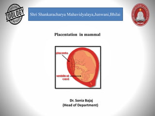
2. Placenta- The placenta is defined as an organ that develops during pregnancy in
mammals. The placenta provides oxygen and nutrients to the growing fetus in the
uterus of the mother.
Circular disc with a diameter of 15-20 cm and thickness of about 2.5 cm at its center.
• It thins off towards the edge.
• It feels spongy and weight about 500 gm.
• Proportion to the weight of the baby being roughly 1:6 at term and occupies about 30% of
the uterine wall.
• It presents two surfaces, fetal and maternal, and a peripheral margin.
Fetal surface
• Covered by the smooth and glistening amnion with the umbilical cord attached at or near its
center.
• Branches of the umbilical vessels are visible beneath the amnion.
• The amnion can be peeled off from the underlying chorion except at the insertion of the
cord.
• At term, about four-fifths of the placenta is of fetal origin.
Maternal surface-
Rough and spongy Dull red colour.
• A thin greyish, somewhat shaggy layer which is remnant of decidua basalis and has come
away wit the placenta, may be visible.
• 15-20 convex polygonal areas known as lobes cotyledons which are limited by fissures.
3. The decidua is the uterine lining (endometrium) during a pregnancy, which forms the
maternal part of the placenta. It is formed under the influence of progesterone and forms
highly characteristic cells.
Structure-
The part of the decidua that interacts with the trophoblast is the decidua basalis (also called
decidua placentalis), while the decidua capsularis grows over the embryo on the luminal
side, enclosing it into the endometrium. The remainder of the decidua is termed the decidua
parietalis or deciduavera, and it will fuse with the decidua capsularis by the fourth month of
gestation.
Three morphologically distinct layers of the decidua basalis can then be described:
Compact outer layer (stratum compactum)
Intermediate layer (stratum spongiosum)
Boundary layer adjacent to the myometrium (stratum basalis)
4. Within the decidua, occasional fibrinoid deposits form where the syncytiotrophoblast is
damaged. The region of fibrinoid deposition where trophoblasts meet the compact
portion of the decidua basalis is called Rohr's layer, while the fibrinoid deposits that
occur between the compact and spongy layer of the decidua basalis is termed
Nitabuch's layer. This layer is absent in placenta accreta.
The decidua has a histologically-
distinct appearance, displaying large polygonal decidual cells in the stroma. These are
enlarged endometrial stromal cells, which resemble epithelium.
Decidualization includes the process of differentiation of the spindle-shape stromal
fibroblasts into the plump secretory decidual cells, which create a pericellular
extracellular matrix rich in fibronectin and laminin (similar to epithelial cells).
Vascularity, as well as vascular permeability, is enhanced in the decidualizing
endometrium.
Its leukocyte population is distinct, with the presence of large endometrial granular
leukocytes being predominant, while polynuclear leukocytes and B cells are scant. The
large granular lymphocytes (CD56 bright) are called "uterine NK cells" or "UNK cell in
human.
5. Development-
After ovulation in placental mammals, the endometrial lining becomes hypertrophic
and vascular under the influence of Estrogens and progesterone.
In animals exhibiting hemochorial placentation, the endometrium undergoes
decidualization following implantation. If implantation does not occur, the secretory
lining will be absorbed estrous cycle or shed (menstrual cycle).
The decidua is shed with the placenta during parturition.
6.
7. Types of Placenta
A. BEHAVIOR DURING PARTURITION-
1.Deciduate type Placenta: The allantochorianic villi penetrate into uterine villi. They are
intimately fused. Hence at the time of birth, the uterus is damaged. Bleeding occurs, the
utrine wall enters into formation of placenta is called deciduas.
2. Non deciduate type placenta: The chorianic villi are simple projections, they lie in
contact with uterus. They have a loose contact. There is no fusion. At the time of birth of
embryo uterus is not damaged.
8. B. ON THE BASIS OF DISTRIBUTION OF VILLI:- According to the distribution of villi five
kinds of placenta are seen.
1.Diffused type placenta: The villi are uniformly distributed on the surface of blastocyst,
except at the extreme ends. Ex: Horse, pig.
2.Cotyledonary placenta: The villi are arranged in groups. Each group is called cotyledon.
Each cotyledon fits into caruncles for uterus. Ex: Sheep, Cow, Deer.
3. Intermediate type Placenta: It is a rare type, it shows free villi on cotyledons. Hence it
is called intermediate type placenta. Ex: Giraffe
In these three types of placenta during perturition the foetus will not damage uterus.
4. Zonary placenta: The placenta takes the form of a complete or incomplete band of
tissue surrounding the fetus.. Ex: Cat, Dog, Carnivores.
5. Discoid type placenta : A single placenta is formed and is discoid in shape. When the
embryo Is growing It move saway from uterus hence the with look like a disc. Ex: Rabbit
9. C. BASING ON HISTOLOGY CONNECTION: According to number of layers of cells present
between foetus and uterus blood supply the placenta Is classified into five types.
a) Epithelio chorion placenta: The foetal chorion Is In contact with epithelium of the
uterus hence it is called epithello chorion placenta. In between foetal, maternal parts six
layers are present. Ex: Pig, Horse
• Endothelium of mother blood vessel.
• Maternal syndesmose connective tissue.
• Epitheliurn of mother
• Chorion of foetus.
• Foetus connective tissue (syndesmose)
• Endothellum of foetal blood vessel.
If all the six layers are present the placenta is called epithellochorlal placenta.
10. b) Syndeumose chorial placenta:
The allanto-chorianic with will pierce into the uterus of the mother, the chorion will come
in contact with syndesmose of mother’s uterus. Hence it is called syndesmose chorial.
Ex: Sheep, Cow.
c) Endotheliochorial placenta: The chàrion of the foetus will come in contact with the
endotheli of mother ‘s uterus, hence it is called endothelio-chorial placenta. Ex: Dog,
Carnivores.
d) Hemo-chorial placenta: The placental connections are more intimate. The chorion of
foetus will float In the blood pools of mother’s uterus. Hence It Is called haemochorIal
placenta. Ex: Bat, Man, Primates
e) Hemo-endothelial placenta: Hence guinea-pig will float In mother’s blood. Hence it
called hemo endothelial placenta. Ex: Rat, Rabbit,
11. D. LAYER THAT FROM YOLK SAC
a) Yolk sac placenta- consisting of the vascular embryonic yolk sac wall intimately
associated with the vascular maternal uterine or oviducal wall, and serving to nourish
the embryo. Ex. Metatheria( Marsupials)
b) Allantois Placenta -the allantois is intimately associated with the chorion,
contributing blood vessels to that structure as it forms—in conjunction with the
endometrium. Ex. Eutharian (Apis)
c) Chorionic Placenta- The fetal part of the placenta is known as the chorion. The
maternal component of the placenta is known as the decidua basalis. Oxygen and
nutrients in the maternal blood in the intervillous spaces diffuse through the walls of
the villi and enter the fetal capillaries. Ex. Primates
12. FUNCTIONS OF PLACENTA:
1.Placenta will form a physiological barrier between mother and foetus. It will possess
foetal and maternal blood mixing.
2.Placenta allows the diffusion of monosaccharaides, amino adds, hormones, vitamins,
oxygen, .carbon dioxide, water and other waste materials, because of this it supplies food,
oxygen to foetus.
3.It works as an excretory organ of foetus. It releases the nitrogenous waste materials Into
mother blood.
4.It works as an endocrine gland. It will secretes lactogen ,progesterone,etc. hormones.
5.The placenta will manufacture fructose from glucose.