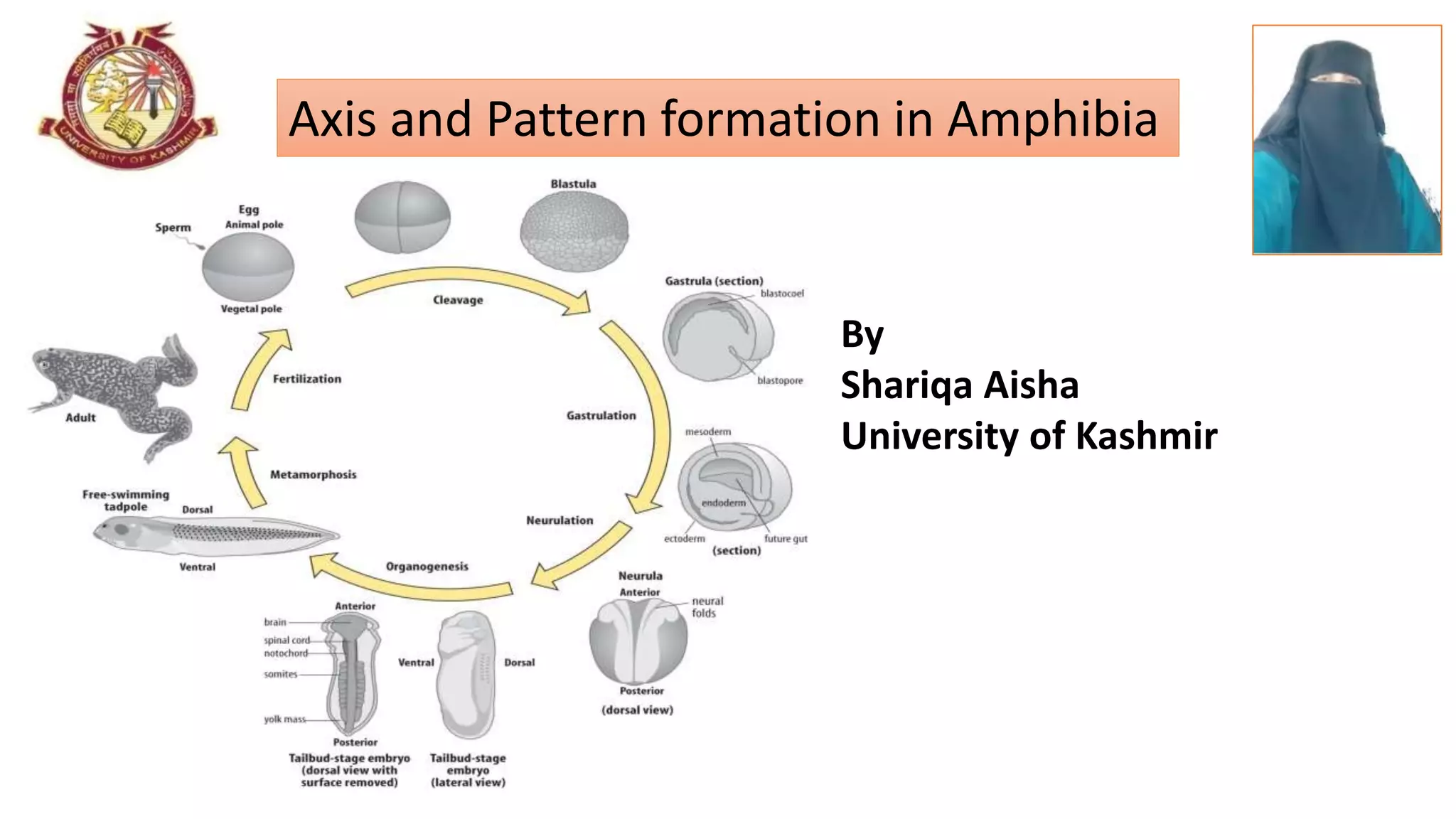
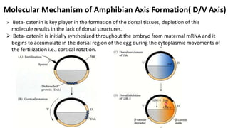
By
Shariqa Aisha
University of Kashmir
Axis and Pattern formation in Amphibia
Picture
representing
the title of
the Topic
LEARNING OBJECTIVES
 To understand the concept of Amphibian development.
 To learn the different developmental processes of Amphibia.
 To know about the various experiments carried on amphibians.
 To find out the various signaling mechanisms in the axis formation of
Amphibia.
Introduction
 A multicellular organism develops from a single cell(the zygote) into a collection of
many different cell types, organized into tissues and organs.
 Development involves cell division, body axis formation, tissue and organ
development, and cell differentiation.
 Frogs and other amphibians have long been model systems to study embryology,
because their eggs are easy to collect and observe during development.
 One such model which scientists studied is Xenopus laevis.
 The major topic we will address with Xenopus is establishment of the vertebrate body
axis.
Introduction
Scientific classification
Kingdom: Animalia
Phylum: Chordata
Class : Amphibia
Order: Anura
Family: Pipidae
Genus: Xenopus
Species: laevis
 Egg type _ Mesolecithal
 Fertilization _ Internal
 Cleavage _ Holoblastic
FERTILIZATION AND CORTICAL ROTATION
 Fertilization can occur anywhere in the animal
hemisphere of the amphibian embryo.
 The point of sperm entry is important as it will mark
the ventral side of the embryo, while the site 180°
degrees opposite the point of sperm entry will mark
the dorsal side.
 After the sperm’s entry , there occurs a cortical
rotation i.e., the cortical cytoplasm rotates 30 degrees
relative to the internal cytoplasm, which results in the
formation of grey crescent.
CORTICAL ROTATION
Reorganisation of the cytoplasm
Before the fertilization, the arrangement
of microtubules are random.
 The sperm centriole organizes the
microtubules of the egg and causes them
to arrange themselves in a parallel array
in the vegetal cytoplasm, separating the
cortical cytoplasm from the yolky internal
cytoplasm.
CLEAVAGE
 Cleavage is unequal, displaced radial, holoblastic.
 First cleavage is meridional cleavage and it begins at the animal pole and slowly
extends down into the vegetal pole.
The second cleavage starts already, while the cleavage furrow is still cleaving the
yolky cytoplasm of the vegetal pole and this cleavage is right angle to the first one
and is also meridional.
 The third cleavage is equational, but it is not actually at the equator but is
displaced toward the animal pole because of the vegetally placed yolk.
 After many cleavages, the morula and then the blastula is formed.
 After cleavage, there is the formation of small numerous cells in the animal pole
and the formation of large few cells in the vegetal pole.
 There is formation of blastocoel cavity, which have two main functions:
1. It allows cell migration during gastrulation.
2. It prevents the cells beneath it from interacting prematurely with the cells above it, if
these cells interact prematurely, they will form Mesoderm.
GASTRULATION ( formation of germinal layers)
 During gastrulation three types of movements occur:
1. Invagination of bottle cells
2. Involution of Dorsal mesoderm
3. Epiboly of ectoderm
 Gastrulation movements in the frog embryos are initiated on the future dorsal side of
the embryo, i.e., the region of grey crescent.
FIRST MOVEMENT STARTS BY INVAGINATION
 here cells invaginate towards inside of the embryo and maintaining contact with the
outside surface by a way of a slender neck.
 These bottle cells form archenteron( primitive gut)
 It is the constriction of these bottle cells that form the blastopore.
 once archenteron is formed, it displace the blastocoel.
SECOND MOVEMENT STARTS BY INVOLUTION:
 During involution the marginal zone cells involute and
form the Dorsal lip of Blastopore.
THIRD MOVEMENT STARTS BY EPIBOLY:
 During gastrulation, the animal cap and noninvoluting
marginal zone cells expand by epiboly to cover the
entire embryo.
 These cells will form the surface ectoderm.
The work of Hans Spemann and Hilde Mangold
Demonstration of nuclear equivalence in newt cleavage
1. When the fertilized egg of the newt Triturus taeniatus was constricted by a ligature,
the nucleus was restricted to one half of the embryo.
2. The cleavage on that side of the embryo reached the 8-cell stage, while the other
side remained undivided.
3. At the 16-cell stage, a single nucleus entered the as-yet undivided half, and the
ligature was further constricted to complete the separation of the two halves.
4. After 140 days, each side has developed into a normal embryo.
Hans Spemann and Hilde Mangold_ Grey Crescent
 When the egg is divided along the plane of first
cleavage into two blastomeres, each of which gets half
of the gray crescent.
 Each experimentally separated cell develops into a
normal embryo.
 When only one of the two blastomeres receives the
entire gray crescent, it alone forms a normal embryo.
 The other blastomere produces a mass of unorganized
tissue lacking dorsal structures.
PHENOMENON OF ORGANISER

The donor tissue invaginates and
forms a second archenteron, and
then a second embryonic axis.
 Both donor and host tissues are
seen in the new neural tube,
notochord, and somites.
Eventually, a second embryo forms
joined to the host.
Molecular Mechanism of Amphibian Axis Formation( D/V Axis)
 Beta- catenin is key player in the formation of the dorsal tissues, depletion of this
molecule results in the lack of dorsal structures.
 Beta- catenin is initially synthesized throughout the embryo from maternal mRNA and it
begins to accumulate in the dorsal region of the egg during the cytoplasmic movements of
the fertilization i.e., cortical rotation.
 GSK3 allows the destruction of Beta- catenin and blocks the dorsal formation.
 Dsh and GBP bind to and block the action of GSK3, and prevent the degradation of Beta-
catenin on the dorsal side of the embryo.
 Wnt11 is also needed to stabilize this reaction, keeping an active source of Dsh.
 The nuclei of the blatomeres in the dorsal region of the embryo receive Beta-catenin,
while the nuclei of those in the ventral region do not.
Specification of Right- Left axis
 The crucial event in left-right axis formation is the expression of a nodal gene in the
lateral plate mesoderm on the left side of the embryo.
In Xenopus, this gene is Xnr1(Xenopus nodal-related 1).
 if the expression of this gene is permitted to occur on the right-hand side, the
position of the heart ( normally found on the left) and the coiling of the gut are
randomized.
 The concentration of the Xnr1 to the left side is caused by the clockwise rotation of
cilia found in the organizer region.
 If rotation of these cilia is blocked, Xnr1 expression fails to occur in the mesoderm
and laterality defects results.
Summary
 Amphibian cleavage is holoblastic, but it is unequal due to the presence of yolk in
the vegetal hemisphere
 Amphibian gastrulation begins with the invagination of the bottle cells, followed
by the coordinated involution of the mesoderm and the epiboly of the ectoderm
 The dorsal lip of the blastopore forms the organizer tissue of the amphibian
gastrula, which consists of pharyngeal endoderm, head mesoderm, notochord,
and dorsal blastopore lip tissue.
 Beta- catenin is key player in the formation of the dorsal tissues, depletion of this
molecule results in the lack of dorsal structures.
 The left-right axis appears to be initiated by the action of a Nodal protein solely
on the left side of the embryo.
List of books and Google links used for title lecture
 Developmental Biology_ SCOTT F. GILBERT
 http://www.ncbi.nllm.nih.gov
 http://www.devbio.biology.gatech.edu
 http://www.embryology.med.unsw.edu.au
 http://www.slideshare.net
http://www.en.m,wikipedia.org
 http://www.sciencedirect.com
Axis and pattern formation in amphibia

Axis and pattern formation in amphibia

  • 1.
    By Shariqa Aisha University ofKashmir Axis and Pattern formation in Amphibia Picture representing the title of the Topic
  • 2.
    LEARNING OBJECTIVES  Tounderstand the concept of Amphibian development.  To learn the different developmental processes of Amphibia.  To know about the various experiments carried on amphibians.  To find out the various signaling mechanisms in the axis formation of Amphibia.
  • 3.
    Introduction  A multicellularorganism develops from a single cell(the zygote) into a collection of many different cell types, organized into tissues and organs.  Development involves cell division, body axis formation, tissue and organ development, and cell differentiation.  Frogs and other amphibians have long been model systems to study embryology, because their eggs are easy to collect and observe during development.  One such model which scientists studied is Xenopus laevis.  The major topic we will address with Xenopus is establishment of the vertebrate body axis.
  • 4.
    Introduction Scientific classification Kingdom: Animalia Phylum:Chordata Class : Amphibia Order: Anura Family: Pipidae Genus: Xenopus Species: laevis  Egg type _ Mesolecithal  Fertilization _ Internal  Cleavage _ Holoblastic
  • 5.
    FERTILIZATION AND CORTICALROTATION  Fertilization can occur anywhere in the animal hemisphere of the amphibian embryo.  The point of sperm entry is important as it will mark the ventral side of the embryo, while the site 180° degrees opposite the point of sperm entry will mark the dorsal side.  After the sperm’s entry , there occurs a cortical rotation i.e., the cortical cytoplasm rotates 30 degrees relative to the internal cytoplasm, which results in the formation of grey crescent.
  • 6.
  • 7.
    Reorganisation of thecytoplasm Before the fertilization, the arrangement of microtubules are random.  The sperm centriole organizes the microtubules of the egg and causes them to arrange themselves in a parallel array in the vegetal cytoplasm, separating the cortical cytoplasm from the yolky internal cytoplasm.
  • 8.
    CLEAVAGE  Cleavage isunequal, displaced radial, holoblastic.  First cleavage is meridional cleavage and it begins at the animal pole and slowly extends down into the vegetal pole. The second cleavage starts already, while the cleavage furrow is still cleaving the yolky cytoplasm of the vegetal pole and this cleavage is right angle to the first one and is also meridional.  The third cleavage is equational, but it is not actually at the equator but is displaced toward the animal pole because of the vegetally placed yolk.  After many cleavages, the morula and then the blastula is formed.  After cleavage, there is the formation of small numerous cells in the animal pole and the formation of large few cells in the vegetal pole.
  • 9.
     There isformation of blastocoel cavity, which have two main functions: 1. It allows cell migration during gastrulation. 2. It prevents the cells beneath it from interacting prematurely with the cells above it, if these cells interact prematurely, they will form Mesoderm.
  • 10.
    GASTRULATION ( formationof germinal layers)  During gastrulation three types of movements occur: 1. Invagination of bottle cells 2. Involution of Dorsal mesoderm 3. Epiboly of ectoderm  Gastrulation movements in the frog embryos are initiated on the future dorsal side of the embryo, i.e., the region of grey crescent. FIRST MOVEMENT STARTS BY INVAGINATION  here cells invaginate towards inside of the embryo and maintaining contact with the outside surface by a way of a slender neck.  These bottle cells form archenteron( primitive gut)  It is the constriction of these bottle cells that form the blastopore.  once archenteron is formed, it displace the blastocoel.
  • 11.
    SECOND MOVEMENT STARTSBY INVOLUTION:  During involution the marginal zone cells involute and form the Dorsal lip of Blastopore. THIRD MOVEMENT STARTS BY EPIBOLY:  During gastrulation, the animal cap and noninvoluting marginal zone cells expand by epiboly to cover the entire embryo.  These cells will form the surface ectoderm.
  • 13.
    The work ofHans Spemann and Hilde Mangold Demonstration of nuclear equivalence in newt cleavage 1. When the fertilized egg of the newt Triturus taeniatus was constricted by a ligature, the nucleus was restricted to one half of the embryo. 2. The cleavage on that side of the embryo reached the 8-cell stage, while the other side remained undivided. 3. At the 16-cell stage, a single nucleus entered the as-yet undivided half, and the ligature was further constricted to complete the separation of the two halves. 4. After 140 days, each side has developed into a normal embryo.
  • 15.
    Hans Spemann andHilde Mangold_ Grey Crescent  When the egg is divided along the plane of first cleavage into two blastomeres, each of which gets half of the gray crescent.  Each experimentally separated cell develops into a normal embryo.  When only one of the two blastomeres receives the entire gray crescent, it alone forms a normal embryo.  The other blastomere produces a mass of unorganized tissue lacking dorsal structures.
  • 16.
    PHENOMENON OF ORGANISER  Thedonor tissue invaginates and forms a second archenteron, and then a second embryonic axis.  Both donor and host tissues are seen in the new neural tube, notochord, and somites. Eventually, a second embryo forms joined to the host.
  • 17.
    Molecular Mechanism ofAmphibian Axis Formation( D/V Axis)  Beta- catenin is key player in the formation of the dorsal tissues, depletion of this molecule results in the lack of dorsal structures.  Beta- catenin is initially synthesized throughout the embryo from maternal mRNA and it begins to accumulate in the dorsal region of the egg during the cytoplasmic movements of the fertilization i.e., cortical rotation.
  • 18.
     GSK3 allowsthe destruction of Beta- catenin and blocks the dorsal formation.  Dsh and GBP bind to and block the action of GSK3, and prevent the degradation of Beta- catenin on the dorsal side of the embryo.  Wnt11 is also needed to stabilize this reaction, keeping an active source of Dsh.  The nuclei of the blatomeres in the dorsal region of the embryo receive Beta-catenin, while the nuclei of those in the ventral region do not.
  • 20.
    Specification of Right-Left axis  The crucial event in left-right axis formation is the expression of a nodal gene in the lateral plate mesoderm on the left side of the embryo. In Xenopus, this gene is Xnr1(Xenopus nodal-related 1).  if the expression of this gene is permitted to occur on the right-hand side, the position of the heart ( normally found on the left) and the coiling of the gut are randomized.  The concentration of the Xnr1 to the left side is caused by the clockwise rotation of cilia found in the organizer region.  If rotation of these cilia is blocked, Xnr1 expression fails to occur in the mesoderm and laterality defects results.
  • 21.
    Summary  Amphibian cleavageis holoblastic, but it is unequal due to the presence of yolk in the vegetal hemisphere  Amphibian gastrulation begins with the invagination of the bottle cells, followed by the coordinated involution of the mesoderm and the epiboly of the ectoderm  The dorsal lip of the blastopore forms the organizer tissue of the amphibian gastrula, which consists of pharyngeal endoderm, head mesoderm, notochord, and dorsal blastopore lip tissue.  Beta- catenin is key player in the formation of the dorsal tissues, depletion of this molecule results in the lack of dorsal structures.  The left-right axis appears to be initiated by the action of a Nodal protein solely on the left side of the embryo.
  • 22.
    List of booksand Google links used for title lecture  Developmental Biology_ SCOTT F. GILBERT  http://www.ncbi.nllm.nih.gov  http://www.devbio.biology.gatech.edu  http://www.embryology.med.unsw.edu.au  http://www.slideshare.net http://www.en.m,wikipedia.org  http://www.sciencedirect.com