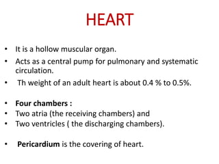
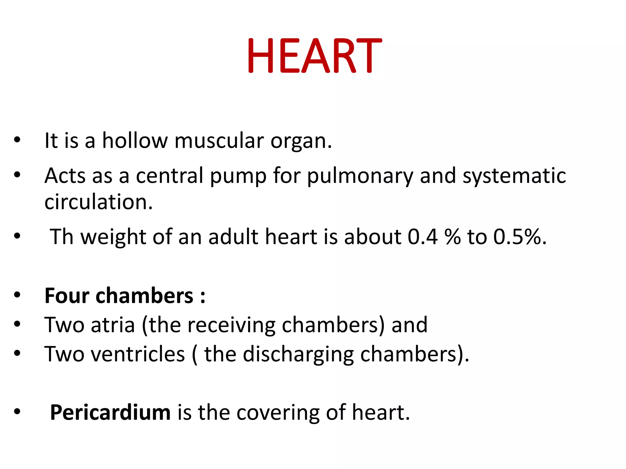
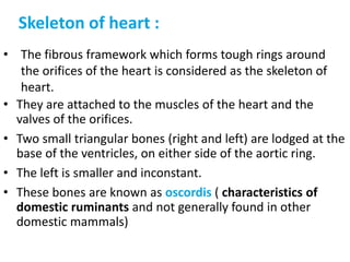
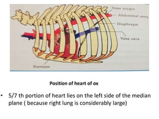
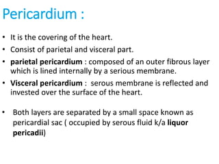
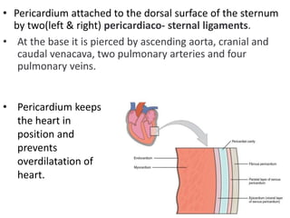
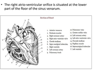
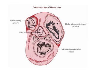
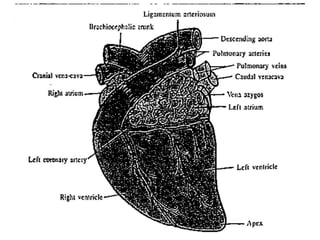
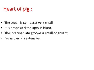
The heart is a hollow muscular organ that acts as the central pump of the circulatory system. It has four chambers - two atria that receive blood and two ventricles that pump blood out. The heart is located in the middle mediastinum in the chest cavity. It has a fibrous pericardium covering and is protected by the pericardial sac that contains serous fluid. The heart has right and left sides, each with an atrium and ventricle, and valves that ensure one-way blood flow.
Right Atrium of human heart
This PPT help to understand the external and internal structures of right atrium.
sulcus terminalis on external surface of rt atrium,
crista terminalis on internal side of rt. atrium,
interior is divided into rough anterior part and smooth posterior part ( sinus venarum)
superior and inferior venae cavae drains deoxygenated blood into rt. atrim
there is Eustachian valve to guard the opening of IVC and Thebesian valve to guard the opening of coronary sinus
septal wall presents fossa ovalis with its border limbus fossa ovalis
THE IMPORTANCE OF MARTIAN ATMOSPHERE SAMPLE RETURN.Sérgio Sacani
The return of a sample of near-surface atmosphere from Mars would facilitate answers to several first-order science questions surrounding the formation and evolution of the planet. One of the important aspects of terrestrial planet formation in general is the role that primary atmospheres played in influencing the chemistry and structure of the planets and their antecedents. Studies of the martian atmosphere can be used to investigate the role of a primary atmosphere in its history. Atmosphere samples would also inform our understanding of the near-surface chemistry of the planet, and ultimately the prospects for life. High-precision isotopic analyses of constituent gases are needed to address these questions, requiring that the analyses are made on returned samples rather than in situ.
Richard's entangled aventures in wonderlandRichard Gill
Since the loophole-free Bell experiments of 2020 and the Nobel prizes in physics of 2022, critics of Bell's work have retreated to the fortress of super-determinism. Now, super-determinism is a derogatory word - it just means "determinism". Palmer, Hance and Hossenfelder argue that quantum mechanics and determinism are not incompatible, using a sophisticated mathematical construction based on a subtle thinning of allowed states and measurements in quantum mechanics, such that what is left appears to make Bell's argument fail, without altering the empirical predictions of quantum mechanics. I think however that it is a smoke screen, and the slogan "lost in math" comes to my mind. I will discuss some other recent disproofs of Bell's theorem using the language of causality based on causal graphs. Causal thinking is also central to law and justice. I will mention surprising connections to my work on serial killer nurse cases, in particular the Dutch case of Lucia de Berk and the current UK case of Lucy Letby.
This pdf is about the Schizophrenia.
For more details visit on YouTube; @SELF-EXPLANATORY;
https://www.youtube.com/channel/UCAiarMZDNhe1A3Rnpr_WkzA/videos
Thanks...!
Slide 1: Title Slide
Extrachromosomal Inheritance
Slide 2: Introduction to Extrachromosomal Inheritance
Definition: Extrachromosomal inheritance refers to the transmission of genetic material that is not found within the nucleus.
Key Components: Involves genes located in mitochondria, chloroplasts, and plasmids.
Slide 3: Mitochondrial Inheritance
Mitochondria: Organelles responsible for energy production.
Mitochondrial DNA (mtDNA): Circular DNA molecule found in mitochondria.
Inheritance Pattern: Maternally inherited, meaning it is passed from mothers to all their offspring.
Diseases: Examples include Leber’s hereditary optic neuropathy (LHON) and mitochondrial myopathy.
Slide 4: Chloroplast Inheritance
Chloroplasts: Organelles responsible for photosynthesis in plants.
Chloroplast DNA (cpDNA): Circular DNA molecule found in chloroplasts.
Inheritance Pattern: Often maternally inherited in most plants, but can vary in some species.
Examples: Variegation in plants, where leaf color patterns are determined by chloroplast DNA.
Slide 5: Plasmid Inheritance
Plasmids: Small, circular DNA molecules found in bacteria and some eukaryotes.
Features: Can carry antibiotic resistance genes and can be transferred between cells through processes like conjugation.
Significance: Important in biotechnology for gene cloning and genetic engineering.
Slide 6: Mechanisms of Extrachromosomal Inheritance
Non-Mendelian Patterns: Do not follow Mendel’s laws of inheritance.
Cytoplasmic Segregation: During cell division, organelles like mitochondria and chloroplasts are randomly distributed to daughter cells.
Heteroplasmy: Presence of more than one type of organellar genome within a cell, leading to variation in expression.
Slide 7: Examples of Extrachromosomal Inheritance
Four O’clock Plant (Mirabilis jalapa): Shows variegated leaves due to different cpDNA in leaf cells.
Petite Mutants in Yeast: Result from mutations in mitochondrial DNA affecting respiration.
Slide 8: Importance of Extrachromosomal Inheritance
Evolution: Provides insight into the evolution of eukaryotic cells.
Medicine: Understanding mitochondrial inheritance helps in diagnosing and treating mitochondrial diseases.
Agriculture: Chloroplast inheritance can be used in plant breeding and genetic modification.
Slide 9: Recent Research and Advances
Gene Editing: Techniques like CRISPR-Cas9 are being used to edit mitochondrial and chloroplast DNA.
Therapies: Development of mitochondrial replacement therapy (MRT) for preventing mitochondrial diseases.
Slide 10: Conclusion
Summary: Extrachromosomal inheritance involves the transmission of genetic material outside the nucleus and plays a crucial role in genetics, medicine, and biotechnology.
Future Directions: Continued research and technological advancements hold promise for new treatments and applications.
Slide 11: Questions and Discussion
Invite Audience: Open the floor for any questions or further discussion on the topic.
Professional air quality monitoring systems provide immediate, on-site data for analysis, compliance, and decision-making.
Monitor common gases, weather parameters, particulates.
1. HEART
• It is a hollow muscular organ.
• Acts as a central pump for pulmonary and systematic
circulation.
• Th weight of an adult heart is about 0.4 % to 0.5%.
• Four chambers :
• Two atria (the receiving chambers) and
• Two ventricles ( the discharging chambers).
• Pericardium is the covering of heart.
2. • They are attached to the muscles of the heart and the
valves of the orifices.
• Two small triangular bones (right and left) are lodged at the
base of the ventricles, on either side of the aortic ring.
• The left is smaller and inconstant.
• These bones are known as oscordis ( characteristics of
domestic ruminants and not generally found in other
domestic mammals)
Skeleton of heart :
• The fibrous framework which forms tough rings around
the orifices of the heart is considered as the skeleton of
heart.
3. Position of heart :
• Cone shaped organ
• Situated at the ventral half of the middle mediastinum with
the base directed upward and apex downward.
• The base extends from 3rd to 6th rib and is placed about
10 to 12 cm below the spine.
• The apex is placed opposite to the 6 th chondrosternal
joint and is very close to the diaphragm.
4. Position of heart of ox
• 5/7 th portion of heart lies on the left side of the median
plane ( because right lung is considerably large)
5. Pericardium :
• It is the covering of the heart.
• Consist of parietal and visceral part.
• parietal pericardium : composed of an outer fibrous layer
which is lined internally by a serious membrane.
• Visceral pericardium : serous membrane is reflected and
invested over the surface of the heart.
• Both layers are separated by a small space known as
pericardial sac ( occupied by serous fluid k/a liquor
pericadii)
6. • Pericardium attached to the dorsal surface of the sternum
by two(left & right) pericardiaco- sternal ligaments.
• At the base it is pierced by ascending aorta, cranial and
caudal venacava, two pulmonary arteries and four
pulmonary veins.
• Pericardium keeps
the heart in
position and
prevents
overdilatation of
heart.
7. Borders and surfaces :
• Both the surfaces ( right & left) are convex.
• The surfaces are traversed by transverse and longitudinal
grooves. ( For occupation of the corresponding coronary
vessels)
• The transverse groove which encircles the heart at the
base of ventricles demarcates the separation b/w auricles
and ventricles.
8. • The longitudinal grooves are mainly two in number (left
& right) , extend from the base of the ventricles and run
towards the apex at the concerned surfaces.
Position : Left ( cranial )
Right ( caudal )
• A small Intermediate longitudinal groove : descends
from the transverse groove along the posterior border of
the heart.
• The left and right longitudinal grooves mark the margin
of the interventricular septum.
9.
10. Right atrium :
• The atrium forms the right anterior part of the base and is
above the right ventricle.
• It consists of a principal cavity called the sinus venarum
and a blind diverticulum called the auricle or auricular
appendix.
• The sinus venarum is the cavity into which the veins open.
(Receives Cranial venacava at the level of 4th rib and
caudal venacava at the level of 5 th rib)
• The auricle curves around the anterior face of the aorta
and its blind end appears in front of the origin of the
pulmonary artery.
11. • Smooth glistening membrane lining the sinus venarum
called the endocardium, which is continuous with that of
the ventricle and the blood vessels entering it.
•
• The opening of the anterior vena cava at the dorso
anterior part of the sinus venarum, which returns the
blood from the anterior parts of the body.
•
• The opening of the posterior venacava returning blood
from the posterior parts of the body and opening in to the
sinus at the dorso-posterior part.
12. • The fossa ovalis is an oval depression in the interatrial
septum at the point of entrance of the posterior vena cava
and is a remnant of the foramen ovale of the foetus.
• The wall of atrium, particularly the appendix presents
some ventrical muscular ridges and looks like a comb and
hence named pectinate muscle.
• The coronary sinus is the opening of the great cardiac vein
and vena hemiazygos into the sinus venarum below the
opening of the posterior vena cava. The orifice is provided
with a semilunar fold of endocardium the coronary valve.
13. • The right atrio-ventricular orifice is situated at the lower
part of the floor of the sinus venarum.
14. Right ventricle :
• The right ventricle forms the anterior part of the ventricular
mass and forms the anterior border of the heart. It does
not reach the apex of the heart.
• It is triangular in outline and crescentric on cross section.
• It communicates above at the base with the right atrium
through the right atrio- ventricular orifice but its left part
projects higher forming the conus arteriosus from which
the pulmonary artery arises.
• The apex of the right ventricle is two inches above the apex
of the heart.
• The septal wall is convex and faces obliquely forward and to
the right.
15.
16. • These are continuous at their bases with the walls of the
ventricle and at the apices they give attachment to the
chordae tendinae which are fibrous cords attached at their
other ends to the cusps of the atrio-ventricular valve.
• The right atrio-ventricular orifice is large oval opening
guarded by the tricuspid valve made up of three cusps.
Of these, one is between the ventricular orifice and the
conus arteriosus; other is against the ventricular septum
and another on the right margin.
• The exit of pulmonary artery is guarded by three
semilunar valves ( thin and convex at their ventricular
surface)
17. • The peripheral edges of the cusps are attached to the
fibrous ring of the atrio-ventricular opening and the
central edges hang down into the ventricle and give
attachment to the chordae tendinae.
• Each cusp receives chordae tendinae from two papillary
muscles. Of the latter, two are on the septum and the third
and the largest springs from the anterior wall.
• The inferior or ventricular surface is convex which is
directed towards the blood when the ventricle contracts.
• A crest-the crista supraventricularis separates cavity of
the conus arteriosus from the atrio-ventricular orifice.
18. Left atrium :
• The left atrium forms the posterior part of the base of the
heart.
• It lies behind the pulmonary artery and aorta and above
the ventricle.
• It consists of a sinus and an auricle.
• The later extends on the left side and its blind end lies
behind the origin of the pulmonary artery.
• The sinus receives the pulmonary veins, about 4 to 7 in
number in two groups.
19. • The following features are seen in the interior :
• The endocardium.
• The openings of pulmonary veins about 4 to 7 in number
into the sinus.
• The musculi pectinate as in the right auricle.
• The left atrio-ventricular orifice.
21. • The edocardium.
• The moderator band commonly two large ones, which are
more tendinous and branched.
• Fewer trabeculae carneae.
• Two large muscular papillaris which are compound and
one on either side.
• The chordae tendinae are fewer but larger than those of
the right ventricle.
• The left atrio-ventricular opening is guarded by the
bicuspid or mitral valve consisting of two cusps (Looks like
bishop's mitre"cap")anterior and posterior.
• Of which, the anterior is between the aortic vestibule and
left atrio-ventricular opening.
22. • The aortic opening is guarded by the aortic valve,
composed of three semi-lunar cusps, one cusp is anterior
and the others right and left posterior in position.
23. Interventricular septum :
• This is situated in between the ventricles in a oblique
manner.
• It's right ventricular surface is convex and left is concave.
• From outside anterior and posterior interventricular
grooves indicate it's attachments with the ventricular walls.
24.
25.
26. Heart of horse :
• It is situated at the level of second intercostal space to 6th
rib.
• The base is completely broader.
• Surface are flatter in comparison to those of ox.
• Apex is placed on 7th sternebra.
• Left and right longitudinal grooves do not meet together.
27. Heart of dog :
• Pericardium is attached to the sternal part of diaphragm
by pericardiaco phrenic ligament.
• Apex is blunt and rounded.
• The organ is placed obliquely at the level between the 3 rd
rib to 6 th Interchondral space.
• The right and left longitudinal grooves meet togather to
the right of the apex.
28. Heart of pig :
• The organ is comparatively small.
• It is broad and the apex is blunt.
• The intermediate groove is small or absent.
• Fossa ovalis is extensive.
29. Heart of fowl :
• Heart is placed at the midline in the cranial part of the
thoraco- abdominal cavity.
• Size is comparatively large.
• Apex is placed between the cranial parts of the two lobes
of the liver.
• Right auriculo ventricular valve is guarded by only one
strong muscular leaf.
• The AV valves are thick.