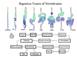
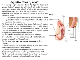
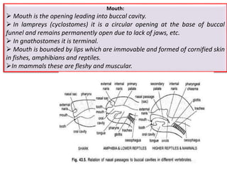
Embryonic Digestive Tract:
Archenteron:
The embryonic archenteron becomes the lining of the adult digestive tract and of all its derivatives.
Splanchnic mesoderm adds layers of connective tissue and smooth muscles around the archenteron.
Ectodermal invagination of the head forms the stomodaeum leading into oral cavity, and a similar mid-ventral ectodermal invagination forms proctodaeum, which leads into the hindgut.
The stomodaeum becomes the adult buccal cavity and gives rise to teeth enamel, epithelial covering of tongue, glands, e.g., mucous, poison and salivary, etc., and Rathke’s pouch of anterior pituitary gland.
The proctodaeum forms either a small terminal part of the cloaca in lower vertebrates and rectum in mammals.
Digestive Tract of Adult:
Following outgrowths arise from the digestive tract- oral glands, Rathke’s pouch, thyroid gland, gill-clefts, tympanic cavity, thymus and other glands of gill-clefts, trachea, lungs, swim bladder, liver, pancreas, yolk sac, and urinary bladder.
Histology: The wall of the alimentary canal is made of four concentric layers.
An outermost visceral peritoneum or serous coat is made of mesothelial cells and thin layer of connective tissue. It is lacking in the oesophagus.
(ii) Below this is a muscular layer formed of smooth muscle fibres arranged in outer longitudinal and inner circular muscle fibres. Between the two layers of muscles is a network of nerve cells and nerve fibres of the autonomic nervous system, known as myenteric plexus or plexus of Auerbach.
(iii) Beneath the muscle layer is a submucosa made of connective tissue having elastic fibres, fat, blood and lymph vessels, nerve cells and fibres glands.
(iv) The innermost layer is a mucosa composed of three regions:
(a) Outer-most narrow muscularis mucosa of outer longitudinal and inner circular smooth muscle fibres.
(b) Middle thin layer of lamina propria of connective tissue, blood vessels, nerves and nodules of lymphatic tissue, and
(c) A basement membrane supporting a layer of columnar epithelial cells which are often glandular and ciliated.
Sense organs are the specialized organs composed of sensory neurons, which help us to perceive and respond to our surroundings. There are five sense organs – eyes, ears, nose, tongue, and skin.
External receptors (exteroceptors): sense organs for touch, smell, taste, sight and hearing.
Internal receptors (interocepyors): these sense organs found in the body which detect the temperature, pain, hunger, thirst, fatigue and muscle position.
INTRODUCTION
The term urogenital refers to something that has both urinary and genital origins. The word urogenital is used because the urinary and reproductive systems in males merge.
These are grouped together because of their proximity to each other, their common embryological origin and the use of common pathways (ex. urethra).
Kidneys and urinary ducts form the urinary system.
The Urinary system performs two important homeostatic processes like excretion and osmoregulation. This system is intimately associated both anatomically, and in terms of embryonic origin with the genital system.
The genital system includes the gonads which generate gametes and the genital ducts that serve as passages for the gametes.
Though functionally different the two organ systems the urinary and the genital system are treated together as the urino- genital system, since both develop from the same segmental blocks of trunk mesoderm or adjacent tissues and share many of the ducts.
Thus although the two systems have nothing common functionally they are closely associated in their use of common ducts and are studied under the broad heading of urinogenital system.
The function of the excretory system is crucial in considering the possible environment of the ‘vertebrate life ’. Several main functions can be attributed to all vertebrate excretory systems:
Excretion of nitrogenous waste products.
Maintaining homeostasis with regard to ions (i.e. salt balance).
Regaining valuable substances (glucose, salts, amino acids, etc.)
Maintaining a physiological osmotic value (i.e. water balance).
The excretory system is formed by a series of paired, segmental nephrons that begin with a nephrostome opening into the coelomic cavity.
A pair of glomeruli per segment, supplied by branches from the aorta, projects into the coelomic cavity close to these nephrostomes.
At a later stage of development, the glomerulus/nephrostome area becomes separated from the rest of the coelomic cavity by an epithelial fold.
The nephrons connect to a duct that is formed by caudal growth of the most anterior nephric tubules. These paired urinary ducts open near the anal region.
In primitive vertebrates, such as the lancelet (petromyzon), the circulating fluid moves without a heart as the central organ of circulation.
In fishes’ single-circuit system, the gills and the heart are placed in series. The two-chambered heart supplies the blood to gills with pressures that exceed those in the arteries. Largely devoid of gravity, fish depend on water for respiration, fluid balance, thermoregulation, reproduction, and fin development.
The amphibians are adapted to life in water only during early stages of their development. Transition to land is marked by loss of fins and gills, and the emergence of tail and limbs.
Adaptation to air respiration introduces a fundamental change in the structure of the cardiovascular system. The heart and the lung are joined by a newly formed pulmonary circulation placed in parallel with the systemic circulation. In contrast to fish, the circulatory loops cross and assume the shape of a lemniscate (figure-eight or ∞-shaped curves).
The heart acquires a new chamber, the left atrium, while a common ventricle is shared between the pulmonary and systemic loops. Amphibians continue to depend for temperature, reproduction, and part of their respiratory needs on water (skin respiration).
Through the development of complicated organ systems such as thermoregulation, respiration, excretion, inner reproduction, and locomotion, mammals have attained a high degree of environmental liberation.
The cardiovascular system consists of two anatomically separate, but functionally unified, parts—the systemic and pulmonary circulations—placed in series.
In addition to an independent inner watery environment, mammals have developed an “inner atmosphere,” reflected primarily in the partial pressure of oxygen and nitrogen in the blood that parallels the atmospheric pressure.
The essential new feature of the mammalian circulation is a pressurized arterial compartment. The similarity of arterial pressure across the mammalian species suggests that the pressure as such does not serve the blood propulsion.
Vertebrate Circulatory Systems:
transport gases, nutrients, waste products, hormones, heat, & various other materials
consist of heart, arteries, capillaries, & veins:
Arteries
carry blood away from the heart
have muscular, elastic walls
terminate in capillary beds
Capillaries
have very thin walls (endothelium only)
are the site of exchange between the blood and body cells
Veins
carry blood back to the heart
have less muscle in their walls than arteries but the walls are very elastic
begin at the end of capillary beds
Heart
a muscular pump (cardiac muscle)
contains a pacemaker to regulate rate but rate can also be influenced by the Autonomic Nervous System
DENTITION IN MAMMALS
The study of arrangement structure and number of types of teeth collectively is called as dentition. Teeth are present in the foetal as well as in adults of mammals, based on the presence of teeth Mammals are two types.
Edentata : In some animals teeth are absent hence called as edentate. e.g., Echidna or spiny ant-eater (Tachyglossus) the teeth are absent in all stages of life.
Dentata : Teeth are present in all mammals though a secon¬dary toothless condition is found in some mammals. Modern turtles and birds lack teeth. The adult platypus (Ornithorhynchus) bears epidermal teeth but no true teeth are present. In platypus embryonic teeth are replaced by horny epidermal teeth in adult.
Classification According to the Shape and Size of the Teeth:
Homodont:
Homodont or Isodont type of teeth is a condition where the teeth are all alike in their shape and size in the toothed whales e.g., Pinnipedians. Fishes, amphibians, reptiles and in the extinct toothed birds.
Heterodont
Heterodont condition is the usual feature in mammals, i.e. the teeth are distinguished according to their shape, size and function. The function is also different at different parts of the tooth row.
According to the Mode of Attachment of Teeth:
Thecodont : The teeth are lodged in bony sockets or alveoli of the jaw bone and capillaries and nerves enter the pulp cavity through the open tips of the hollow roots e.g., mammals, crocodiles and in some fishes.
Acrodont: The teeth are fused to the surface of the underlying jawbone. They have no roots and are attached to the edge of the jawbone by fibrous membrane e.g., fishes, amphibians and some reptiles.
Pleurodont:
The teeth are attached to the inner-side of the jawbone. The tooth touches the bone only with the outer surface of its root. In acrodont and pleurodont types of dentition, there are no roots, and nerves and blood vessels do not enter the pulp cavity at the base, e.g., Necturus (Amphibia) and some reptiles.
According to the Succession or Replace¬ment of Teeth:
looking after the eggs or young until they are independent to defend from predators is known as parental care.
Amphibians show great diversity in Parental care.
Evolutionary change in heart of vertebrates
Heart is situated ventral to the oseophagus in the pericardial section of the coelom.
Heart is a highly muscular pumping organ that pumps blood into arteries and sucks it back through the veins.
In vertebrates it has undergone transformation by twisting from a straight tube to a complex multi-chambered organ.
. There has been an increase in the number of chambers in heart during evolution of vertebrates.
The heart is covered by a transparent protective covering, called pericardium. It is a single layer in fish.
Within pericardium there is a pericardial fluid, protects the heart from the external injury.
The evolution of the heart is based on the separation of oxygenated blood from deoxygenated blood for efficient oxygen transport.
Affinities of Dipnoi or lungfishes towards fishes and amphibians and their phylogenetic relationship and position with respect to Chordates diversification.
They are not the father of amphibians rather they are the uncle of amphibians.
They might have originated from Latimaria like ancestor.
Moreover it is now confirmed that Dipnoi, Crossopterygii and Labirynthodint amphibians are originated from the common ancestor.
Why do animals need to breathe?
Breathing is important to organisms because cells require energy (oxygen) to move, reproduce and function. Breath also expels carbon dioxide, which is a by-product of cellular processes within the bodies of animals.
Respiration is the process of releasing energy from food and this takes place inside the cells of the body.
The process of respiration involves taking in oxygen (of air) into cells, using it for releasing energy by burning food, and then eliminating the waste products (carbon dioxide and water) from the body.
Respiration is essential for life because it provides energy for carrying out all the life processes which are necessary to keep the organisms alive.
The energy produced during respiration is stored in the form of ATP (Adenosine Tri- Phosphate) molecules in the cells of the body and used by the organism as when required.
KEY POINTS
Life started in an anaerobic environment in the so called ‘primodial broth’ (a mixture of organic molecules.
Subsequently, oxygen strangely enough became an crucial factor for aerobic metabolism especially in the higher life forms.
The rise of an oxygenic environment was an important event in the diversification of life.
It evoked a dramatic shift from inefficient to sophisticated oxygen dependent oxidizing ecosystems.
Anaerobic fermentation, the metabolic process that prevailed for the first about 2 billion years of the evolution of life, was a very inefficient way of extracting energy from organic molecules. Ex: A molecule of glucose, e.g., produces only two molecules of ATP (≈ 15 kCal) compared with 36 ATP molecules (≈ 263 kCal) in oxygenic respiration.
Aerobic metabolism must have developed at a critical point when the partial pressure of oxygen rose from an initial level to one adequately high to drive it passively across the cell membrane.
Respiration is a complex and highly integrated biomechanical, physiological, and behavioral processes.
The transfer of O2 occurs through a flow of tissue barriers and compartments by diffusion down a partial pressure gradient, which drops to about zero at the mitochondrial level.
Acquisition of molecular oxygen (O2) from the external fluid media (water and air) and the discharge of carbon dioxide (CO2) into the same milieu is the primary role of respiration.
The respiratory system is a biological system consisting of specific organs and structures.
INTRODUCTION
The jaw (Upper and lower) is any opposable articulated structure at the entrance of the mouth.
It is typically used for grasping and manipulating food.
Jaw suspension means the fusion of upper jaw and lower jaw or skull for efficient biting.
There are different ways in which these attachments are attained depending upon the modifications in visceral arches in vertebrates.
In most vertebrates, the jaws are bony or cartilaginous and oppose vertically.
The vertebrate jaw is derived from the most anterior two pharyngeal arches supporting the gills, and usually bears numerous teeth.
The vertebrate jaw probably originally evolved in the Silurian period and appeared in the Placoderm fish which further diversified in the Devonian.
It is believed that the hyoid system suspends the jaw from the brain case of the skull, permitting great mobility of the jaws.
The original selective advantage offered by the jaw may not be related to feeding, but rather to increased respiration efficiency.
The jaws were used in the buccal pump (observable in modern fish and amphibians) that pumps water across the gills of fish or air into the lungs in the case of amphibians.
Over evolutionary time the more familiar use of jaws (to humans), in feeding, was selected for and became a very important function in vertebrates. Many teleost fish have substantially modified jaws for suction feeding and jaw protrusion, resulting in highly complex jaws with dozens of bones involved.
Jaw Suspension or Suspensoria:
The method by which the upper and lower jaws are suspended or attached from the chondrocranium is known as jaw suspension or suspensorium.
Amongst the visceral arches, the first (mandibular) arch consists of
= a dorsal palato pterygoquadrate bar forming the upper jaw,
= and ventral Meckel’s cartilage forms the lower jaw.
The second (hyoid) arch consists of = a dorsal hyomandibular supporting and suspending the jaws with the cranium, and a ventral hyoid.
The remaining visceral arches support the gills and are, hence, called branchial arches. Thus, splanchnocranium forms the jaws and suspends them with the chondrocranium.
Development of tongue and its salivary glands /certified fixed orthodontic co...Indian dental academy
The Indian Dental Academy is the Leader in continuing dental education , training dentists in all aspects of dentistry and offering a wide range of dental certified courses in different formats.
Aquatic mammals & their adaptation.fully aquatic mammal and amphibian aquatic...Anand P P
this slide animation and videos work mainly in power point 2013 version.the slide contain aquatic mammals and their evolutions.mainly evolutions and their adaptive mechanisms are also listed
Development of tongue and its salivary glands /cosmetic dentistry coursesIndian dental academy
The Indian Dental Academy is the Leader in continuing dental education , training dentists in all aspects of dentistry and
offering a wide range of dental certified courses in different formats.for more details please visit
www.indiandentalacademy.com
Development of tongue and its salivary glands/prosthodontic coursesIndian dental academy
The Indian Dental Academy is the Leader in continuing dental education , training dentists in all aspects of dentistry and
offering a wide range of dental certified courses in different formats.for more details please visit
www.indiandentalacademy.com
The Indian Dental Academy is the Leader in continuing dental education , training dentists in all aspects of dentistry and
offering a wide range of dental certified courses in different formats.for more details please visit
www.indiandentalacademy.com
Crash-Course for AIPMT & Other Medical Exams 2016 (Essentials cockroach)APEX INSTITUTE
Dear Students/Parents
We at 'Apex Institute' are committed to provide our students best quality education with ethics. Moving in this direction, we have decided that unlike other expensive and 5star facility type institutes who are huge investors and advertisers, we shall not invest huge amount of money in advertisements. It shall rather be invested on the betterment, enhancement of quality and resources at our center.
We are just looking forward to have 'word-of-mouth' publicity instead. Because, there is only a satisfied student and his/her parents can judge an institute's quality and it's faculty members coaching.
Those coaching institutes, who are investing highly on advertisements, are actually, wasting their money on it, in a sense. Rather, the money should be invested on highly experienced faculty members and on teaching gears.
We all at 'Apex' are taking this initiative to improve the quality of education along-with each student's development and growth.
Committed to excellence...
With best wishes.
S . Iqbal
( Motivator & Mentor)
Development of tongue and its salivary glands / dental implant coursesIndian dental academy
The Indian Dental Academy is the Leader in continuing dental education , training dentists in all aspects of dentistry and
offering a wide range of dental certified courses in different formats.
Water pollution is a threat to aquatic organisms, including fish, and is a global concern for aquatic biodiversity and ecosystem integrity. This study evaluates the effect of waterborne pollutants on Gangetic Mystus (Mystus cavasius) collected from Chambal River at Nagda, Ujjain. Mystus appeared to be a useful biomarker to assess the impact of toxicity of water pollution.
Zooplankton are the animal component of the plankton community. They are heterotrophic, meaning they can't make their own food and must eat other organisms. In particular, they eat phytoplankton, which are generally smaller than zooplankton.11 species of zooplankton were found in the Shivna River. The most abundant species were copepods Oithona similis, Paracalanus sp., and Calanus sinicus.The species composition of zooplankton varies by season. The highest number of species were found in winter, followed by autumn, summer, and spring. The highest abundance of zooplankton was found in summer, and the lowest in post-monsoon.
This presentation explores how climate change alters the pursuit of economic development: the transformation of poor economies and their people into prosperous ones.
This is hardly the first attempt to reconcile the climate agenda with that of economic development. The United Nations’ Sustainable Development Goals are significant for defining a dual agenda where development targets for people and planet sit alongside each other in a unifying framework.1 Much commentary focuses on the compatibility of the two agendas. A radical and specious view pits progress on climate change and economic development as strict substitutes and calls for no less than the unravelling of economic development to save the planet.2 Cooler heads point instead to their complementarity: the critical role of economic development in supporting adaptation and the recognition that investments in the green transition will propel economies rather than sacrifice living standards.3
In contrast, this essay takes as its starting point that the goals and salience of economic development are immutable. The question posed here is how the quest for economic development changes in a world gripped by a changing climate. The essay argues that climate change will force three major changes: a reappraisal of the causes of and prospects for development, the rebirth of the economics of transition, and a reformulation of the problem development is trying to solve. In a final section, it asks what these changes could mean for international security and for the community of national and global actors who set policy and strategy in this field.
Climate change refers to long-term shifts in temperatures and weather patterns. Such shifts can be natural, due to changes in the sun’s activity or large volcanic eruptions. But since the 1800s, human activities have been the main driver of climate change, primarily due to the burning of fossil fuels like coal, oil and gas.
Burning fossil fuels generates greenhouse gas emissions that act like a blanket wrapped around the Earth, trapping the sun’s heat and raising temperatures.
The main greenhouse gases that are causing climate change include carbon dioxide and methane. These come from using gasoline for driving a car or coal for heating a building, for example. Clearing land and cutting down forests can also release carbon dioxide. Agriculture, oil and gas operations are major sources of methane emissions. Energy, industry, transport, buildings, agriculture and land use are among the main sectors causing greenhouse gases.
In India, bacteria that cause common infections, such as urinary tract and bloodstream infections, are becoming resistant to nearly all antibiotics. This resistance is due to a combination of factors: uncontrolled access to antibiotics, gaps in infection prevention and control (IPC) practices, and high rates of communicable diseases. Antibiotic resistance, or AR, is a serious problem throughout the country, and threatens to reduce the usefulness of antibiotics both in India and around the world.
Because of this emerging threat, India is committed to slowing the spread of AR. Two institutions within India’s Ministry of Health – the Indian Council of Medical Research and National Centre for Disease Control – each developed national networks of public and private hospitals to measure AR trends, prevent healthcare-associated infections (HAIs), and enhance appropriate use of antibiotics. The All India Institute of Medical Sciences is coordinating HAI measurement and prevention efforts in both networks. In addition, efforts in the state of Tamil Nadu focus on building district-level IPC capacity to prevent HAIs, focusing on maternal and neonatal patients.
The Indian Governamnet is is working closely with partners at the national and state level to:
Detect AR pathogens, including novel strains, by developing lab networks and lab expertise.
Use standardized surveillance to monitor and track AR infections in healthcare to learn how often these infections occur and to help develop strategies to prevent them.
Implement focused IPC activities and training.
Optimize use and reduce misuse of critical antibiotics through antibiotic stewardship programs.
Physiological and histopathological effects of Bisphenol A .Bisphenol A is less soluble in water. For that reason, dimethyl sulfoxide (DMSO) was used as a medium to obtain proper distribution in the test solution (Chen, J., et al, 2015). Working solution of commercial grade Bisphenol A (97% pure) was prepared by dilution of stock solution double distilled water immediately prior to experimental use. Serial dilutions of the stock solution were prepared using previously aerated, copper free and stored tap water. The water was continuously aerated. This was prepared by dissolving BPA (50mg) in 100ml of DMSO and the desired concentrations of BPA in tap water were prepared by adding appropriate volumes of this stock solution into test aquarium. A static non-renewable bioassay was conducted in triplicate for each concentration with four animals in each tub. No water exchange was done and the fishes were not fed during the period of the experiment. Percentage mortality was recorded at 12, 24, 48, 72 and 96 h interval. Control group was subjected to acetone at the maximum acetone volume used in the dilution of the dose concentrations. The range of LC50 for H.fossilis (mean wt. 36.78 g) under given conditions was determined to lie between 5 and 10 mg/L for BPA. Hence, for the definitive test, concentrations such as 2, 4, 6, 8, 10, 12, and 14 mg/L of BPA concentration were selected. The test was conducted in triplicate for each concentration with 10 fishes in each tank. At the end of 96 h, the fishes that had survived were anesthetized with clove oil at 100 mg/L, sampled for blood, and processed for hematological analysis. The data obtained from the experiment was processed by probit analysis using a Microsoft Excel computer program.
When pollutants are discharged from a specific location such as a drain pipe carrying industrial effluents discharged directly into a water body it represents point source pollution.
In contrast, non-point sources include discharge of pollutants from diffused sources or from a larger area such as runoff from agricultural fields, grazing lands, construction sites, abandoned mines and pits, etc.
Targets of Sustainable Development Goal 3
WHO Framework Convention on Tobacco Control; support research, development and universal access to affordable vaccines and medicines; increase health financing and support health workforce in developing countries
INTRODUCTION
Toxicology is the science of the poisons. It also studies the nature, effects, detection, assessment and treatment of their effects on biological material.
Toxicology is a multidisciplinary science. The ultimate objective of the combined research is to determine how an organism is affected by exposure to an agent.
This includes an understanding of:
How the agent moves and interact with living cells and tissues of the organism;
What parts of the organism are affected by its presence and health outcomes of this exposure.
Evaluation of the toxicity of substances whose biological effects may not have been well characterized.
The influence of chemical toxicity is mainly
determined by the dosage, duration of exposure,
route of exposure, species, age, sex, and environment.
The goal of toxicology is to contribute to the
general knowledge and harmful actions of
chemical substances.
2. to study their mechanisms of action,
3. and to estimate their possible risks to humans
HISTORY
Dioscorides, a Greek physician in the court of the Roman emperor Nero, made the first attempt to classify plants according to their toxic and therapeutic effect. Poisonous plants and animals were recognized and their extracts used for hunting or in warfare.
In 1500 BC people used hemlock, opium, arrow poisons, and certain metals to poison enemies or for state executions.
Theophrastus Phillipus Auroleus Bombastus von Hohenheim (1493–1541) (also referred to as Paracelsus, a Roman physician from the first century) is considered "the father" of toxicology.
He stated that "All things are poisonous and nothing is without poison; only the dose makes a thing not poisonous.“
Mathieu Orfila (1813) is considered the modern father of toxicology.
In 1850, Jean Stas became the first person to successfully isolate plant poisons from human tissue.
Hippolyte Visart de Bocarmé used nicotine to kill his brother-in-law. He extracted nicotine from tobacco leaves.
The 20th and 21st Centuries have marked by great advancements in the level of understanding of toxicology. DNA and various biochemicals that maintain body functions have been discovered. Our level of knowledge of toxic effects on organs and cells has expanded to the molecular level.
Introduction
Gnathostomata are the jawed vertebrates. (gnathos= "jaw" + (stoma)="mouth".
It comprises roughly 60,000 species. (99% of all living vertebrates).
Living gnathostomes have teeth, and paired appendages.
A horizontal semicircular canal is present in the inner ear.
Myelin sheaths is present on the neurons.
Adaptive immune system uses V(D) J recombination ( it is the mechanism of somatic recombination that occurs only in developing lymphocytes during the early stages of T and B cell maturation. VDJ recombination is the process by which T cells and B cells randomly assemble different gene segments – known as variable (V), diversity (D) and joining (J) genes – in order to generate unique receptors (known as antigen receptors) that can collectively recognize many different types of molecule. While Agnatha (petromyzon and hagfish) use genetic recombination in the variable lymphocyte receptor gene.
It is now assumed that Gnathostomata evolved from ancestors that already possessed a pair of both pectoral and pelvic fins.
In addition to this, some placoderms were shown to have a third pair of paired appendages, that had been modified to claspers in males and basal plates in females—a pattern not seen in any other vertebrate group.
It is believed that the jaws evolved from anterior gill support arches that had acquired a new role, being modified to pump water over the gills by opening and closing the mouth more effectively – the buccal pump mechanism.
Presence of Calcified, bony skull and vertebra are the characteristic features of Gnathostomata (fishes, amphibians, reptiles, birds and mammals).
Pelvic fins are situated just in front of the anus.
Interventrals and basiventrals present in the backbone. These are the elements of the backbone which lie under the notochord, and match the basidorsals and interdorsals respectively.
Gill arches which lie internally to the gills and branchial blood vessels, contrary to the gill arches of all jawless craniates, which are external to the gills and blood vessels.
A horizontal semicircular canal in the inner ear.
Paired nasal sacs which are independent from the hypophysial tube.
There are numerous other characteristics of the soft anatomy and physiology (e.g. myelinated nerve fibres, sperms passing through urinary ducts, etc.), which are unique to the gnathostomes among extant craniates, but cannot by observed in fossils.
Introduction
Ostracoderms (shell-skinned) are of several groups of extinct, primitive, jawless fishes that were covered in an armour of bony plates.
They appeared in the Cambrian, about 510 million years ago, and became extinct towards the end of the Devonian, about 377 million years ago. They were quite abundant during the upper Silurian and Devonian periods. Most of fossils of Ostracodermi were preserved in the bottom sediments of freshwater streams.
However, the opinion is sharply divided as to whether their habitat was freshwater or marine.
The first fossil fishes that were discovered were ostracoderms.
The Swiss anatomist Louis Agassiz received some fossils of bony armored fish from Scotland in the 1830s.
The ostracoderms resembled the present day cyclostomes (lampreys and hagfishes) in many respects and together with them constitute a special group of jawless vertebrates, the Agnatha.
Characteristics: They use gills exclusively for respiration but not for feeding . Earlier chordates with gills used them for both respiration and feeding. Ostracoderms had separate pharyngeal gill pouches along the side of the head, which were permanently open with no protective operculum. mostly small to medium-sized fishes, protected by a heavy, bony dermal (derived from skin) armor. bottom-dwellers; filter-feeders or grazers. no paired fins, but many with stabilizing paired flaps on either side of head.
(1) Ostracoderms were the first vertebrates.
(2) They were popularly called armoured fishes.
(4) They lived in freshwater.
(5) They were bottom dwellers.
(6) Their body was fish-like and did not exceed 30 cm in size.
(7) Paired fins were absent.
(8) Median and caudal fins were present.
(9) The caudal fin was of heterocercal type.
(10) The head and thorax were covered by heavy armour of bones. It protected ostracoderms from the giant scorpion like arthropods, eurypterids.
(11) Bony skull was well developed.
(12) Mouth was mostly present on the ventral side.
(13) They were having large number of gill slits.
(14) The nervous system had 10 pairs of cranial nerves.
(15) The head had a pair of lateral eyes, and a median pineal eye.
(16) They were filter feeders, feeding like a vacuum cleaner.
(17) The endoskeleton was either bony or cartilaginous.
Introduction:
Adaptation to environment is one of the basic characteristics of the living organisms. Living organisms are plastic and posses the inherent properties to respond to a particular environment.
It is a facet of evolution and involve structural diversities amongst living organisms that are heritable. Organisms exhibit numerous structural and functional adaptations that help them to survive as species and to overcome the tremendous competition in nature.
All classes of vertebrates have their representatives leading to partial or total aquatic life.
Water is a homogenous medium for animals.
As a medium, it is heavy in concentration than air.
Stable gaseous and osmotic concentration in a specific region.
Temperature fluctuation is minimum for a particular region.
Water bodies generally have very rich food resources.
Characters of an Aquatic Animal:
An aquatic animal should have the ability to swim to overcome the resistance of the surrounding medium.
Therefore, it should have a streamlined body with an organ or ability to float.
The animal should also have to overcome the problem of osmoregulation.
There are two types of animals living in the present day water, which have undergone aquatic adaptation.
According to their origin, they are primary and secondary aquatic animals.
Adaptations to water habitat are of two types:
Primary aquatic adaptations which includes primitive gill-breathing vertebrates (fishes); Those animals, whose ancestors and themselves are living in the water from the very beginning of their evolution, are called primary aquatic animals. In other words, primary aquatic animals never had a terrestrial ancestry. They exhibit perfect aquatic adaptations. All fishes are primary aquatic animals.
Secondary aquatic adaptations which are acquired as in reptiles, birds and mammals. Those animals whose ancestors were lung breathing land animals, migrated to the water for some reason and ultimately got adapted to live in aquatic habitat, are called secondary aquatic animals. Some of them live partially while others live totally in the water. All aquatic reptiles, aves and mammals are representatives of secondary aquatic animals. Amphibians are in a transitional form between primary and secondary aquatic life.
Sensory adaptations like, electroreception for electrolocation and electro communication, olfaction (vomeronasal system), balance (spatial orientation, movement perception), vision (cornea curvature, retinal topography), and hearing (acoustics, ear anatomy) under the underwater sound reception mechanisms in various aquatic amniotes are well developed.
Origin of the Lateral Line System
Lateral line is a canal along the side of a fish containing pores that open into tubes supplied with sense organs sensitive to low vibrations.
Robert H. Denison explained the origin of the lateral line system. He explained that early vertebrates had a pore-canal system in the dermis which functioned as a primitive sensory system in detecting water movement.
Through the evidences from fossils, embryology and comparative anatomy, Denison (1966) established that the inner ear is closely related to the lateral line system. He found a distinct relationship between the pore canal system and the lateral line in Osteotraci.
The inner ear and the lateral line are developed from ectodermal thickenings, called dorso-lateral placodes. These have a number of similarities, including receptors with sensory hairs, and are both innervated by fibers in the acoustico-lateral area of the brain.
The pore canal system is present and developed in Osteostraci (ostracoderm).
It is also present in Heterostraci which is another group of ostracoderms and includes early vertebrates such as lungfishes and crossopterygians.
As its presence is extensive, it is reasonable to suggest that the pore canal system was a primitive character in early vertebrates .
In transverse sections also , it is very difficult to differentiate the pore canal system from a lateral line canal.
Structure of the Lateral Line System
Epidermal structures called neuromasts form the peripheral area of the lateral line.
Neuromasts consist of two types of cells, hair cells and supporting cells.
Hair cells have an epidermal origin and each hair cell has one high kynocyle (5-10 μm) and 30 to 150 short stereocilia (2-3 μm).
The number of hair cells in each neuromast depends on its size, and they can range from dozens to thousands.
Hair cells can be oriented in two opposite directions with each hair cell surrounded by supporting cells.
At the basal part of each hair cell, there are synaptic contacts with afferent and efferent nerve fibers. Afferent fibers, transmit signals to the neural centres of the lateral line and expand at the neuromast base. The regulation of hair cells is achieved by the action of efferent fibers.
Stereocilia and kinocilium of hair cells are immersed into a cupula and are located above the surface of the sensory epithelium.
The cupula is created by a gel-like media, which is secreted by non-receptor cells of the neuromast.
Central nervous system: The central nervous system consists of the brain and spinal cord. The brain plays a central role in the control of most bodily functions, including awareness, movements, sensations, thoughts, speech, and memory. Some reflex movements can occur via spinal cord pathways without the participation of brain structures. The spinal cord is connected to a section of the brain called the brainstem and runs through the spinal canal.
Peripheral Nervous System: Nerve fibers that exit the brainstem and spinal cord become part of the peripheral nervous system. Cranial nerves exit the brainstem and function as peripheral nervous system mediators of many functions, including eye movements, facial strength and sensation, hearing, and taste.
The autonomic nervous system: The autonomic nervous system is a control system that acts largely unconsciously and regulates bodily functions, such as the heart rate, digestion, respiratory rate, pupillary response, urination, and sexual arousal. This system is the primary mechanism in control of the fight-or-flight response.
The autonomic nervous system comprises two antagonistic sets of nerves, the sympathetic and parasympathetic nervous systems. The hypothalamus is the key brain site for central control of the autonomic nervous system, and the paraventricular nucleus is the key hypothalamic site for this control.
Divisions of Nervous System:
The vertebrate nervous system has three divisions:
(i) A central nervous system comprising the brain and spinal cord. Its function is to receive the stimulus from the receptors and transmit its response to the effectors. Thus, it coordinates all the functions of the body.
(ii) A peripheral nervous system consisting of cranial and spinal nerves arising from the brain and spinal cord respectively. It forms a connecting link between the receptors, central nervous system (CNS) and effectors.
(iii) An autonomic nervous system made of two ganglionated sympathetic nerves, ganglia in the head and viscera, and their connecting nerves. The autonomic nervous system is often regarded as a part of the peripheral nervous system because the two are connected. But all the three divisions of the nervous system are connected intimately both structurally and functionally.
Remote Sensing and Computational, Evolutionary, Supercomputing, and Intellige...University of Maribor
Slides from talk:
Aleš Zamuda: Remote Sensing and Computational, Evolutionary, Supercomputing, and Intelligent Systems.
11th International Conference on Electrical, Electronics and Computer Engineering (IcETRAN), Niš, 3-6 June 2024
Inter-Society Networking Panel GRSS/MTT-S/CIS Panel Session: Promoting Connection and Cooperation
https://www.etran.rs/2024/en/home-english/
Professional air quality monitoring systems provide immediate, on-site data for analysis, compliance, and decision-making.
Monitor common gases, weather parameters, particulates.
Phenomics assisted breeding in crop improvementIshaGoswami9
As the population is increasing and will reach about 9 billion upto 2050. Also due to climate change, it is difficult to meet the food requirement of such a large population. Facing the challenges presented by resource shortages, climate
change, and increasing global population, crop yield and quality need to be improved in a sustainable way over the coming decades. Genetic improvement by breeding is the best way to increase crop productivity. With the rapid progression of functional
genomics, an increasing number of crop genomes have been sequenced and dozens of genes influencing key agronomic traits have been identified. However, current genome sequence information has not been adequately exploited for understanding
the complex characteristics of multiple gene, owing to a lack of crop phenotypic data. Efficient, automatic, and accurate technologies and platforms that can capture phenotypic data that can
be linked to genomics information for crop improvement at all growth stages have become as important as genotyping. Thus,
high-throughput phenotyping has become the major bottleneck restricting crop breeding. Plant phenomics has been defined as the high-throughput, accurate acquisition and analysis of multi-dimensional phenotypes
during crop growing stages at the organism level, including the cell, tissue, organ, individual plant, plot, and field levels. With the rapid development of novel sensors, imaging technology,
and analysis methods, numerous infrastructure platforms have been developed for phenotyping.
Deep Behavioral Phenotyping in Systems Neuroscience for Functional Atlasing a...Ana Luísa Pinho
Functional Magnetic Resonance Imaging (fMRI) provides means to characterize brain activations in response to behavior. However, cognitive neuroscience has been limited to group-level effects referring to the performance of specific tasks. To obtain the functional profile of elementary cognitive mechanisms, the combination of brain responses to many tasks is required. Yet, to date, both structural atlases and parcellation-based activations do not fully account for cognitive function and still present several limitations. Further, they do not adapt overall to individual characteristics. In this talk, I will give an account of deep-behavioral phenotyping strategies, namely data-driven methods in large task-fMRI datasets, to optimize functional brain-data collection and improve inference of effects-of-interest related to mental processes. Key to this approach is the employment of fast multi-functional paradigms rich on features that can be well parametrized and, consequently, facilitate the creation of psycho-physiological constructs to be modelled with imaging data. Particular emphasis will be given to music stimuli when studying high-order cognitive mechanisms, due to their ecological nature and quality to enable complex behavior compounded by discrete entities. I will also discuss how deep-behavioral phenotyping and individualized models applied to neuroimaging data can better account for the subject-specific organization of domain-general cognitive systems in the human brain. Finally, the accumulation of functional brain signatures brings the possibility to clarify relationships among tasks and create a univocal link between brain systems and mental functions through: (1) the development of ontologies proposing an organization of cognitive processes; and (2) brain-network taxonomies describing functional specialization. To this end, tools to improve commensurability in cognitive science are necessary, such as public repositories, ontology-based platforms and automated meta-analysis tools. I will thus discuss some brain-atlasing resources currently under development, and their applicability in cognitive as well as clinical neuroscience.
Travis Hills' Endeavors in Minnesota: Fostering Environmental and Economic Pr...Travis Hills MN
Travis Hills of Minnesota developed a method to convert waste into high-value dry fertilizer, significantly enriching soil quality. By providing farmers with a valuable resource derived from waste, Travis Hills helps enhance farm profitability while promoting environmental stewardship. Travis Hills' sustainable practices lead to cost savings and increased revenue for farmers by improving resource efficiency and reducing waste.
Richard's aventures in two entangled wonderlandsRichard Gill
Since the loophole-free Bell experiments of 2020 and the Nobel prizes in physics of 2022, critics of Bell's work have retreated to the fortress of super-determinism. Now, super-determinism is a derogatory word - it just means "determinism". Palmer, Hance and Hossenfelder argue that quantum mechanics and determinism are not incompatible, using a sophisticated mathematical construction based on a subtle thinning of allowed states and measurements in quantum mechanics, such that what is left appears to make Bell's argument fail, without altering the empirical predictions of quantum mechanics. I think however that it is a smoke screen, and the slogan "lost in math" comes to my mind. I will discuss some other recent disproofs of Bell's theorem using the language of causality based on causal graphs. Causal thinking is also central to law and justice. I will mention surprising connections to my work on serial killer nurse cases, in particular the Dutch case of Lucia de Berk and the current UK case of Lucy Letby.
hematic appreciation test is a psychological assessment tool used to measure an individual's appreciation and understanding of specific themes or topics. This test helps to evaluate an individual's ability to connect different ideas and concepts within a given theme, as well as their overall comprehension and interpretation skills. The results of the test can provide valuable insights into an individual's cognitive abilities, creativity, and critical thinking skills
The use of Nauplii and metanauplii artemia in aquaculture (brine shrimp).pptxMAGOTI ERNEST
Although Artemia has been known to man for centuries, its use as a food for the culture of larval organisms apparently began only in the 1930s, when several investigators found that it made an excellent food for newly hatched fish larvae (Litvinenko et al., 2023). As aquaculture developed in the 1960s and ‘70s, the use of Artemia also became more widespread, due both to its convenience and to its nutritional value for larval organisms (Arenas-Pardo et al., 2024). The fact that Artemia dormant cysts can be stored for long periods in cans, and then used as an off-the-shelf food requiring only 24 h of incubation makes them the most convenient, least labor-intensive, live food available for aquaculture (Sorgeloos & Roubach, 2021). The nutritional value of Artemia, especially for marine organisms, is not constant, but varies both geographically and temporally. During the last decade, however, both the causes of Artemia nutritional variability and methods to improve poorquality Artemia have been identified (Loufi et al., 2024).
Brine shrimp (Artemia spp.) are used in marine aquaculture worldwide. Annually, more than 2,000 metric tons of dry cysts are used for cultivation of fish, crustacean, and shellfish larva. Brine shrimp are important to aquaculture because newly hatched brine shrimp nauplii (larvae) provide a food source for many fish fry (Mozanzadeh et al., 2021). Culture and harvesting of brine shrimp eggs represents another aspect of the aquaculture industry. Nauplii and metanauplii of Artemia, commonly known as brine shrimp, play a crucial role in aquaculture due to their nutritional value and suitability as live feed for many aquatic species, particularly in larval stages (Sorgeloos & Roubach, 2021).
Nucleophilic Addition of carbonyl compounds.pptxSSR02
Nucleophilic addition is the most important reaction of carbonyls. Not just aldehydes and ketones, but also carboxylic acid derivatives in general.
Carbonyls undergo addition reactions with a large range of nucleophiles.
Comparing the relative basicity of the nucleophile and the product is extremely helpful in determining how reversible the addition reaction is. Reactions with Grignards and hydrides are irreversible. Reactions with weak bases like halides and carboxylates generally don’t happen.
Electronic effects (inductive effects, electron donation) have a large impact on reactivity.
Large groups adjacent to the carbonyl will slow the rate of reaction.
Neutral nucleophiles can also add to carbonyls, although their additions are generally slower and more reversible. Acid catalysis is sometimes employed to increase the rate of addition.
The ability to recreate computational results with minimal effort and actionable metrics provides a solid foundation for scientific research and software development. When people can replicate an analysis at the touch of a button using open-source software, open data, and methods to assess and compare proposals, it significantly eases verification of results, engagement with a diverse range of contributors, and progress. However, we have yet to fully achieve this; there are still many sociotechnical frictions.
Inspired by David Donoho's vision, this talk aims to revisit the three crucial pillars of frictionless reproducibility (data sharing, code sharing, and competitive challenges) with the perspective of deep software variability.
Our observation is that multiple layers — hardware, operating systems, third-party libraries, software versions, input data, compile-time options, and parameters — are subject to variability that exacerbates frictions but is also essential for achieving robust, generalizable results and fostering innovation. I will first review the literature, providing evidence of how the complex variability interactions across these layers affect qualitative and quantitative software properties, thereby complicating the reproduction and replication of scientific studies in various fields.
I will then present some software engineering and AI techniques that can support the strategic exploration of variability spaces. These include the use of abstractions and models (e.g., feature models), sampling strategies (e.g., uniform, random), cost-effective measurements (e.g., incremental build of software configurations), and dimensionality reduction methods (e.g., transfer learning, feature selection, software debloating).
I will finally argue that deep variability is both the problem and solution of frictionless reproducibility, calling the software science community to develop new methods and tools to manage variability and foster reproducibility in software systems.
Exposé invité Journées Nationales du GDR GPL 2024
4. Embryonic Digestive Tract:
Archenteron:
The embryonic archenteron becomes the
lining of the adult digestive tract and of all
its derivatives.
Splanchnic mesoderm adds layers of
connective tissue and smooth muscles
around the archenteron.
Ectodermal invagination of the head forms
the stomodaeum leading into oral cavity, and
a similar mid-ventral ectodermal
invagination forms proctodaeum, which
leads into the hindgut.
The stomodaeum becomes the adult
buccal cavity and gives rise to teeth enamel,
epithelial covering of tongue, glands, e.g.,
mucous, poison and salivary, etc., and
Rathke’s pouch of anterior pituitary gland.
The proctodaeum forms either a small
terminal part of the cloaca in lower
vertebrates and rectum in mammals.
The alimentary canal in embryos from
stomach to cloaca is attached to the dorsal
body wall by a double fold of peritoneum,
called the dorsal mesentery, and to ventral
body wall by a ventral mesentery.
In adults, dorsal mesentery persists but the
ventral mesentery disappears leaving only in
the region of liver and urinary bladder.
5. Digestive Tract of Adult:
Following outgrowths arise from the digestive tract- oral
glands, Rathke’s pouch, thyroid gland, gill-clefts, tympanic
cavity, thymus and other glands of gill-clefts, trachea, lungs,
swim bladder, liver, pancreas, yolk sac, and urinary bladder.
Histology: The wall of the alimentary canal is made of four
concentric layers.
i. An outermost visceral peritoneum or serous coat is made
of mesothelial cells and thin layer of connective tissue. It is
lacking in the oesophagus.
(ii) Below this is a muscular layer formed of smooth muscle
fibres arranged in outer longitudinal and inner circular muscle
fibres. Between the two layers of muscles is a network of nerve
cells and nerve fibres of the autonomic nervous system, known
as myenteric plexus or plexus of Auerbach.
(iii) Beneath the muscle layer is a submucosa made of
connective tissue having elastic fibres, fat, blood and lymph
vessels, nerve cells and fibres glands.
(iv) The innermost layer is a mucosa composed of three
regions:
(a) Outer-most narrow muscularis mucosa of outer longitudinal
and inner circular smooth muscle fibres.
(b) Middle thin layer of lamina propria of connective tissue,
blood vessels, nerves and nodules of lymphatic tissue, and
(c) A basement membrane supporting a layer of columnar
epithelial cells which are often glandular and ciliated.
6. Mouth:
Mouth is the opening leading into buccal cavity.
In lampreys (cyclostomes) it is a circular opening at the base of buccal
funnel and remains permanently open due to lack of jaws, etc.
In gnathostomes it is terminal.
Mouth is bounded by lips which are immovable and formed of cornified skin
in fishes, amphibians and reptiles.
In mammals these are fleshy and muscular.
7. Buccal Cavity:
The space between the lips and the jaws is a vestibule.
It may be bounded on the outside by cheeks and on the inside by the
gums.
Mucous glands of cheeks open into the vestibule.
The mouth opens into a buccal cavity, which is a space between the
mouth and the pharynx. The exact point where the stomodaeal ectoderm
and pharyngeal endoderm merge is variable and not easy to discern.
In elasmobranchs and most bony fishes the nasal cavities do not open into
the buccal cavity.
In birds, this palate is cleft due to which nasal and buccal cavities
communicate with each other.
In mammals, secondary palate is continued posteriorly as a membranous
soft palate.
In human beings soft palate hangs into the laryngeal pharynx in the form
of fleshy process, called uvula.
8. Derivatives of Buccal Cavity:
1. Oral Glands:
There are two kinds of integumentary multicellular glands opening into the buccal cavity.
They are mucous glands and enzymatic glands.
Fishes and aquatic amphibians have only mucous glands.
Reptiles have glands in groups, such as palatine, lingual, sublingual, and labial glands named
according to location, they also produce mucus.
In poisonous snakes the upper labial glands are modified to secrete venom, while in the Gila
monster the sublingual glands produce poison.
Birds have sublingual glands and a gland in the angle of the mouth.
Mammals have many small mucous glands besides true and enlarged salivary glands which
are enzymatic. They are parotid, sublingual, sub maxillary and infraorbital salivary glands,
secreting mucin and ptyalin.
2. Tongue:
Tongue in vertebrates show much diversity and are not homologous.
In cyclostomes, there is a muscular, fleshy, rasping tongue with horny teeth for rasping the
skin and muscles of their prey.
Fishes have a primary tongue formed of a fleshy fold of the buccal floor. It has no muscles, but
receptors and teeth are present on the tongue in some bony fishes. The tongue is covered with
mucous membrane.
In some amphibians the tongue is either lacking or immovable. Most amphibians, however,
have a protrusible tongue and in some frogs and toads it may be folded back on itself when not
in use.
9. The tongue in lizards and snakes is often highly developed. In chameleons, it
is very extensible used to capture insects. The tip is thickened and sticky. The
forked tip of the tongue in snakes serves as a means of transferring chemical
stimuli from the external environment to the paired vomero-nasal organs on
the roof of the mouth.
In turtles and crocodilians, the tongue cannot be extended.
The amniote tongue has voluntary muscles, it receives the hypoglossal nerve
and has glands and taste buds. It also develops intrinsic muscles which move
the tongue.
In birds, the tongue is slender and has a horny covering. In some birds the
tongue is immobile, while in some birds it is long, protractile and often used for
capturing the food.
In most mammals, except whales, the tongue is highly developed and
capable of considerable movement, in addition to extension and retraction, due
to the presence of a number of intrinsic muscles.
In mammals the mucous membrane below the tongue forms a median fold,
called frenulum which joins the tongue to the floor of the mouth.
In mammals the upper surface of the tongue bears four kinds of papillae,
(filiform, fungiform, foliate and circumvallate), bearing taste buds except
filiform papillae.
10. 3. Teeth:
Vertebrates have two types of teeth attached to jaw bones- epidermal teeth and true
teeth.
Epidermal teeth are best developed in cyclostomes. They are hard, conical, horny
structures derived from the stratum corneum.
In lampreys they are found on the walls of the buccal funnel and on the tongue.
Tadpole larvae of frogs and toads have serrated epidermal teeth in rows on the lips.
In mammals the adult duckbill platypus has epidermal teeth.
True Teeth:
Teeth are not found in baleen whales and anteaters in mammals, and agnathans,
sturgeons, some toads, sirenians, turtles and modern birds, etc.
In lower vertebrates (such as fish, amphibians and most reptiles) teeth may be
replaced continually an indefinite number of times, such teeth are called polyphyodont.
These teeth are homodont (similar type) and acrodont. (with the jaw bones).
In most mammals teeth are diphyodont, thecodont and heterodont. In some
mammals these are monophyodont having only one set of teeth, e.g., moles, Indian
squirrel.
Teeth are similar in structure to the placoid scales of sharks, formed of a central pulp
cavity, around which is present a thick but soft layer, the dentine, which is externally
covered by a thin, extremely hard enamel.
These are supposed to have derived from bony scales of ostracoderms and
placoderms. For details readers may see the Dentition in Mammals.
11. 4. Adenohypophysis:
The anterior lobe of pituitary gland develops as a dorsal evagination of stomodaeum,
called Rathke’s pouch, which constricts off to form the anterior and middle lobes of
pituitary gland (Adenohypophysis). The posterior lobe of pituitary or neurohypophysis is
the ventral evagination of diencephalon, called infundibulum. Thus, it is nervous part.
Pharynx:
The part of the alimentary canal immediately behind the buccal cavity is a pharynx, lined
with endoderm. It is a common passage serving both for the digestion and respiration.
In fishes, the pharynx is large and laterally perforated for gill-slits, while in tetrapoda, it is
short and bears openings of nostrils.
In embryos, the wall of pharynx gives off a number evaginations which develop into
spiracles, gill-clefts, air bladders, lungs, tonsils and a few endocrine glands (e.g., thymus,
thyroid and parathyroids).
Oesophagus:
The oesophagus is short in most fishes and amphibians because they lack neck, but it is
longer in amniotes due to presence of neck.
The oesophagus of reptiles is more elongate than that of fishes and amphibians. In many
birds, a portion of the oesophagus is enlarged into a crop to store the food. The crop is lacking
in digestive glands but secreted ‘pigeon milk’ to feed the young.
In mammals, the oesophagus is long, lacks glands and varies in relation to the length of
neck. It passes through the diaphragm, the portion below the diaphragm is covered with
visceral peritoneum which is lacking from the upper part.
Oesophagus has mucous glands. Its lining forms longitudinal folds, or finger-like fleshy
papillae (elasmobranchs) or horny papillae in marine turtles.
12. Histologically, the oesophagus differs from the rest of the
alimentary canal in three facts:
(i) It has no visceral peritoneum because it lies outside the
coelom, its outermost covering layer is a thin tunica
adventitia.
(ii) The muscle fibres in its anterior part are striped, middle
part has both striped and unstriped muscles, and the
posterior part has only unstriped muscles. But there are
exceptions in ruminating mammals, all the muscles are
striped or voluntary.
(iii) The mucous membrane lining is made of stratified
squamous epithelial cells and not of columnar cells.
13. Stomach:
There is practically no stomach in cyclostomes, chimeras, lung fishes and some primitive
teleost fishes, since it has no gastric glands.
In most fishes and tetrapoda it is dilated for storage and maceration of solid food, and
digestion of food because it contains gastric glands.
The first part of the stomach, next to the oesophagus, is the cardiac region and the lower
end near the intestine is the pyloric region, which has a pylorus or pyloric valve in which the
mucous membrane lining is surrounded by a thick sphincter muscle which regulates the
opening and closing of the pyloric stomach into the intestine.
Stomach is straight in cyclostomes, gar, Belone, etc., and spindle-shaped in Proteus,
Necturus, some lizards and snakes. In turtles and tortoises, it is a wide curved tube, and in
elasmobranchs the stomach is J-shaped.
In crocodiles and birds the stomach has two parts, a proventriculus with gastric glands,
and a highly muscular gizzard, which represents the pyloric region and has a hard, cornified
lining for grinding food.
In mammals the stomach lies transversely and may be a simple sac or divided into 3
regions, namely cardiac, fundic and pyloric and each region has its gastric glands.
In many ruminants the stomach has four chambers- a rumen, reticulum, omasum and an
abomasum. It is claimed that the first three chambers are modifications of the oesophagus,
and abomasum is the true stomach representing the cardiac, fundic, and pyloric parts of the
stomach.
In camels, there is no omasum, the rumen and reticulum have pouch-like water cells which
were once believed to store water, but they are probably digestive.
14.
15. Histologically, the stomach has the typical parts of the alimentary canal,
but it has two peculiarities.
The muscularis mucosa is made of an outer longitudinal layer and an
inner circular layer of muscles.
The epithelium lining is thick with several types of glandular cells
forming gastric glands of three types called cardiac, fundic, and pyloric
gastric glands.
The cardiac and pyloric glands secrete only mucus from their surface
cells. Fundic glands (or cardiac glands in some) have three kinds of cells,
mucous neck cells produce mucus, oxyntic cells produce hydrochloric acid,
they may be present in the cardiac region also, zymogen cells or peptic cells
secrete pepsin.
In most animals the zymogen cells also secrete two proenzymes called
propepsin and prorennin which are converted by hydrochloric acid into
pepsin and rennin respectively. The secretions of all stomach cells form a
mixture called gastric juice.
16. Small Intestine:
Small intestine is long, narrow and coiled tube after the pylorus.
It is the most important part of the digestive tract because the digestion and
absorption of food take place in it.
In cyclostomes, the intestine is a short straight tube with a spirally arranged
longitudinal flap extending into it.
In elasmobranchs, it is divided into small and large portions, and the small portion
has a spiral valve which greatly increases the absorptive surface.
A spiral valve is also present in the small intestine of a few more primitive bony
fishes, but is lacking in higher forms in which the intestine is long and coiled.
In caecilians, (Amphibia) it is little coiled and not differentiated into a small and
large tract.
In frogs and toads it is relatively long and coiled.
In reptiles, it is more coiled than in amphibians.
For the first time in vertebrates a caecum or blind diverticulum arises at the
junction of small and large intestines. However, this is not permanent in all reptiles.
In birds, the small intestine is coiled or looped and one or two colic caeca are also
present at the junction of small and large intestines.
In most mammals also the small intestine is proportionately long and coiled. Its
length, however, is correlated with food habits. In herbivores it is relatively more
longer in comparison to insectivores and carnivores.
17.
18. There is a blind pocket or caecum at the junction of the colon and small intestine
which is generally small in carnivorous species and quite long in many herbivores.
The first part of the small intestine is the duodenum, which is short beginning
from pylorus and terminates beyond the entrance of pancreatic and hepatic ducts.
It has many folded villi and contains branching Brunner’s glands in the
submucosa which secrete mucus, some alkaline watery-fluid, and a little enzyme.
Duodenum also produces two hormones called secretin and cholecystokinin
which stimulate the pancreas and gall bladder to liberate their juices. Ducts from
the gall bladder and pancreas open into the duodenum.
Behind the duodenum is an ileum, which only in mammals is differentiated into
an anterior smaller jejunum and posterior longer ileum. A large number of small
digestive glands are present in the small intestine. They are tubular glands or crypts
of Lieberkuhn found through the entire length, they secrete mucus and a succus
entericus which has several enzymes.
The lining of the small intestine is folded to form small villi, which increase the
surface area for secretion and absorption. The villi are covered densely by minute
finger-like projections, called microvilli which assist in absorption into the villi. In
mammals nodules of lymphoid tissue called Peyer’s patches are found on the ileum.
19. Large Intestine:
Large intestine has a larger diameter than the small intestine.
It is generally short in fishes, amphibians, reptiles, and birds, but in mammals it is
long.
In lower forms the large intestine forms a rectum, but in tetrapoda it has a colon
and terminal rectum.
In most fishes and amphibians, the terminal part of the rectum leads into a cloaca
formed by the proctodaeum.
The rectum, excretory ducts, and genital ducts open into the cloaca, and it opens to
the exterior by a cloacal aperture. But in many bony fishes and all mammals (except
monotremes) the rectum and urinogenital ducts have separate openings to the
exterior; the opening of the former is an anus.
Rectum of mammals is not homologous with the rectum of vertebrates since in
mammals it is derived by partitioning of embryonic cloaca.
In elasmobranchs the large intestine bears a pair of rectal glands which secrete
mucus and sodium chloride.
In amniotes there is an ileocolic valve at the junction of small and large intestines,
which is absent in fishes. It prevents bacteria to enter into ileum from colon.
In amniotes from this junction arises an ileocolic caecum which is two in birds. It
contains cellulose digesting bacteria. It is very long in herbivorous mammals (rabbit,
horse, cow, etc.). In primates caecum is small having a vestigial vermiform appendix.
20. Digestive Glands:
1. Liver:
The liver arises as a single or double outgrowth from the ventral wall of the embryonic
archenteron.
This outgrowth forms a hollow hepatic diverticulum, which soon differentiates into an
anterior part, which proliferates to become the liver and its bile ducts, and a posterior part,
which gives rise to the gall bladder and cystic duct. The bile ducts join to form a hepatic duct
which unites with the cystic duct to form a common bile duct or ductus choledochus. The region
of the archenteron from which the liver arises becomes the duodenum.
The liver is the largest lobed gland in the body, suspended by a double layer of peritoneum
from the transverse septum or its representative.
A gall bladder is for storage of bile secreted by the hepatic cells, lies in the liver and drains
into the duodenum through common bile duct formed by the union of cystic duct and hepatic
duct. A gall bladder is not indispensable and is lacking in many birds and mammals.
A liver is present in all vertebrates.
In cyclostomes, it is small, single lobed (lampreys) and two lobed in hagfishes.
It is bilobed in elasmobranchs, two or three lobed in bony fishes, amphibians, reptiles and
birds and many lobed in mammals.
Liver is long, narrow and cylindrical in fishes, urodeles and snakes.
It is short, broad and flattened in birds and mammals.
A gall bladder and bile duct are present in larval cyclostomes but they are absent in the adult.
Fishes, amphibians, and reptiles generally have a gall bladder, but it is lacking in many birds.
Most mammals have a gall bladder, but it is absent in Cetacea and Ungulata.
The liver secretes a watery, alkaline bile but have no enzymes. It neutralises the acidity of
21. 2. Pancreas:
Pancreas is formed from the endoderm of the embryonic archenteron.
A single dorsal diverticulum from embryonic duodenum and one or two
ventral outgrowths from the liver form pancreatic diverticula.
The distal parts of diverticula undergo budding to form the main mass of
pancreatic cells to which mesodermal derivatives are added. Thus, a single
gland is made which has several lobes forming either a diffuse or a compact
pancreas.
The pancreas is both an exocrine and endocrine gland, bound together by
delicate strands of connective tissue. The exocrine part secretes digestive
enzymes which are poured into the duodenum through pancreatic ducts.
Whereas the endocrine part secretes hormones such as insulin and glucagon.
A pancreas is present in all vertebrates.
In lampreys, some bony fishes, lungfishes and lower tetrapods, it is a
diffuse organ embedded in the liver, mesenteries and intestinal wall.
Hagfishes have a small pancreas.
Elasmobranchs have a well defined bilobed pancreas. In higher tetrapoda it
is generally a compact gland. One or two pancreatic ducts open into the
duodenum.