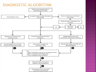
Dr. Md. Ashiqur Rahman
Phase-B resident
Dept. of Gastroenterology
 Author : Mazyar Malakouti, Archish Kataria, Sayed
K. Ali and Steven Schenker
 Published : Journal of Clinical and Translational
Hepatology
 Year : 2017
 Type : Review Article
 Evaluation of abnormal liver enzymes level in
asymptomatic individuals is challenging.
 Serious investigations to find the causes may
lead to high procedural risk and expenses.
 Failure to investigate early may result in life
threatening diagnosis.
 Approx. 1% to 9% asymptomatic patients have
elevated liver enzymes when screened routinely.
 In a US from 1999 to 2002, 8.9% of the study
population show elevated alanine
aminotransferase level.
 Elevated ALT or AST above ULN ( 30 IU/L for men
and 20 IU/L for women ) without any identifiable
risk factors should be evaluated.
 Physiological causes of raise
 Pregnency ( ALP)
 Vigorous Exercise ( ALT,AST)
 Detail History
 Clinical Examinations
 Relevants Investigations
Blood tests
Liver Imaging
Liver Histology
Elevated Liver Enzymes
Repeat the tests
Search for Drug exposure
If Still Persist
Mild
< 5x ULN
Moderate
5-10x ULN
Severe
> 10x ULN
Hepatocellular Injury(
ALT/AST ± bilirubin )
Cholestasis ( ALP ±
bilirubin )
Mixed ( both ALT/AST and
ALP)
Isolated Hyperbilirubineia
1. Patient age and ethnicity;
2. Presence of signs and symptoms of chronic
liver disease
3. Risk factors for viral hepatitis
4. Presence of comorbid conditions like
diabetes, obesity, hyperlipidemia, neurologic
manifestations in Wilson’s disease,
emphysema in alpha-1-antitrypsin deficiency;
5. History of alcohol consumption, medication use,
and toxin exposure
6. Family history of genetic conditions such as
hemochromatosis and WD
7. History of chronic diarrhea, indicating
extrahepatic causes like celiac sprue, thyroid
disorders, IBD etc.
8. Presence of signs and symptoms of heart failure,
9. History of other autoimmune disorders
 Stigmata of acute and chronic liver disease
 Hemochomatosis and WD
 Arthritis
 Acne
 Skin color change
 K-F ring
 Clubbing
 CCF
 Raised JVP
 Hepatomegaly
 Lung basal cracles
 First indicator is AST:ALT >2:1
 GGT is sensitive, but non-specific
 GGT >2x ULN with AST:ALT >2:1 strongly indicate
ARHI
 ALT may be normal
 Raised bilirubin in severe acute alcoholic hepatitis
 Risk of raised transaminase more in overweight or
obesity
 Early and empiric testing of HBV and HCV even in
absence of risk factors for pts
 HEV considered in endemic area
 Most chronic patient ALT,AST <100
 ALT:AST >1
 AST:ALT >1 indicate progress to cirrhosis (79%)
 Simple steatosis to NASH to cirrhosis
 High risk group (T2DM, morbid obesity)
 ALT >AST making AST:ALT<1
 Reversal of ratio that means AST:ALT>1 indicate advanced fibrosis
 GGT upto 3x ULN in 50%
 Bilirubin, Albumin preserved in early stage
 Leucopenia, thrombocytopenia – suspicion of cirrhosis or occult portal
HTN
 Biopsy – Gold standard
 Histology- Fatty infiltration, periportal fibrosis, inflamation and
hepatic necrosis
 Considerderd in men in north European descent
 Screening
 Iron level
 TSAT
 +ve screening test – TSAT>45%
 Liver biopsy (Gold standard) – Hepatic iron >1.9
indicate HH
 Genetic test has lack of sensitivity
 Ferritin <1000µg/L, normal AST, No hepatomegaly –
No cirrhosis
 Raised liver enzymes without clinical symptoms
 Initial screening is s.ceruloplasmin (85%)
 Slit-lamp exam for K-F ring is clinical clue
 24 urinary cu >100µg/day suggestive
 Cu conc. >250µg/g dry liver wt on biopsy
 Genetic test is not useful to make Dx
 Young to middle age women with concominent
autoimmuno disease
 80% pt have hyperGglobinemia
 IgG >2x suggestive
 <40 years, ANA/ASMA +ve – type1
 2-14 years, Anti-LKM +ve – type2
 SLA- type3
 Intermittent flare mimicking acute hepatitis
 Query for common drugs
 Liver biopsy to determine severity
History & Examination
 Fatigue, arthalgia, low-grade fever, jaundice >
Viral Hepatitis
 Abdominal pain, Fever, jaundice > Acute biliary
obstruction
 Presence of shock > Ischemic Hepatitis
 Differential diagnosis - minor hepatitis viruses (EBV, CMV
etc.), WD, hemochromatosis, and autoimmune,
extrahepatic and congenital causes
 Up to 49% of patients with AIH present with moderate
increase in aminotransferase levels and bilirbin
 Acute extrahepatic biliary obstruction - high AST levels (up
to 10x ULN, with peak >50x ULN in 1–2% of patients)
 USG/MRCP usually provide definitive diagnosis
 If Dx evaluation is negative for moderate to severe
aminotransferase elevation, a liver biopsy can be
considered
 Elevated ALP
 Could be physiological
 Liver and bone diseases are pathological cause
 Elevated GGT with raised ALP points to
hepatocellular injury
 Drug-induced injury shows cholestatic pattern
with normal USG
 ALP repeat after discontinuation of drugs at 6-
8wks
 Disease specific markers are –ve, but ALP raised,
then MRCP, liver biopsy should perform
 PBC - no extrahepatic biliary obstruction with at
least 2 of the following criteria
ALP of at least 1.5x ULN
AMA at a titer of 1:40 or higher
Histological evidence of disease
 PSC is a chronic progressive disease with
radiographic findings of abnormal bile ducts with
wall thickening, dilation and stricture
 Unconjgated
 Hemolysis – markers of hemolysis
 Gilbert’s syndrome
 Drugs – rifampicin
 Crigler-najjer syndrome
 Conjugated
 Dubin-johnson syndrome
 Rotor syndrome
 Elevation of liver enzymes is one of the most
common problems in the primary care setting
 History-taking and physical examination is very
important for diagnosis
 Laboratory testing can be based on the pattern
and degree of the elevation
 A systematic approach is help the clinician to
find the cause of elevation
Thank You
Evaluation of liver enzyme elevation.pptx

Evaluation of liver enzyme elevation.pptx

  • 1.
    Dr. Md. AshiqurRahman Phase-B resident Dept. of Gastroenterology
  • 2.
     Author :Mazyar Malakouti, Archish Kataria, Sayed K. Ali and Steven Schenker  Published : Journal of Clinical and Translational Hepatology  Year : 2017  Type : Review Article
  • 4.
     Evaluation ofabnormal liver enzymes level in asymptomatic individuals is challenging.  Serious investigations to find the causes may lead to high procedural risk and expenses.  Failure to investigate early may result in life threatening diagnosis.
  • 5.
     Approx. 1%to 9% asymptomatic patients have elevated liver enzymes when screened routinely.  In a US from 1999 to 2002, 8.9% of the study population show elevated alanine aminotransferase level.
  • 6.
     Elevated ALTor AST above ULN ( 30 IU/L for men and 20 IU/L for women ) without any identifiable risk factors should be evaluated.  Physiological causes of raise  Pregnency ( ALP)  Vigorous Exercise ( ALT,AST)
  • 7.
     Detail History Clinical Examinations  Relevants Investigations Blood tests Liver Imaging Liver Histology
  • 8.
    Elevated Liver Enzymes Repeatthe tests Search for Drug exposure If Still Persist Mild < 5x ULN Moderate 5-10x ULN Severe > 10x ULN Hepatocellular Injury( ALT/AST ± bilirubin ) Cholestasis ( ALP ± bilirubin ) Mixed ( both ALT/AST and ALP) Isolated Hyperbilirubineia
  • 10.
    1. Patient ageand ethnicity; 2. Presence of signs and symptoms of chronic liver disease 3. Risk factors for viral hepatitis 4. Presence of comorbid conditions like diabetes, obesity, hyperlipidemia, neurologic manifestations in Wilson’s disease, emphysema in alpha-1-antitrypsin deficiency;
  • 11.
    5. History ofalcohol consumption, medication use, and toxin exposure 6. Family history of genetic conditions such as hemochromatosis and WD 7. History of chronic diarrhea, indicating extrahepatic causes like celiac sprue, thyroid disorders, IBD etc. 8. Presence of signs and symptoms of heart failure, 9. History of other autoimmune disorders
  • 12.
     Stigmata ofacute and chronic liver disease  Hemochomatosis and WD  Arthritis  Acne  Skin color change  K-F ring  Clubbing  CCF  Raised JVP  Hepatomegaly  Lung basal cracles
  • 14.
     First indicatoris AST:ALT >2:1  GGT is sensitive, but non-specific  GGT >2x ULN with AST:ALT >2:1 strongly indicate ARHI  ALT may be normal  Raised bilirubin in severe acute alcoholic hepatitis  Risk of raised transaminase more in overweight or obesity
  • 15.
     Early andempiric testing of HBV and HCV even in absence of risk factors for pts  HEV considered in endemic area  Most chronic patient ALT,AST <100  ALT:AST >1  AST:ALT >1 indicate progress to cirrhosis (79%)
  • 16.
     Simple steatosisto NASH to cirrhosis  High risk group (T2DM, morbid obesity)  ALT >AST making AST:ALT<1  Reversal of ratio that means AST:ALT>1 indicate advanced fibrosis  GGT upto 3x ULN in 50%  Bilirubin, Albumin preserved in early stage  Leucopenia, thrombocytopenia – suspicion of cirrhosis or occult portal HTN  Biopsy – Gold standard  Histology- Fatty infiltration, periportal fibrosis, inflamation and hepatic necrosis
  • 17.
     Considerderd inmen in north European descent  Screening  Iron level  TSAT  +ve screening test – TSAT>45%  Liver biopsy (Gold standard) – Hepatic iron >1.9 indicate HH  Genetic test has lack of sensitivity  Ferritin <1000µg/L, normal AST, No hepatomegaly – No cirrhosis
  • 18.
     Raised liverenzymes without clinical symptoms  Initial screening is s.ceruloplasmin (85%)  Slit-lamp exam for K-F ring is clinical clue  24 urinary cu >100µg/day suggestive  Cu conc. >250µg/g dry liver wt on biopsy  Genetic test is not useful to make Dx
  • 19.
     Young tomiddle age women with concominent autoimmuno disease  80% pt have hyperGglobinemia  IgG >2x suggestive  <40 years, ANA/ASMA +ve – type1  2-14 years, Anti-LKM +ve – type2  SLA- type3  Intermittent flare mimicking acute hepatitis
  • 20.
     Query forcommon drugs  Liver biopsy to determine severity
  • 22.
    History & Examination Fatigue, arthalgia, low-grade fever, jaundice > Viral Hepatitis  Abdominal pain, Fever, jaundice > Acute biliary obstruction  Presence of shock > Ischemic Hepatitis
  • 24.
     Differential diagnosis- minor hepatitis viruses (EBV, CMV etc.), WD, hemochromatosis, and autoimmune, extrahepatic and congenital causes  Up to 49% of patients with AIH present with moderate increase in aminotransferase levels and bilirbin  Acute extrahepatic biliary obstruction - high AST levels (up to 10x ULN, with peak >50x ULN in 1–2% of patients)  USG/MRCP usually provide definitive diagnosis  If Dx evaluation is negative for moderate to severe aminotransferase elevation, a liver biopsy can be considered
  • 25.
     Elevated ALP Could be physiological  Liver and bone diseases are pathological cause  Elevated GGT with raised ALP points to hepatocellular injury  Drug-induced injury shows cholestatic pattern with normal USG  ALP repeat after discontinuation of drugs at 6- 8wks  Disease specific markers are –ve, but ALP raised, then MRCP, liver biopsy should perform
  • 26.
     PBC -no extrahepatic biliary obstruction with at least 2 of the following criteria ALP of at least 1.5x ULN AMA at a titer of 1:40 or higher Histological evidence of disease  PSC is a chronic progressive disease with radiographic findings of abnormal bile ducts with wall thickening, dilation and stricture
  • 27.
     Unconjgated  Hemolysis– markers of hemolysis  Gilbert’s syndrome  Drugs – rifampicin  Crigler-najjer syndrome  Conjugated  Dubin-johnson syndrome  Rotor syndrome
  • 28.
     Elevation ofliver enzymes is one of the most common problems in the primary care setting  History-taking and physical examination is very important for diagnosis  Laboratory testing can be based on the pattern and degree of the elevation  A systematic approach is help the clinician to find the cause of elevation
  • 29.