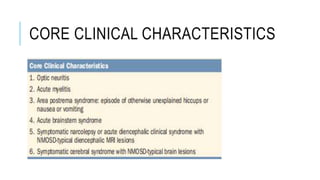
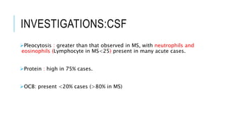
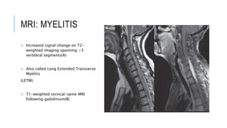
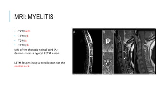
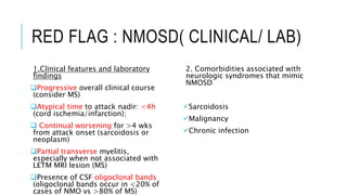
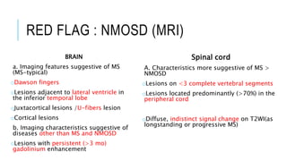
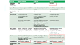
NEUROMYELITIS OPTICA
Tareq Esteak
Resident(Neurology)
NINS
DEFINITION
Neuromyelitis optica (NMO; Devic’s disease) is an aggressive
inflammatory disorder characterized by-
Severe immune mediated demyelination and axonal damage
Predominantly targeting the Optic nerve and Spinal Cord
distinct from multiple sclerosis (MS) that is associated with serum
aquaporin-4 immunoglobulin G antibodies (AQP4-IgG).
NMO SPECTRUM DISORDER
(NMOSD)
A more inclusive term NMO Spectrum Disorder (NMOSD) has been
proposed to incorporate individuals with partial forms, and also those
with involvement of additional structures in the central nervous
system
EPIDEMIOLOGY
NMO is more frequent in women than men (>3:1)
Typically begins in adulthood (30-40Y) but can arise at any age
Incidence: .05-0.4 per100,000 population.
Prevelance: .5-4.4 per 100,000 population.
PATHPHOGENESIS
IL-6 mediated active inflammation , astrocyte injury, demyelination
After crossing the BBB
AQP4-Ig binds with the foot process of astrocyte
Following an infection/Trigger
AQP4-Ig is synthesized in serum by mature B-cell
AQP4 is expressed at the BBB on the foot processes of Astrocyte
as well as at paranodal regions near nodes of Ranvier
CLINICAL COARSE
NMO is typically a recurrent disease; quite disabling over time
The course is monophasic in <10% of patients. Individuals who test
negative for AQP-4 antibody are more likely to have a monophasic
course.
Respirator failure from cervical myelitis may occur over time
8 years after onset, 60% of patients may end up blind and more than
half
will have permanent paralysis of one or more limbs. –if untreated
CLINICAL FEATURE
Hallmarks :
Optic neuritis
Transverse Myelitis
CLINICAL FEATURE: OPTIC
NEURITIS(1)
Usually bilateral
More severe
If unilateral sequential ON in other eye within quick succesion
CLINICAL FEATURE:TRANSVERSE
MYELITIS(2)
Myelitis can be severe and transverse (rare in MS)
Typically longitudinally extensive involving three or
more contiguous vertebral segments.
Also termed as LETM /Longitudinal Extensive Transverse Myelitis
Myelopathic symptoms like Bowel-bladder disturbance ,sensory level
are common.
Lhermitte sign can also be observed.(also present in MS)
CLINICAL FEATURE: CEREBRAL
INVOLVEMENT(3)
Encephalopathy
Seizure
Cognitive impairment
Hemiperesis
Aphasia
Homonymous hemianopia
CLINICAL FEATURE:BRAIN
STEM(4,5)
Commonly area postrema in lower medulla presenting is involved.
Patient present with as intractable hiccoughs or vomiting
May present with acute brainstem syndromes.
May present with bulbar palsy or cranial nerve palsies.
CLINICAL
FEATURE:DIENCEPHALON(6)
1. Endocrinopathies :
Diabetes Insipidus
Hypotohyridiosm
Hypothalamic dysfunctin: Amenorrhoea, Galactorrhoea,
Hyperphagia,weight gain.
2. Narcolepsy
CORE CLINICAL CHARACTERISTICS
INVESTIGATIONS
NO single diagnostic test
Diagnosis on the basis of fulfillment of diagnostic criteria.
DIAGNOSTIC CRITERIA: AQP4
POSITIVE
DIAGNOSTIC CRITERIA:AQP4
NEGATIVE/UNAVAILABLE
DIAGNOSTIC CRITERIA: ADDITIONAL MRI
REQUIREMENT
INVESTIGATIONS:CSF
Pleocytosis : greater than that observed in MS, with neutrophils and
eosinophils (Lymphocyte in MS<25) present in many acute cases.
Protein : high in 75% cases.
OCB: present <20% cases (>80% in MS)
INVESTIGATIONS:SERUM
S.AQP4 IgG: Aquaporin4 antibody
Serum testing >is more sensitive than in csf
Positive in 70% cases
Anti-AQP4 seropositive patients have a high risk for future relapses
More than half will relapse within 1 year if untreated.
NEUROIMAGING
MRI is the choice of Imaging
Commonly lesion found in Optic nerve & Spinal cord
The brain MRI was earlier thought to be normal in NMO
But it is now recognized that in many cases brain lesions are present,
including areas of nonspecific signal changes. E.g. Cerebrum, Area
postrema, Brain stem, Hypothalamus
MRI: MYELITIS
 Increased signal change on T2-
weighted imaging spanning >3
vertebral segments(A)
 Also called Long Extended Transverse
Myelitis
(LETM)
 T1-weighted cervical-spine MRI
following gadolinium(B)
MRI: MYELITIS
• T2WI:A,D
• T1WI+:E
• T2WI:B
• T1WI+:C
MRI of the thoracic spinal cord (A)
demonstrates a typical LETM lesion
LETM lesions have a predilection for the
central cord
MRI:OPTIC NEURITIS
T1WI+: enhancement
of the left optic nerve.
T2WI:coronal (J) planes shows increased
signal in left optic nerve, especially its
posterior portion
T1WI+: Optic chiasma enhancement
MRI: AREA POSTREMA
FLAIR(A), T2(B), T1+: MRI shows
a lesion in the dorsal medulla
(AP) arrow
Axial, FLAIR (D; arrows) and T1-
weighted MRI with gadolinium (E;
arrowheads) show dorsal medulla
involvement in a patient with acute
area postrema
MRI:BRAIN STEM
FLAIR MRI shows dorsal midbrain
(G; arrow);Sagittal section shows
increased signal surrounding the
fourth ventricle(H)
FLAIR MRI shows
periependymal lesions
involving the pon
MRI:CEREBRAL LESION
FLAIR : A-thalamic, B-
Hypothalamic, C- diffuse white
matter lesion
FLAIR: Corpus callosal lesion
RED FLAG : NMOSD
RED FLAG : NMOSD( CLINICAL/ LAB)
1.Clinical features and laboratory
findings
Progressive overall clinical course
(consider MS)
Atypical time to attack nadir: <4h
(cord ischemia/infarction);
 Continual worsening for >4 wks
from attack onset (sarcoidosis or
neoplasm)
Partial transverse myelitis,
especially when not associated with
LETM MRI lesion (MS)
Presence of CSF oligoclonal bands
(oligoclonal bands occur in <20% of
cases of NMO vs >80% of MS)
2. Comorbidities associated with
neurologic syndromes that mimic
NMOSD
Sarcoidosis
Malignancy
Chronic infection
RED FLAG : NMOSD (MRI)
BRAIN
a. Imaging features suggestive of MS
(MS-typical)
oDawson fingers
oLesions adjacent to lateral ventricle in
the inferior temporal lobe
oJuxtacortical lesions /U-fibers lesion
oCortical lesions
b. Imaging characteristics suggestive of
diseases other than MS and NMOSD
oLesions with persistent (>3 mo)
gadolinium enhancement
Spinal cord
A. Characteristics more suggestive of MS >
NMOSD
oLesions on <3 complete vertebral segments
oLesions located predominantly (>70%) in the
peripheral cord
oDiffuse, indistinct signal change on T2WI(as
longstanding or progressive MS)
DISEASE ASSOCIATED WITH
NMOSD
Autoimmune: Up to 40% of NMO patients have a systemic
autoimmune disorder,
E.g. SLE, Sjögren’s syndrome, (p-ANCA)–associated vasculitis,
myasthenia gravis, Hashimoto’s thyroiditis, or MCTD.
Infective:In others, onset may be associated with acute
infection with varicella zoster virus, Epstein-Barr virus, HIV, or
tuberculosis.
Malignancy: Rarely paraneoplastic and associated with
breast, lung, or other cancers.6
TREATMENT: ACUTE ATTACK
Acute attack: high dose GC
IV methylprednisolone 1 g/d for 5–10 d
If effective:
then oral prednisone slow taper
Ineffective:
Plasma exchange(every alt
day for 7 exchange)
TREATMENT: PREVENTION OF
RELAPSE
• Oral
mycophenolate
mofetil (1000 mg
bid);
• Inhibit B-cell &
T-cell
proliferation
• IV Injection
• Rituximab
• CD-20 inhibitor
which inhibit B-
cell proliferation
• Combination
• Glucocorticoid+
• Azathioprine
• Inhibit purin
synthesis which
inhibit inhibit B-
cell proliferation
DMTS: INEFFECTIVE IN NMOSD
Available evidence suggests that interferon beta is ineffective and
paradoxically may increase the risk of NMO relapses
Based on limited data glatiramer acetate, fingolimod, natalizumab,
and alemtuzumab also appear to be ineffective.
IT highlights the need for efficient diagnosis of this disorder.
DURATION OF THE ATTACK
oDMT at least for 5years
oIf severe ,recurrent relapses lifelong
oLength can be tailored according to the disease severity ,and
disability.
EMERGING THERAPIES
 Anti-CD19 monoclonal antibody (inebilizumab)
The terminal complement inhibitor (eculizumab),
DEMYELINATION ASSOCIATED
WITH
ANTI-MOG ANTIBODIES
No single term is widely accepted to describe this disease.
 Most recently, the term MOG-antibody (MOG-IgG) disease has been
suggested
Approximately 20% to 25% of patients with NMOSD are AQP4-IgG
negative.
Up to 25% of patients with seronegative NMOSD will have
antibodies to MOG-IgG.
Anti-MOG antibodies are at risk for bilateral, synchronous optic
neuritis and myelitis
Also associated with cases of acute disseminated encephalomyelitis
(ADEM)
The treatment approach to AQP4-IgG–seronegative NMOSD is
Thank You………..

Neuromyelitis optica

  • 1.
  • 2.
    DEFINITION Neuromyelitis optica (NMO;Devic’s disease) is an aggressive inflammatory disorder characterized by- Severe immune mediated demyelination and axonal damage Predominantly targeting the Optic nerve and Spinal Cord distinct from multiple sclerosis (MS) that is associated with serum aquaporin-4 immunoglobulin G antibodies (AQP4-IgG).
  • 3.
    NMO SPECTRUM DISORDER (NMOSD) Amore inclusive term NMO Spectrum Disorder (NMOSD) has been proposed to incorporate individuals with partial forms, and also those with involvement of additional structures in the central nervous system
  • 4.
    EPIDEMIOLOGY NMO is morefrequent in women than men (>3:1) Typically begins in adulthood (30-40Y) but can arise at any age Incidence: .05-0.4 per100,000 population. Prevelance: .5-4.4 per 100,000 population.
  • 5.
    PATHPHOGENESIS IL-6 mediated activeinflammation , astrocyte injury, demyelination After crossing the BBB AQP4-Ig binds with the foot process of astrocyte Following an infection/Trigger AQP4-Ig is synthesized in serum by mature B-cell AQP4 is expressed at the BBB on the foot processes of Astrocyte as well as at paranodal regions near nodes of Ranvier
  • 6.
    CLINICAL COARSE NMO istypically a recurrent disease; quite disabling over time The course is monophasic in <10% of patients. Individuals who test negative for AQP-4 antibody are more likely to have a monophasic course. Respirator failure from cervical myelitis may occur over time 8 years after onset, 60% of patients may end up blind and more than half will have permanent paralysis of one or more limbs. –if untreated
  • 7.
    CLINICAL FEATURE Hallmarks : Opticneuritis Transverse Myelitis
  • 8.
    CLINICAL FEATURE: OPTIC NEURITIS(1) Usuallybilateral More severe If unilateral sequential ON in other eye within quick succesion
  • 9.
    CLINICAL FEATURE:TRANSVERSE MYELITIS(2) Myelitis canbe severe and transverse (rare in MS) Typically longitudinally extensive involving three or more contiguous vertebral segments. Also termed as LETM /Longitudinal Extensive Transverse Myelitis Myelopathic symptoms like Bowel-bladder disturbance ,sensory level are common. Lhermitte sign can also be observed.(also present in MS)
  • 10.
    CLINICAL FEATURE: CEREBRAL INVOLVEMENT(3) Encephalopathy Seizure Cognitiveimpairment Hemiperesis Aphasia Homonymous hemianopia
  • 11.
    CLINICAL FEATURE:BRAIN STEM(4,5) Commonly areapostrema in lower medulla presenting is involved. Patient present with as intractable hiccoughs or vomiting May present with acute brainstem syndromes. May present with bulbar palsy or cranial nerve palsies.
  • 12.
    CLINICAL FEATURE:DIENCEPHALON(6) 1. Endocrinopathies : DiabetesInsipidus Hypotohyridiosm Hypothalamic dysfunctin: Amenorrhoea, Galactorrhoea, Hyperphagia,weight gain. 2. Narcolepsy
  • 13.
  • 14.
    INVESTIGATIONS NO single diagnostictest Diagnosis on the basis of fulfillment of diagnostic criteria.
  • 15.
  • 16.
  • 17.
  • 18.
    INVESTIGATIONS:CSF Pleocytosis : greaterthan that observed in MS, with neutrophils and eosinophils (Lymphocyte in MS<25) present in many acute cases. Protein : high in 75% cases. OCB: present <20% cases (>80% in MS)
  • 19.
    INVESTIGATIONS:SERUM S.AQP4 IgG: Aquaporin4antibody Serum testing >is more sensitive than in csf Positive in 70% cases Anti-AQP4 seropositive patients have a high risk for future relapses More than half will relapse within 1 year if untreated.
  • 20.
    NEUROIMAGING MRI is thechoice of Imaging Commonly lesion found in Optic nerve & Spinal cord The brain MRI was earlier thought to be normal in NMO But it is now recognized that in many cases brain lesions are present, including areas of nonspecific signal changes. E.g. Cerebrum, Area postrema, Brain stem, Hypothalamus
  • 21.
    MRI: MYELITIS  Increasedsignal change on T2- weighted imaging spanning >3 vertebral segments(A)  Also called Long Extended Transverse Myelitis (LETM)  T1-weighted cervical-spine MRI following gadolinium(B)
  • 22.
    MRI: MYELITIS • T2WI:A,D •T1WI+:E • T2WI:B • T1WI+:C MRI of the thoracic spinal cord (A) demonstrates a typical LETM lesion LETM lesions have a predilection for the central cord
  • 23.
    MRI:OPTIC NEURITIS T1WI+: enhancement ofthe left optic nerve. T2WI:coronal (J) planes shows increased signal in left optic nerve, especially its posterior portion T1WI+: Optic chiasma enhancement
  • 24.
    MRI: AREA POSTREMA FLAIR(A),T2(B), T1+: MRI shows a lesion in the dorsal medulla (AP) arrow Axial, FLAIR (D; arrows) and T1- weighted MRI with gadolinium (E; arrowheads) show dorsal medulla involvement in a patient with acute area postrema
  • 25.
    MRI:BRAIN STEM FLAIR MRIshows dorsal midbrain (G; arrow);Sagittal section shows increased signal surrounding the fourth ventricle(H) FLAIR MRI shows periependymal lesions involving the pon
  • 26.
    MRI:CEREBRAL LESION FLAIR :A-thalamic, B- Hypothalamic, C- diffuse white matter lesion FLAIR: Corpus callosal lesion
  • 27.
  • 28.
    RED FLAG :NMOSD( CLINICAL/ LAB) 1.Clinical features and laboratory findings Progressive overall clinical course (consider MS) Atypical time to attack nadir: <4h (cord ischemia/infarction);  Continual worsening for >4 wks from attack onset (sarcoidosis or neoplasm) Partial transverse myelitis, especially when not associated with LETM MRI lesion (MS) Presence of CSF oligoclonal bands (oligoclonal bands occur in <20% of cases of NMO vs >80% of MS) 2. Comorbidities associated with neurologic syndromes that mimic NMOSD Sarcoidosis Malignancy Chronic infection
  • 29.
    RED FLAG :NMOSD (MRI) BRAIN a. Imaging features suggestive of MS (MS-typical) oDawson fingers oLesions adjacent to lateral ventricle in the inferior temporal lobe oJuxtacortical lesions /U-fibers lesion oCortical lesions b. Imaging characteristics suggestive of diseases other than MS and NMOSD oLesions with persistent (>3 mo) gadolinium enhancement Spinal cord A. Characteristics more suggestive of MS > NMOSD oLesions on <3 complete vertebral segments oLesions located predominantly (>70%) in the peripheral cord oDiffuse, indistinct signal change on T2WI(as longstanding or progressive MS)
  • 30.
    DISEASE ASSOCIATED WITH NMOSD Autoimmune:Up to 40% of NMO patients have a systemic autoimmune disorder, E.g. SLE, Sjögren’s syndrome, (p-ANCA)–associated vasculitis, myasthenia gravis, Hashimoto’s thyroiditis, or MCTD. Infective:In others, onset may be associated with acute infection with varicella zoster virus, Epstein-Barr virus, HIV, or tuberculosis. Malignancy: Rarely paraneoplastic and associated with breast, lung, or other cancers.6
  • 31.
    TREATMENT: ACUTE ATTACK Acuteattack: high dose GC IV methylprednisolone 1 g/d for 5–10 d If effective: then oral prednisone slow taper Ineffective: Plasma exchange(every alt day for 7 exchange)
  • 32.
    TREATMENT: PREVENTION OF RELAPSE •Oral mycophenolate mofetil (1000 mg bid); • Inhibit B-cell & T-cell proliferation • IV Injection • Rituximab • CD-20 inhibitor which inhibit B- cell proliferation • Combination • Glucocorticoid+ • Azathioprine • Inhibit purin synthesis which inhibit inhibit B- cell proliferation
  • 33.
    DMTS: INEFFECTIVE INNMOSD Available evidence suggests that interferon beta is ineffective and paradoxically may increase the risk of NMO relapses Based on limited data glatiramer acetate, fingolimod, natalizumab, and alemtuzumab also appear to be ineffective. IT highlights the need for efficient diagnosis of this disorder.
  • 34.
    DURATION OF THEATTACK oDMT at least for 5years oIf severe ,recurrent relapses lifelong oLength can be tailored according to the disease severity ,and disability.
  • 35.
    EMERGING THERAPIES  Anti-CD19monoclonal antibody (inebilizumab) The terminal complement inhibitor (eculizumab),
  • 36.
    DEMYELINATION ASSOCIATED WITH ANTI-MOG ANTIBODIES Nosingle term is widely accepted to describe this disease.  Most recently, the term MOG-antibody (MOG-IgG) disease has been suggested Approximately 20% to 25% of patients with NMOSD are AQP4-IgG negative. Up to 25% of patients with seronegative NMOSD will have antibodies to MOG-IgG. Anti-MOG antibodies are at risk for bilateral, synchronous optic neuritis and myelitis Also associated with cases of acute disseminated encephalomyelitis (ADEM) The treatment approach to AQP4-IgG–seronegative NMOSD is
  • 39.