Downloaded 276 times
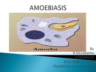


Entamoeba histolytica is a protozoan parasite that causes amoebiasis through fecal-oral transmission. It has a lifecycle involving an infective cyst stage and pathogenic trophozoite stage. Trophozoites cause intestinal and extra-intestinal disease through virulence factors like cysteine proteases. Symptoms range from mild diarrhea to severe colitis, liver abscesses, or other extra-intestinal complications. Diagnosis involves microscopy, antigen detection in stool, or serology. Treatment involves luminal agents like diloxanide furoate or tissue agents like metronidazole. Prevention relies on proper hygiene and sanitation practices.


![Entamoeba_histolytica[1].pptparasitology](https://cdn.slidesharecdn.com/ss_thumbnails/entamoebahistolytica1-251110165959-534d5627-thumbnail.jpg?width=640&height=640&fit=bounds)


![Immunosuppressants [autosaved]](https://cdn.slidesharecdn.com/ss_thumbnails/immunosuppressantsautosaved-131028200143-phpapp01-thumbnail.jpg?width=640&height=640&fit=bounds)


