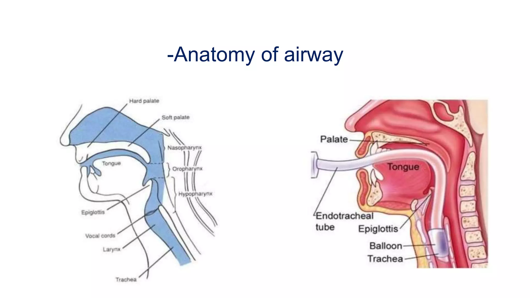
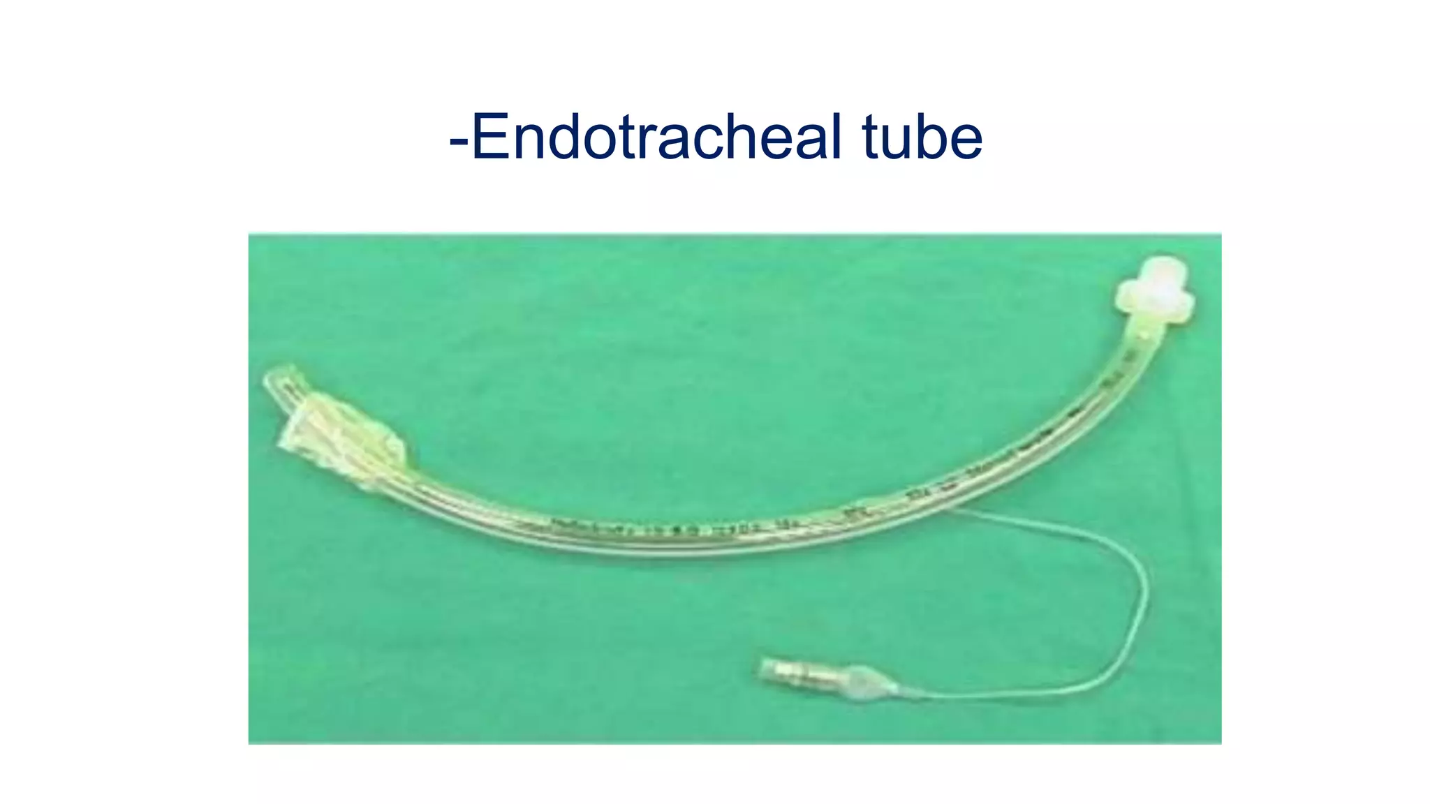
7. -Conditions associated with difficult intubation
-Congenital anomalies
Pierre Robin Syndrome, Down’s Syndrome
-Infection in airway
Retropharyngeal abscess, epiglottis
-Tumor in oral cavity or Larynx
-Enlarge thyroid causing compression / displacement
of trachea
8. -Conditions associated with difficult intubation
--Maxillofacial cervical of laryngeal trauma
--Temporomandibular joint dysfunction
--Burn scar at Face and Neck
--Morbidly obese or pregnancy
15. -EQUIPMENTS
--Laryngoscope with relevant size blade
--Magill's Forceps
--Flexible Introducer
--10 to 20 ml syringe
--Oropharyngeal airways –All sizes
--Tape or adhesive Plaster
--E.T tubes--relevant sizes
--Bag—Valve—Mask with oxygen connected
--Suction unit with Yankauer nozzle and
endotracheal suction catheter
20. -Endotracheal Tube: Size( mm Internal Diameter)
--New Born—3 Months = 3.0 mm ID
--3 to 9 Months = 3.5 mm ID
--9 to 18 Months = 4.0 mm ID
--2 Years to 6 Years = (Age/3) + 3.5
-- >6 Years = (Age/4) + 4.5
--Adult Male = 8 to 8.5 mm ID
--Adult Female =7.0 to 7.5 mm ID
21. -Depth of endotracheal tube : should be placed at
Mid trachea or below vocal cords = 2 cms
--Adult -> Male = 23 cms
--Adult -> Female = 21 cms
--Children
a- Oral endotracheal tube = (Age/2) + 12 cms
b- Nasal endotracheal tube = (Age/2) + 15 cms
25. -Technique of endotracheal Intubation
--Position the patient supine, open the airway with
a head- tilt-chin-lift maneuver.
(suspected spinal injury, attempt Nasotracheal intubation
spine in neutral position
--Open mouth by separating the lips and pulling on
upper jaw with the index finger
26. -Technique of endotracheal Intubation
--Hold laryngoscope in the left hand, insert scope into the mouth with blade directed to right tonsil.
--Once right tonsil is reached, sweep the blade to the midline keeping the tongue on the left.
--This brings the Epiglottis into view. “DO NOT LOOSE SITE OF IT “
--Advance the blade until it reaches the angle between the base of the tongue and
epiglottis (Vallecular space)
--Lift the laryngoscope upwards and away from the Nose – towards the chest. This should
bring the vocal cords into view. It may be necessary for a colleague to press on the trachea
to improve the view of the larynx
27. -Technique of endotracheal Intubation cont.
--Place the ETT in the right hand. Keep the concavity of the tube facing the right side
of the mouth
--Insert the tube watching it enter through the cords.
--Insert the tube just so the cuff has passed the cords and then inflate the cuff
--Listen for air entry at both apices and both axillae to ensure correct placement
using stethoscope
28. -CONFIRMATION OF PROPER TUBE PLACEMENT
--PLACEMENT UNDER VISION
--FOUR QUADRANT AUSCULTATION
--CAPNOMETERY / CAPNOGRAPHY
--VENTILATOR GRAPHS
29. --GOLDEN RULES OF
INTUBATION
--Always have a suction unit available
--An intubation attempt should never exceed 30 seconds
--Oxygenate the patient Pre and Post intubation with a Bag-valve-Mask
and monitor SpO2 continuously
--Have sedative / analgesic medicines available
--Always confirm tube placement by more then one methods
--Do not attempt intubation unless you are totally skilled, rather perform
Bag-Valve-Mask Ventilation
--Always confirm tube placement from time to time