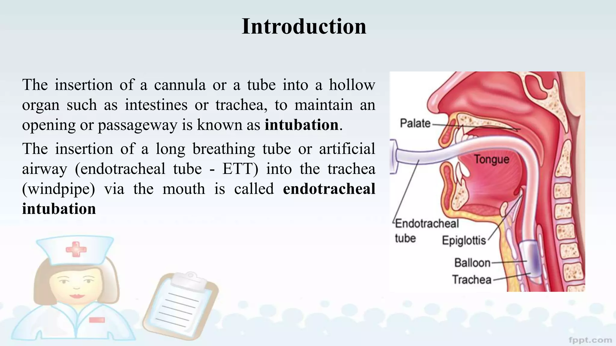
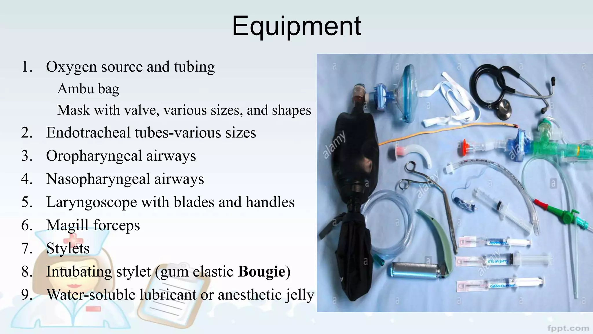
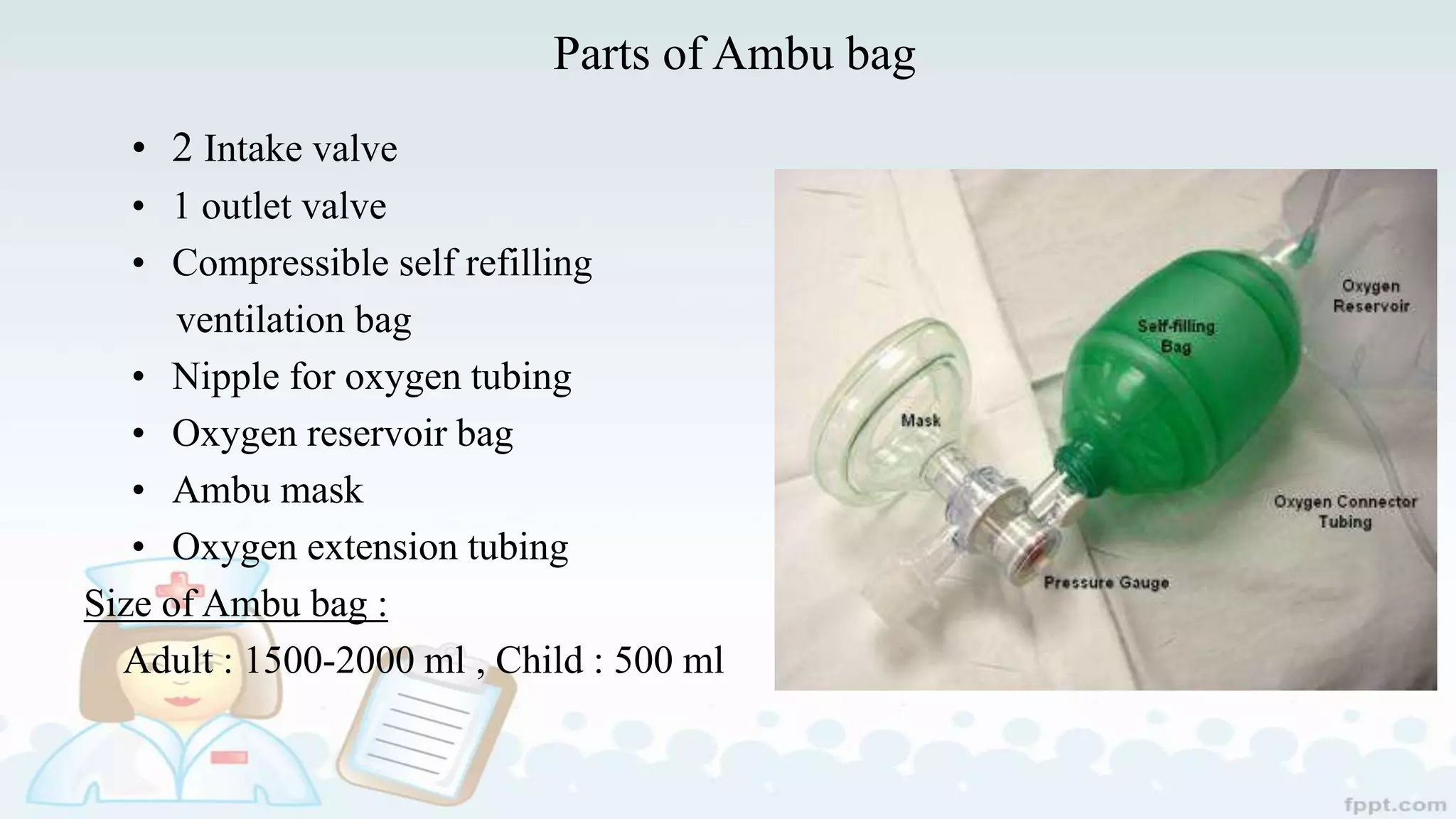
Endotracheal Intubation
Prepared by : Aasma Poudel
Critical Care Nursing
BNS 3rd Year
BHNC, NAMS
Introduction
The insertion of a cannula or a tube into a hollow
organ such as intestines or trachea, to maintain an
opening or passageway is known as intubation.
The insertion of a long breathing tube or artificial
airway (endotracheal tube - ETT) into the trachea
(windpipe) via the mouth is called endotracheal
intubation
Cont…
Rapid-sequence intubation (RSI) is the sequential administration of an
induction agent and neuromuscular blocking agent to facilitate endotracheal
intubation.
• It is the method of choice.
• RSI allows the highest intubation success rate .
• Not all patients targeted for intubation are best managed with RSI; patients
deeply comatose and those in cardiac or respiratory arrests
Cont…
• Intubation can be via Oral or mouth.
Oral intubation
Advantages of Oral Intubation
– Larger tube can be inserted
– Tube can be inserted usually with more speed and ease with less
trauma
– Easier suctioning
– Less airflow resistance
– Reduced risk of tube kinking
Cont…
Disadvantages of Oral Intubation
– Gagging, coughing, salivation, and irritation can be induced with intact
airway reflexes Tube fixation is difficult, self-extubation
– Gastric distention from frequent swallowing of air
– Mucosal irritation and ulcerations of mouth (change tube position)
Cont…
Nasal Intubation
Advantages of Nasal Intubation
- More comfort long term
- Decreased gagging
- Less salivation, easier to swallow
- Improved mouth care
- Better tube fixation
- Improved communication
Cont…
Disadvantages of Nasal Intubation
– Pain and discomfort
– Nasal and paranasal complications, i.e., epistaxis, sinusitis,
– More difficult procedure
– Smaller tube needed
– Increased airflow resistance
– Difficult suctioning
– Bacteremia
Indication
 Insufficient Oxygenation
 Insufficient Ventilation
 Loss of Airway Protection
 Impending Airway Problems
 Control and removal of pulmonary secretions
 Supporting ventilation during general anesthesia
 Supporting ventilation with pathologic disease
Contraindication
Neck immobility or increased risk of neck trauma
Inability to open mouth
Potential Complications during Intubation
• Inability to view vocal cords
• Breaking teeth/dislodging bridgework
• Damage to gums
• Faulty cuff
• Unrecognized esophageal intubation
• Unrecognized right main stem intubation
• Laryngospasm
• Failure to complete intubation
Preparation
• Clinical assessment.
• Pulse oximetry.
• Capnography (the monitoring of the concentration or partial pressure of
carbon dioxide in the respiratory gases).
• The expected course of the patient all collectively guide decisions regarding
the need for tracheal intubation.
Equipment
1. Oxygen source and tubing
Ambu bag
Mask with valve, various sizes, and shapes
2. Endotracheal tubes-various sizes
3. Oropharyngeal airways
4. Nasopharyngeal airways
5. Laryngoscope with blades and handles
6. Magill forceps
7. Stylets
8. Intubating stylet (gum elastic Bougie)
9. Water-soluble lubricant or anesthetic jelly
Cont…
10. Tongue blade
11. Suction catheter &
Suction source
12. Pulse oximetry
13. Carbon dioxide
detector
14. Syringes (10 cc ) : To
inflate cuff
15. ET tube plaster
16. Stethoscope
17. EKG electrodes
18. Medicine :
I. Ketamine
II. Ketofol
III. Etomide
IV. Propofol
V. Succinylcholine
VI. Rocuronium
VII.Vecuronium
VIII.Fentanyl
IX. Midazolam
X. Thiopental
Ambu (Assisted Manual breathing unit) Bag
• Ambu bag or generically as a manual
resuscitator or "self-inflating bag”
• A hand-held device commonly used to
provide positive pressure ventilation.
• It is also known as a bag mask valve
(BMV), manual
Parts of Ambu bag
• 2 Intake valve
• 1 outlet valve
• Compressible self refilling
ventilation bag
• Nipple for oxygen tubing
• Oxygen reservoir bag
• Ambu mask
• Oxygen extension tubing
Size of Ambu bag :
Adult : 1500-2000 ml , Child : 500 ml
Checking manual resuscitator
• Inspect for wear and tear
• Check for leak
• Intake Valve
• Patient valve
• Patient Valve with Pressure Relief
• Reservoir Flap Valves (located in the Intake Valve assy.)
Decontamination
• Washing and rinsing
• Disinfection/sterilization
Endotracheal tube
• Endotracheal tubes are curved tubes used for intubation.
• ET tube can be cuff or uncuff
• The ET tube has the following components:
Proximal end
Central portion
A vocal cord guide (black line)
A radio-opaque marker
The distance indicator (marked in centimeters)
 A cuff- incase of cuff ET tube
 Distal end - has Murphy’s eye
Fig : Endotrachial Tube
Size of ET tube
Adult male : 7.5-8 mm
Adult female : 7-7.5 mm
Pediatric : (16 + AGE)/4
Nasal intubation : Size reduced 1-2 mm
Depth of insertion:
• The tube should be in the middle third of the trachea with the head in neutral
position. The following calculations can be used.
– Length in cm = age/2 + 12 OR weight in kg/5 + 12 OR height in
cm/10 + 5 OR 3 × ID (mm)
• In adults, the tube should be passed until the cuff is 2.25 to 2.5cm below the
vocal cord
• In average size adult patients, securing the tube at the anterior incisor at 23cm
in males and 21cm in females will usually avoid endo bronchial intubation.
• For nasal intubation, 5cm should be added to this length for positioning at the
nares.
Duration
• Endotracheal tube can erode the trachea after being in place longer than 2
weeks, the tracheostomy is necessary.
Laryngoscope
• It is a metallic medical device with in-built light source, which is used, for
direct laryngoscopy.
• This device is of two types, simple laryngoscope & fiber-optic
laryngoscope.
• It is for direct inspection of larynx. It has two separate parts; handle and
interchangeable blades.
• Macintosh (curved) and Miller (straight) blade.
Cont…
• Size used:
In adults, the curved Macintosh #3 is popular, and #4 is more useful in large
patients. The straight Miller #2 and #3 are popular for the same purposes
Cont…
Fig : Macintosh
Fig : Miller
Parts to laryngoscope
• Beak/Tip
• Web/Vertical step
• Light source
• Horizontal flange
• Tongue spatula
• Hook on base
• Handle
Oropharyngeal Airways
• It extends from lips to pharynx, fitting between tongue & posterior pharyngeal
wall. Made up of elastomeric or plastic materials. Parts are
– Flange:
– Bite Portion:
– Curved portion:
Size used
• 000  Small premature infant
• 00 Premature infant
• 0 Neonate
• 1 Small child
• 2 Child
• 3 ( 80 mm) Adult
• 4(90 mm) Large adult
• 5(100mm) Extra-large adult
Technique of insertion:
• Correct size estimated by holding the airway
next to the patient's mouth. The tip should
rest cephalad to the angle of the mandible.
• The airway is inserted with its concave side
facing the upper lip.
• When the junction of the bite portion and the
curved section is near the incisors, the airway
is rotated 180°and slipped behind the tongue
into the final position.
The Laryngeal Mask Airway (LMA)
• New device to maintain airway during anesthesia when
TI is not desired.
• It’s easier in insertion and has high rate of success
• It is made in 8 sizes to suite neonates, children and
adults.
• Better inserted with propofol (that depresses laryngeal
reflex) or deep inhalation anesthesia.
• After adequate anesthesia, LMA is inserted to mouth
blindly without laryngoscope and pushed downward till
resistance is felt. The cough is then inflated
Cont…
• Uses
 In short procedures
 Life-saving difficult intubation
 Conduit for smooth emergence
 Way of intubation in difficult
cases
Contraindications
 Increased risk of aspiration
 Full stomach
Magill forceps:
• It is designed for guiding tip of ETT through larynx
during nasal intubation.
• Also helpful during insertion of nasogastric tubes,
removal of foreign body in mouth of putting
pharyngeal pack.
Procedure of intubation
• Preparatory phase
– Assess the patient's heart rate, level of consciousness, and respiratory status
• Performance phase
– Remove the patient's dental bridgework and plates
– Remove the headboard from the bed (optional).
– Prepare equipment.
Cont…
- Aspirate the stomach contents if a nasogastric tube .
- If the patient is confused, it may be necessary to apply soft wrist .
- Put on gloves and face shield this Prevents contact with patient's oral
secretions.
- During oral intubation if cervical spine is not injured, place patient's head in a
position (extended at the junction of the neck and thorax and flexed at the
junction of the spine and skull).
- Spray the back of the patient's throat with anesthetic spray.
Cont…
‒ Ventilate and oxygenate the patient with the resuscitation bag and mask before
intubation.
‒ Hold the handle of the laryngoscope in the left hand and hold the patient's
mouth open with the right hand by placing crossed fingers on the teeth.
‒ Insert the curved blade of the laryngoscope along the right side of the tongue,
push the tongue to the left, and use right thumb and index finger to pull
patient's lower lip away from lower teeth.
‒ Lift the laryngoscope forward (toward ceiling) to expose the epiglottis. Do not
use teeth as a fulcrum.
Cont…
̶ Lift the laryngoscope upward and forward at a 45-degree angle to expose the
glottis and visualize vocal cords.
̶ As the epiglottis is lifted forward (toward ceiling), the vertical opening of the
larynx between the vocal cords will come into view.
̶ Once the vocal cords are visualized, insert the tube into the right corner of the
mouth and pass the tube while keeping vocal cords in constant view.
̶ Gently push the tube through the triangular space formed by the vocal cords
and back wall of trachea.
Cont…
̶ Stop insertion just after the tube cuff has disappeared from view beyond the
cords.
̶ Withdraw laryngoscope while holding ET tube in place. Disassemble mask
from resuscitation bag, attach bag to ET tube, and ventilate the patient.
̶ Inflate the cuff with the minimal amount of air required to occlude the trachea
̶ Ascertain expansion of both sides of the chest by observation and auscultation
of breath sounds.
Cont…
̶ Record distance from proximal end of tube to the point where the tube reaches
the teeth.
̶ Secure the tube to the patient's face with adhesive tape or apply a commercially
available endotracheal tube stabilization device. Obtain a chest X-ray to verify
tube position.
̶ Document and monitor tube distance from lips to end of ET tube
Cont…
• After procedure
̶ Record tube type and size, cuff pressure, and patient tolerance of the
procedure.
̶ Auscultate breath sounds every 2 hours or if signs and symptoms of
respiratory distress occur.
̶ Assess ABGs after intubation if requested by the health care provider.
ABGs may be prescribed to ensure adequacy of ventilation and
oxygenation. Tube displacement may result in extubation (cuff above vocal
cords), tube touching carina (causing paroxysmal coughing), or intubation
of a mainstem bronchus (resulting in collapse of the unventilated lung).
Cont…
– Measure cuff pressure with manometer; adjust pressure. Make adjustment
in tube placement based on the chest X-ray results. The tube may be
advanced or removed several centimeters for proper placement based on the
chest X-ray results.
Cuff maintenance:
– ET tube cuffs should be inflated continuously and deflated only during
intubation, extubation, and tube repositioning
– Tracheostomy tube cuffs also should be inflated continuously in patients on
mechanical ventilation or continuous positive airway pressure (CPAP).
– Tracheostomies patients who are breathing spontaneously may have the
cuff inflated continuously (in the patient with decreased level of
consciousness without ability to fully protect airway), deflated
continuously, or inflated only for feeding if the patient is at risk of
aspiration.
– Monitor cuff pressure every 4 hours
Care of patient with an endotracheal tube
Immediate care after intubation:
• Check symmetry of chest expansion.
• Auscultate breath sounds of anterior and lateral chest bilaterally.
• Obtain order for chest x-ray to verify proper tube placement.
• Check cuff pressure every 6-8 hours.
• Monitor for signs and symptoms of aspiration.
Cont…
• Ensure high humidity; a visible mist should appear in the T-piece or ventilator
tubing.
• Administer oxygen concentration as prescribed by physician.
• Secure the tube to the patient’s face with tape, and mark the proximal end for
position maintenance.
• Cut proximal end of tube if it is longer than 7.5 cm (3 inches) to prevent
kinking.
Cont…
• Insert an oral airway or mouth device to prevent the patient from biting and
obstructing the tube.
• Use sterile suction technique and airway care to prevent iatrogenic
contamination and infection.
• Continue to reposition patient every 2 hours and as needed to prevent
atelectasis and to optimize lung expansion
• Provide oral hygiene and suction the oropharynx when necessary.
Extubation (Removal of Endotracheal Tube)
• Explain procedure.
• Have self-inflating bag and mask ready in case ventilat -assistance is required
immediately after extubation.
• Suction the tracheobronchial tree and oropharynx, rem tape, and then deflate the
cuff.
• Give 100% oxygen for a few breaths, and then insert a new sterile suction
catheter inside tube.
Cont…
• Have the patient inhale. At peak inspiration, remove the tube, suctioning the
airway through the tube as it is pulled out.
Care of Patient Following Extubation
• Give heated humidity and oxygen by facemask and maintain the patient in a
sitting or high Fowler's position.
• Monitor respiratory rate and quality of chest excursions. Note stridor, color
change, and change in mental alertness or behavior.
• Monitor the patient's oxygen level using a pulse oximetry
• Keep NPO or give only ice chips for next few hours.
• Provide mouth care and teach how to perform coughing and deep- breathing
exercises. 、
References
Rajapakse , S. (2009). Handbook of Critical care medicine : Srilanka
Tintinalli, J.E. (2016). Tintinalli’s Emergency Medicine. USA: The MClinew
M, Sandra.(2006). Lippincott Manual Of Nursing Practice (8th ed). : Lippincott
Williams & Wilkins
Smeltzer.,S.C., Hinkle,J.L., Bare,B.G.,& Cheever, K.H.(2012). Medical Surgical
Nursing (12th ed):Wolters Kluwer: India
Derr.P., Mcevoy, mike (2014) Emergency And Critical Care (8th ) : Jones &
Bartle learning
http.//www.slideshare.com
endotrachial intubation

endotrachial intubation

  • 1.
    Endotracheal Intubation Prepared by: Aasma Poudel Critical Care Nursing BNS 3rd Year BHNC, NAMS
  • 2.
    Introduction The insertion ofa cannula or a tube into a hollow organ such as intestines or trachea, to maintain an opening or passageway is known as intubation. The insertion of a long breathing tube or artificial airway (endotracheal tube - ETT) into the trachea (windpipe) via the mouth is called endotracheal intubation
  • 3.
    Cont… Rapid-sequence intubation (RSI)is the sequential administration of an induction agent and neuromuscular blocking agent to facilitate endotracheal intubation. • It is the method of choice. • RSI allows the highest intubation success rate . • Not all patients targeted for intubation are best managed with RSI; patients deeply comatose and those in cardiac or respiratory arrests
  • 4.
    Cont… • Intubation canbe via Oral or mouth. Oral intubation Advantages of Oral Intubation – Larger tube can be inserted – Tube can be inserted usually with more speed and ease with less trauma – Easier suctioning – Less airflow resistance – Reduced risk of tube kinking
  • 5.
    Cont… Disadvantages of OralIntubation – Gagging, coughing, salivation, and irritation can be induced with intact airway reflexes Tube fixation is difficult, self-extubation – Gastric distention from frequent swallowing of air – Mucosal irritation and ulcerations of mouth (change tube position)
  • 6.
    Cont… Nasal Intubation Advantages ofNasal Intubation - More comfort long term - Decreased gagging - Less salivation, easier to swallow - Improved mouth care - Better tube fixation - Improved communication
  • 7.
    Cont… Disadvantages of NasalIntubation – Pain and discomfort – Nasal and paranasal complications, i.e., epistaxis, sinusitis, – More difficult procedure – Smaller tube needed – Increased airflow resistance – Difficult suctioning – Bacteremia
  • 8.
    Indication  Insufficient Oxygenation Insufficient Ventilation  Loss of Airway Protection  Impending Airway Problems  Control and removal of pulmonary secretions  Supporting ventilation during general anesthesia  Supporting ventilation with pathologic disease
  • 9.
    Contraindication Neck immobility orincreased risk of neck trauma Inability to open mouth
  • 10.
    Potential Complications duringIntubation • Inability to view vocal cords • Breaking teeth/dislodging bridgework • Damage to gums • Faulty cuff • Unrecognized esophageal intubation • Unrecognized right main stem intubation • Laryngospasm • Failure to complete intubation
  • 11.
    Preparation • Clinical assessment. •Pulse oximetry. • Capnography (the monitoring of the concentration or partial pressure of carbon dioxide in the respiratory gases). • The expected course of the patient all collectively guide decisions regarding the need for tracheal intubation.
  • 12.
    Equipment 1. Oxygen sourceand tubing Ambu bag Mask with valve, various sizes, and shapes 2. Endotracheal tubes-various sizes 3. Oropharyngeal airways 4. Nasopharyngeal airways 5. Laryngoscope with blades and handles 6. Magill forceps 7. Stylets 8. Intubating stylet (gum elastic Bougie) 9. Water-soluble lubricant or anesthetic jelly
  • 13.
    Cont… 10. Tongue blade 11.Suction catheter & Suction source 12. Pulse oximetry 13. Carbon dioxide detector 14. Syringes (10 cc ) : To inflate cuff 15. ET tube plaster 16. Stethoscope 17. EKG electrodes 18. Medicine : I. Ketamine II. Ketofol III. Etomide IV. Propofol V. Succinylcholine VI. Rocuronium VII.Vecuronium VIII.Fentanyl IX. Midazolam X. Thiopental
  • 14.
    Ambu (Assisted Manualbreathing unit) Bag • Ambu bag or generically as a manual resuscitator or "self-inflating bag” • A hand-held device commonly used to provide positive pressure ventilation. • It is also known as a bag mask valve (BMV), manual
  • 15.
    Parts of Ambubag • 2 Intake valve • 1 outlet valve • Compressible self refilling ventilation bag • Nipple for oxygen tubing • Oxygen reservoir bag • Ambu mask • Oxygen extension tubing Size of Ambu bag : Adult : 1500-2000 ml , Child : 500 ml
  • 16.
    Checking manual resuscitator •Inspect for wear and tear • Check for leak • Intake Valve • Patient valve • Patient Valve with Pressure Relief • Reservoir Flap Valves (located in the Intake Valve assy.)
  • 17.
    Decontamination • Washing andrinsing • Disinfection/sterilization
  • 18.
    Endotracheal tube • Endotrachealtubes are curved tubes used for intubation. • ET tube can be cuff or uncuff • The ET tube has the following components: Proximal end Central portion A vocal cord guide (black line) A radio-opaque marker The distance indicator (marked in centimeters)  A cuff- incase of cuff ET tube  Distal end - has Murphy’s eye
  • 19.
  • 20.
    Size of ETtube Adult male : 7.5-8 mm Adult female : 7-7.5 mm Pediatric : (16 + AGE)/4 Nasal intubation : Size reduced 1-2 mm
  • 21.
    Depth of insertion: •The tube should be in the middle third of the trachea with the head in neutral position. The following calculations can be used. – Length in cm = age/2 + 12 OR weight in kg/5 + 12 OR height in cm/10 + 5 OR 3 × ID (mm) • In adults, the tube should be passed until the cuff is 2.25 to 2.5cm below the vocal cord • In average size adult patients, securing the tube at the anterior incisor at 23cm in males and 21cm in females will usually avoid endo bronchial intubation. • For nasal intubation, 5cm should be added to this length for positioning at the nares.
  • 22.
    Duration • Endotracheal tubecan erode the trachea after being in place longer than 2 weeks, the tracheostomy is necessary.
  • 23.
    Laryngoscope • It isa metallic medical device with in-built light source, which is used, for direct laryngoscopy. • This device is of two types, simple laryngoscope & fiber-optic laryngoscope. • It is for direct inspection of larynx. It has two separate parts; handle and interchangeable blades. • Macintosh (curved) and Miller (straight) blade.
  • 24.
    Cont… • Size used: Inadults, the curved Macintosh #3 is popular, and #4 is more useful in large patients. The straight Miller #2 and #3 are popular for the same purposes
  • 25.
  • 26.
    Parts to laryngoscope •Beak/Tip • Web/Vertical step • Light source • Horizontal flange • Tongue spatula • Hook on base • Handle
  • 27.
    Oropharyngeal Airways • Itextends from lips to pharynx, fitting between tongue & posterior pharyngeal wall. Made up of elastomeric or plastic materials. Parts are – Flange: – Bite Portion: – Curved portion:
  • 28.
    Size used • 000 Small premature infant • 00 Premature infant • 0 Neonate • 1 Small child • 2 Child • 3 ( 80 mm) Adult • 4(90 mm) Large adult • 5(100mm) Extra-large adult
  • 29.
    Technique of insertion: •Correct size estimated by holding the airway next to the patient's mouth. The tip should rest cephalad to the angle of the mandible. • The airway is inserted with its concave side facing the upper lip. • When the junction of the bite portion and the curved section is near the incisors, the airway is rotated 180°and slipped behind the tongue into the final position.
  • 30.
    The Laryngeal MaskAirway (LMA) • New device to maintain airway during anesthesia when TI is not desired. • It’s easier in insertion and has high rate of success • It is made in 8 sizes to suite neonates, children and adults. • Better inserted with propofol (that depresses laryngeal reflex) or deep inhalation anesthesia. • After adequate anesthesia, LMA is inserted to mouth blindly without laryngoscope and pushed downward till resistance is felt. The cough is then inflated
  • 31.
    Cont… • Uses  Inshort procedures  Life-saving difficult intubation  Conduit for smooth emergence  Way of intubation in difficult cases Contraindications  Increased risk of aspiration  Full stomach
  • 32.
    Magill forceps: • Itis designed for guiding tip of ETT through larynx during nasal intubation. • Also helpful during insertion of nasogastric tubes, removal of foreign body in mouth of putting pharyngeal pack.
  • 33.
    Procedure of intubation •Preparatory phase – Assess the patient's heart rate, level of consciousness, and respiratory status • Performance phase – Remove the patient's dental bridgework and plates – Remove the headboard from the bed (optional). – Prepare equipment.
  • 34.
    Cont… - Aspirate thestomach contents if a nasogastric tube . - If the patient is confused, it may be necessary to apply soft wrist . - Put on gloves and face shield this Prevents contact with patient's oral secretions. - During oral intubation if cervical spine is not injured, place patient's head in a position (extended at the junction of the neck and thorax and flexed at the junction of the spine and skull). - Spray the back of the patient's throat with anesthetic spray.
  • 35.
    Cont… ‒ Ventilate andoxygenate the patient with the resuscitation bag and mask before intubation. ‒ Hold the handle of the laryngoscope in the left hand and hold the patient's mouth open with the right hand by placing crossed fingers on the teeth. ‒ Insert the curved blade of the laryngoscope along the right side of the tongue, push the tongue to the left, and use right thumb and index finger to pull patient's lower lip away from lower teeth. ‒ Lift the laryngoscope forward (toward ceiling) to expose the epiglottis. Do not use teeth as a fulcrum.
  • 36.
    Cont… ̶ Lift thelaryngoscope upward and forward at a 45-degree angle to expose the glottis and visualize vocal cords. ̶ As the epiglottis is lifted forward (toward ceiling), the vertical opening of the larynx between the vocal cords will come into view. ̶ Once the vocal cords are visualized, insert the tube into the right corner of the mouth and pass the tube while keeping vocal cords in constant view. ̶ Gently push the tube through the triangular space formed by the vocal cords and back wall of trachea.
  • 37.
    Cont… ̶ Stop insertionjust after the tube cuff has disappeared from view beyond the cords. ̶ Withdraw laryngoscope while holding ET tube in place. Disassemble mask from resuscitation bag, attach bag to ET tube, and ventilate the patient. ̶ Inflate the cuff with the minimal amount of air required to occlude the trachea ̶ Ascertain expansion of both sides of the chest by observation and auscultation of breath sounds.
  • 38.
    Cont… ̶ Record distancefrom proximal end of tube to the point where the tube reaches the teeth. ̶ Secure the tube to the patient's face with adhesive tape or apply a commercially available endotracheal tube stabilization device. Obtain a chest X-ray to verify tube position. ̶ Document and monitor tube distance from lips to end of ET tube
  • 39.
    Cont… • After procedure ̶Record tube type and size, cuff pressure, and patient tolerance of the procedure. ̶ Auscultate breath sounds every 2 hours or if signs and symptoms of respiratory distress occur. ̶ Assess ABGs after intubation if requested by the health care provider. ABGs may be prescribed to ensure adequacy of ventilation and oxygenation. Tube displacement may result in extubation (cuff above vocal cords), tube touching carina (causing paroxysmal coughing), or intubation of a mainstem bronchus (resulting in collapse of the unventilated lung).
  • 40.
    Cont… – Measure cuffpressure with manometer; adjust pressure. Make adjustment in tube placement based on the chest X-ray results. The tube may be advanced or removed several centimeters for proper placement based on the chest X-ray results.
  • 41.
    Cuff maintenance: – ETtube cuffs should be inflated continuously and deflated only during intubation, extubation, and tube repositioning – Tracheostomy tube cuffs also should be inflated continuously in patients on mechanical ventilation or continuous positive airway pressure (CPAP). – Tracheostomies patients who are breathing spontaneously may have the cuff inflated continuously (in the patient with decreased level of consciousness without ability to fully protect airway), deflated continuously, or inflated only for feeding if the patient is at risk of aspiration. – Monitor cuff pressure every 4 hours
  • 42.
    Care of patientwith an endotracheal tube Immediate care after intubation: • Check symmetry of chest expansion. • Auscultate breath sounds of anterior and lateral chest bilaterally. • Obtain order for chest x-ray to verify proper tube placement. • Check cuff pressure every 6-8 hours. • Monitor for signs and symptoms of aspiration.
  • 43.
    Cont… • Ensure highhumidity; a visible mist should appear in the T-piece or ventilator tubing. • Administer oxygen concentration as prescribed by physician. • Secure the tube to the patient’s face with tape, and mark the proximal end for position maintenance. • Cut proximal end of tube if it is longer than 7.5 cm (3 inches) to prevent kinking.
  • 44.
    Cont… • Insert anoral airway or mouth device to prevent the patient from biting and obstructing the tube. • Use sterile suction technique and airway care to prevent iatrogenic contamination and infection. • Continue to reposition patient every 2 hours and as needed to prevent atelectasis and to optimize lung expansion • Provide oral hygiene and suction the oropharynx when necessary.
  • 45.
    Extubation (Removal ofEndotracheal Tube) • Explain procedure. • Have self-inflating bag and mask ready in case ventilat -assistance is required immediately after extubation. • Suction the tracheobronchial tree and oropharynx, rem tape, and then deflate the cuff. • Give 100% oxygen for a few breaths, and then insert a new sterile suction catheter inside tube.
  • 46.
    Cont… • Have thepatient inhale. At peak inspiration, remove the tube, suctioning the airway through the tube as it is pulled out.
  • 47.
    Care of PatientFollowing Extubation • Give heated humidity and oxygen by facemask and maintain the patient in a sitting or high Fowler's position. • Monitor respiratory rate and quality of chest excursions. Note stridor, color change, and change in mental alertness or behavior. • Monitor the patient's oxygen level using a pulse oximetry • Keep NPO or give only ice chips for next few hours. • Provide mouth care and teach how to perform coughing and deep- breathing exercises. 、
  • 49.
    References Rajapakse , S.(2009). Handbook of Critical care medicine : Srilanka Tintinalli, J.E. (2016). Tintinalli’s Emergency Medicine. USA: The MClinew M, Sandra.(2006). Lippincott Manual Of Nursing Practice (8th ed). : Lippincott Williams & Wilkins Smeltzer.,S.C., Hinkle,J.L., Bare,B.G.,& Cheever, K.H.(2012). Medical Surgical Nursing (12th ed):Wolters Kluwer: India Derr.P., Mcevoy, mike (2014) Emergency And Critical Care (8th ) : Jones & Bartle learning http.//www.slideshare.com