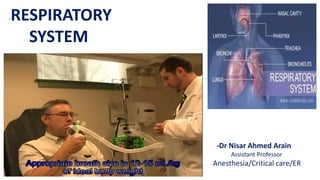
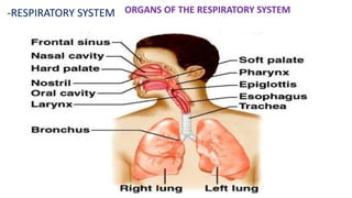
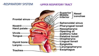
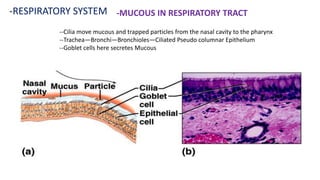
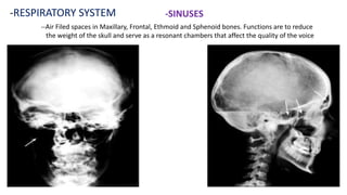
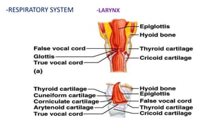
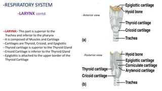
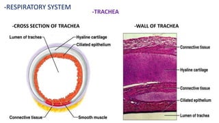
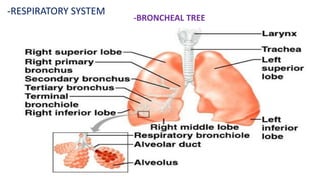
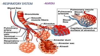
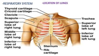
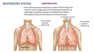
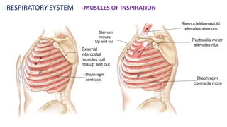
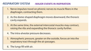
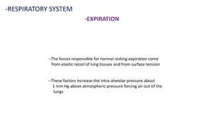
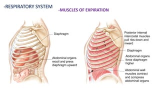
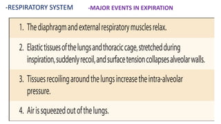
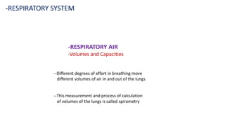
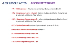
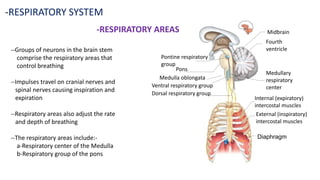
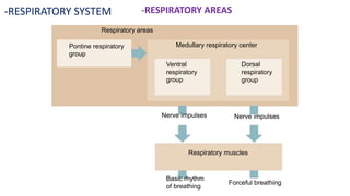
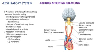
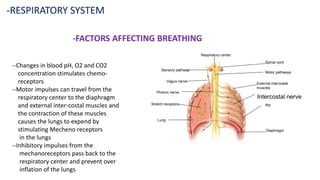
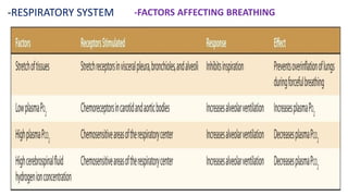
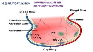
The document provides information on the respiratory system, including its structures and functions. It discusses the processes of ventilation, external respiration, transport of gases, and cellular respiration. It describes the structures of the upper respiratory tract such as the nose, pharynx and larynx. It also details the trachea, bronchi, bronchioles, and alveoli. Furthermore, it examines the muscles involved in inspiration and expiration, respiratory volumes and capacities, and the control of breathing.
The document describes the anatomy and mechanism of the human respiratory system. It outlines the main organs - nose, pharynx, larynx, trachea, and lungs. It explains that the nose warms and filters air before it reaches the lungs. The lungs contain bronchi and bronchioles that facilitate the exchange of oxygen and carbon dioxide between inhaled air and blood. The document also describes the process of inspiration where intercostal muscles and the diaphragm expand the chest cavity to inhale air, and expiration where they relax to exhale air from the lungs.
The respiratory system is made up of organs responsible for oxygen intake and carbon dioxide removal through inhalation and exhalation. It is divided into the upper respiratory tract consisting of the nose, sinuses and larynx, and the lower respiratory tract containing the trachea, bronchi and lungs. In the lungs, oxygen passes into the bloodstream and carbon dioxide moves out through tiny air sacs called alveoli. The diaphragm and intercostal muscles control inhalation and exhalation, expanding and contracting the lungs and chest cavity.
The respiratory system works to deliver oxygen to the body and remove carbon dioxide. It includes the mouth, nose, pharynx, larynx, lungs, trachea, bronchi, and diaphragm. Air enters through the nose or mouth and travels down the trachea to the lungs, where oxygen passes into blood vessels and carbon dioxide is removed. The lungs, trachea, and bronchi form a branching tree structure ending in tiny air sacs called alveoli that facilitate gas exchange with blood in the pulmonary circulation. The diaphragm and intercostal muscles help drive breathing by expanding the lungs and lowering their pressure.
The document summarizes the key components and functions of the respiratory system. It describes the upper respiratory tract including the nose, nasal passages, pharynx and larynx. It then details the lower respiratory tract including the trachea, bronchi, bronchioles, alveolar ducts and alveoli. It explains the processes of inspiration, expiration, gas exchange and the control systems that regulate breathing.
The respiratory system has three major organs: the airways, lungs, and muscles of respiration. The airways carry air from the outside into the lungs and include the nose, mouth, throat, voice box, windpipe, and airways. The lungs are the functional units that pass oxygen into the body and carbon dioxide out. The diaphragm and intercostal muscles work together as a pump to push air in and out of the lungs during breathing.
Anatomy of respiratory system with special reference to anatomy of lungs,
mechanism of respiration, regulation of respiration
Lung Volumes and capacities transport of respiratory gases, artificial respiration,
and resuscitation methods.
1st science 12 describes the function of each respiratory organShirley Sison
The document describes the parts and functions of the respiratory system. It discusses the upper respiratory tract including the nose, mouth and throat and their functions of warming, filtering and moistening air. It then discusses the lower respiratory tract including the lungs, bronchi, trachea and their functions of gas exchange and transporting air. It also describes how the ribs, rib cage and diaphragm protect the lungs and how the lungs are made up of bronchi, bronchioles and alveoli where gas exchange occurs through capillaries.
The respiratory system functions to oxygenate tissues and remove carbon dioxide through gas exchange. It consists of the upper respiratory tract including the nose and pharynx, and the lower respiratory tract including the larynx, trachea, bronchi, bronchioles and alveoli in the lungs. Oxygen diffuses into the blood in the alveoli while carbon dioxide diffuses out. Breathing is controlled by respiratory centers in the brain and involves inspiration through contraction of the diaphragm and expiration through relaxation.
The respiratory system provides oxygen to the body's cells while removing carbon dioxide through the processes of inhalation and exhalation. It consists of an upper respiratory tract including the nose, mouth and larynx, and a lower respiratory tract including the trachea, bronchi, bronchioles and alveoli in the lungs. The lungs contain volumes of air that can be measured including tidal volume, inspiratory reserve volume, expiratory reserve volume and residual volume. Gas exchange occurs through diffusion between the alveoli and blood plasma, and between plasma and tissues throughout the body, facilitated by factors like partial pressures and surface area.
This document discusses the human respiratory system, including its main components and functions. It describes the upper respiratory tract which filters and conditions incoming air, and the lower respiratory tract which includes the lungs and facilitates gas exchange. Additionally, it examines the process of breathing and some common respiratory disorders like asthma and bronchitis.
This document summarizes the respiratory system. It begins with an introduction and then discusses the developmental anatomy, functions including ventilation and gas exchange, and regulation of respiration through neural control centers in the brainstem. It also covers applied topics like effects of altitude and diving. The respiratory system works to oxygenate tissues and remove carbon dioxide through four main functions: ventilation, gas diffusion, transport, and regulation. Respiration is regulated by chemoreceptors sensitive to oxygen and carbon dioxide levels, as well as pH levels, to maintain homeostasis.
Respiration involves three main functions: ventilation, gas exchange, and oxygen utilization. Ventilation is the mechanical process of breathing which moves air in and out of the lungs. Gas exchange occurs through diffusion between the alveoli and capillaries, allowing oxygen to diffuse from the air into the blood and carbon dioxide to diffuse out. Oxygen is then utilized in cellular respiration throughout the body. The document discusses the anatomy and physiology of respiration including the conducting and respiratory zones of the lungs, gas pressures, lung volumes, and disorders that can affect respiration.
This document summarizes key aspects of respiration, including the blood-air barrier where gas exchange occurs, muscles involved like the diaphragm and intercostal muscles, the mechanisms of inspiration and expiration, pulmonary volumes and capacities, oxygen and carbon dioxide transport, and control of breathing by the respiratory center in the medulla oblongata. Control is influenced by chemoreceptors responding to changes in oxygen and carbon dioxide levels, as well as other factors like speech, emotions, drugs, and sleep.
The human respiratory system takes in oxygen and expels carbon dioxide using the lungs and other organs. The lungs carry out the exchange of gases as we breathe. Red blood cells collect oxygen from the lungs and distribute it throughout the body. During inhalation, the diaphragm and intercostal muscles contract and expand the rib cage and lungs, increasing their volume and allowing more air to enter. Exhalation is the movement of air out of the bronchial tubes and lungs to the external environment during breathing.
The respiratory system has three main functions: gas exchange, pulmonary ventilation, and transport of respiratory gases. It has two main zones - the conducting zone which includes the nose, pharynx, larynx, trachea, and bronchi, and the respiratory zone where gas exchange occurs in the alveoli. Gas exchange takes place across the alveolar-capillary membrane, which is only one cell thick. Oxygen moves from the alveoli into the blood in the pulmonary capillaries and carbon dioxide moves out of the blood into the alveoli.
The document summarizes key aspects of the human respiratory system, including its functions, organization, physiology, and some common respiratory diseases. It describes how the respiratory system draws air into the lungs and releases it, supplying blood with oxygen and removing carbon dioxide. It also outlines the main parts of the respiratory system like the nose, pharynx, lungs and their roles in gas exchange and transport.
The document provides an overview of the respiratory system and control of respiration. It describes the key organs involved, including the nose, pharynx, larynx, trachea, bronchi, lungs and alveoli. It explains the mechanics of breathing through inspiration and expiration. Gas exchange occurs as oxygen passes from the alveoli into the blood and carbon dioxide passes from the blood into the alveoli to be exhaled. The lungs, diaphragm, ribs and autonomic nervous system work together to regulate breathing and ventilation.
The document summarizes key aspects of the respiratory system including:
1. The three major steps of respiration: pulmonary ventilation, external respiration, and internal respiration.
2. The major organs of the respiratory system including the nose, pharynx, larynx, trachea, bronchi, lungs, and alveoli.
3. The processes of breathing including inhaling via the diaphragm and intercostal muscles and exhaling via relaxation of these muscles.
This document provides an overview of respiratory system terminology in Chinese. It discusses key terms related to respiration, the nose, trachea, bronchi, lungs, pleura, and the two phases of breathing - inspiration and expiration. Respiration is carried out by the lungs and other respiratory structures that facilitate the flow of air to and from the lungs, such as the trachea and bronchi.
The document provides information on the respiratory system including its structures, functions, and mechanisms of breathing. Some key points:
1. The respiratory system includes the nose, pharynx, larynx, trachea, bronchi, lungs, and muscles of respiration. Gas exchange occurs in the alveoli of the lungs.
2. Breathing is controlled by respiratory centers in the brain and involves inspiration and expiration. Inspiration is driven by contraction of the diaphragm and intercostal muscles which increase the thoracic cavity. Expiration occurs passively.
3. The lungs contain alveoli where oxygen and carbon dioxide diffuse between the air and blood through the respiratory membrane. Various factors can impact
The respiratory system allows us to breathe by taking in oxygen and expelling carbon dioxide. The lungs and nose or mouth are used to inhale air which travels through the trachea into the lungs. When we breathe in, the diaphragm contracts pulling air into the lungs, and when breathing out it relaxes pushing air out. Oxygen passes from the lungs into the blood while carbon dioxide passes from the blood to the lungs.
The respiratory system brings oxygen into the body and removes carbon dioxide. It includes the nose, pharynx, larynx, trachea, bronchi, bronchioles, and lungs. The circulatory system transports blood throughout the body using the heart, arteries, veins and capillaries. It has two circuits - pulmonary and systemic. Diseases can affect both systems like asthma, pneumonia, anemia and atherosclerosis.
Respiratory system, physiology of respiratory system and neural control sunil JMI
The respiratory system includes the nose, pharynx, larynx, trachea, bronchi, and lungs. Gas exchange occurs in the alveoli of the lungs where oxygen enters the blood and carbon dioxide leaves. Inspiration is an active process involving contraction of the diaphragm and intercostal muscles which increases the thoracic cavity volume. Expiration is usually a passive process involving relaxation of these muscles and elastic recoil of the lungs. The respiratory centers in the medulla control breathing rhythm and depth via nervous and chemical feedback mechanisms.
The respiratory system allows for oxygen intake and carbon dioxide elimination through the process of breathing, which consists of inspiration and expiration. The organs of the respiratory system include the nose, pharynx, larynx, trachea, bronchi, bronchioles and alveoli. The upper respiratory tract functions to warm, moisten and filter air, while the lower respiratory tract transports air to the lungs for gas exchange to occur between the blood and alveoli.
The respiratory system consists of a conducting zone and respiratory zone. The conducting zone includes the nose, pharynx, larynx, trachea, and bronchi which are responsible for conducting air to the respiratory zone. The respiratory zone includes bronchioles, alveolar ducts and 300 million alveoli which are the primary sites of gas exchange between air and blood.
The respiratory system allows for the exchange of gases in the body. It includes the nose, mouth, throat, trachea, bronchi, lungs, diaphragm and alveoli. The lungs and alveoli work to exchange oxygen and carbon dioxide between the air and blood. Common respiratory disorders include asthma, bronchitis, emphysema and pneumonia which can involve inflammation or infection of the lungs, bronchi or alveoli.
This document provides an overview of the physiology of the respiratory system. It begins with the anatomical structures of the respiratory tract, including the nasal cavity, pharynx, larynx, trachea, bronchi, bronchioles, alveolar ducts and alveoli. It then describes the mechanics of ventilation, including how the diaphragm and intercostal muscles cause inhalation and exhalation through changes in thoracic cavity volume. Finally, it discusses pulmonary volumes such as tidal volume, vital capacity, functional residual capacity, and their clinical measurements using spirometry.
Appropriate airway equipment and techniques.Nisar Arain
This document provides an overview of airway anatomy, equipment, and techniques for airway management. It discusses:
1. The importance of airway control and the development of advanced cardiac life support.
2. The objectives of reviewing upper and lower airway anatomy, basic and advanced airway techniques, equipment for difficult airways, and clinical management of the airway.
3. Details of upper airway structures including the nose, oral cavity, pharynx, and larynx. It also reviews lower airway structures like the trachea and lungs.
appropriate airway equipment and techniques-AnesthesiaNISAR ARAIN
The document discusses airway anatomy, physiology, and management techniques. It covers the structures of the upper and lower airways, including the nose, pharynx, larynx, trachea, and lungs. It emphasizes that airway management involves ensuring patency, adequate ventilation, and oxygenation through various basic and advanced techniques, not just intubation. Maintaining an open airway is vital for gas exchange and preventing hypoxia.
The respiratory system has several functions including distributing air throughout the body, exchanging gases, warming and humidifying air, and allowing for smell. It consists of upper and lower tracts. The upper tract includes the nose, nasal cavity, sinuses, pharynx and larynx. The lower tract includes the trachea, bronchial tree and lungs. During breathing, pressure changes caused by the diaphragm and intercostal muscles moving air in and out of the lungs, allowing for gas exchange in the alveoli. Various volumes of air are involved including tidal volume, expiratory reserve volume, and residual volume.
This document provides an overview of respiratory physiology, including:
1. The functions of the respiratory system involve gas exchange through pulmonary ventilation, alveolar ventilation, gas transport in the blood, and exchange in tissues.
2. The anatomical components of the respiratory system include the nose, pharynx, larynx, trachea, bronchi, bronchioles, and alveoli.
3. Pulmonary ventilation is achieved through inspiration and expiration driven by changes in pressure and the contraction of respiratory muscles like the diaphragm and intercostals.
This document summarizes the mechanics of breathing. It describes normal breathing rates and types of abnormal breathing. It discusses the boundaries of the thoracic cage and the two pleura layers. Breathing involves both positive pressure from inspiration and negative pressure from expiration. Inspiration is an active process using the diaphragm and intercostal muscles while expiration is usually passive. Gas exchange occurs through pressure gradients in the lungs. The document outlines the muscles, pressures, and mechanics involved in inspiration and expiration.
ANATOMY AND PHYSIOLOGY OF RESPIRATORY SYSTEM.pptxAyurgyan2077
Anatomy and physiology of respiratory system basics. The structural and functional unit of life are called cells. The group of cells with similar structure and function constitute a tissue and similar group of tissues constitute an organ. Likewise, the similar functioning organs constutute the body system.
The respiratory system has three main functions: gas exchange between the atmosphere and blood, filtering and warming of inspired air, and sound production. It has three basic steps: pulmonary ventilation (breathing), external (pulmonary) respiration involving gas exchange in the lungs, and internal (tissue) respiration involving gas exchange in tissues. Inspiration is an active process using inspiratory muscles like the diaphragm and external intercostals to expand the thoracic cavity and lower lung pressure, allowing air to flow in. Expiration is usually a passive process involving elastic recoil of the lungs and chest wall. Other factors like alveolar surface tension, lung compliance, and airway resistance also influence ventilation.
These lecture notes were prepared by Dr. Hamdi Turkey- Pulmonologist- Department of internal medicine - Taiz university
It contains :
- Notes on Embryology, Anatomy and Physiology of respiratory system
- Cardinal symptoms in respiratory diseases
- Diagnostic procedures
Do Not Forget To Visit Our Pages On Facebook on the following Links:
https://www.facebook.com/groups/569435236444761/
AND
https://www.facebook.com/groups/690331650977113/
The respiratory system works with the cardiovascular system to oxygenate the blood and remove carbon dioxide. It is composed of the nose, pharynx, larynx, trachea, bronchi, lungs, and related structures. The nose warms and moisturizes inhaled air before it reaches the lungs. Gas exchange occurs in the alveoli, where oxygen diffuses into blood and carbon dioxide diffuses out. The respiratory center controls breathing and is sensitive to carbon dioxide and oxygen levels in the blood.
it explains the structure of respiratory system, organs involved, volume of air exchanges and review questions to check students knowledge after the chapter is over
The respiratory system allows the body to produce energy through the process of respiration, which requires oxygen and glucose. Mechanical respiration involves the inhalation and exhalation of air through the lungs using muscle movements in the chest and rib cage. During inhalation, the diaphragm contracts and the rib cage expands, decreasing air pressure in the lungs and allowing air to flow in through the nose and trachea. Exhalation occurs when the diaphragm relaxes and the rib cage contracts, increasing air pressure in the lungs and forcing the used air out.
The respiratory system allows air to enter the body through the nose or mouth and then passes through multiple structures down to the lungs. Air is warmed, cleaned, and moistened as it passes through the nasal cavity. It then travels through the pharynx, larynx, trachea, bronchi and bronchioles before reaching the alveoli in the lungs. In the alveoli, oxygen from the air enters the bloodstream and carbon dioxide moves from the blood into the air to be exhaled. Smoking damages cilia and alveoli, impairing gas exchange and increasing risks of diseases like emphysema and lung cancer.
This document summarizes respiration in animals. It begins with an overview of the topics to be covered, including the anatomy of the respiratory system, external respiration (gas exchange between the lungs and environment), and internal respiration (gas exchange between blood and body tissues). It then defines respiration and describes the modes. The anatomy section details the structures of the airways, lungs, and respiratory muscles. External respiration involves inhaling oxygen and exhaling carbon dioxide between the lungs and air. Internal respiration is gas exchange between blood in the pulmonary capillaries and tissues via diffusion. The cycle of respiration continuously supplies oxygen to tissues and removes carbon dioxide.
The respiratory system includes the nose, mouth, pharynx, larynx, trachea, bronchi, bronchioles, lungs, diaphragm, and intercostal muscles. It works to oxygenate the blood and remove carbon dioxide through a process of inspiration and expiration. During inspiration, muscles contract to expand the chest cavity and draw air into the lungs. Expiration is a passive process where muscles relax and the chest cavity returns to its original size, pushing air out. Gas exchange occurs in alveoli, where oxygen diffuses into blood and carbon dioxide diffuses out. The respiratory system regulates breathing through neural and chemical controls that sense carbon dioxide and oxygen levels.
The document discusses the anatomy and physiology of the respiratory system. It describes the functions of the respiratory system as air distribution, gas exchange, filtering and warming air, influencing speech, and allowing for smell. It then outlines the divisions of the respiratory system into the upper respiratory tract including the nose, nasal cavity, sinuses, pharynx and larynx, and the lower respiratory tract within the thorax including the trachea, bronchial tree and lungs. Several structures of the upper and lower respiratory tract are defined such as the larynx, trachea, bronchi and lungs. Pulmonary ventilation and the mechanics of breathing including inspiration and expiration are explained.
Respiration involves the exchange of oxygen and carbon dioxide between the environment and body through breathing. It is a catabolic process where oxygen is used to oxidize glucose to produce energy in mitochondria. The principal organs of respiration include the nasal passages, pharynx, larynx, trachea, bronchi, lungs, and associated muscles. In humans, breathing is driven by the contraction and relaxation of the diaphragm and intercostal muscles, causing the volume of the thoracic cavity to alternately increase and decrease. This draws air in and out of the lungs, facilitating gas exchange between alveolar air and blood in the pulmonary capillaries via simple diffusion.
This document discusses airway local blocks and awake intubation. It describes the indications for awake intubation including comorbidities, risk of aspiration, difficult airway assessment, and emergencies. It provides details on the pharmacological agents, equipment, personnel, and techniques used for airway local blocks and awake intubation. Specifically, it outlines common methods for anesthetizing different areas of the airway using lidocaine, including dosage calculations and risks of lidocaine toxicity. The goal is to safely anesthetize the airway to allow for awake intubation.
This document discusses endotracheal tubes and intubation. It covers indications for intubation including airway protection, optimizing gas exchange, decreasing metabolic demand, and reducing work of breathing. Conditions associated with difficult intubation are described such as congenital anomalies, infections, tumors, injuries, and obesity. Proper equipment, tube sizing, intubation technique including positioning and confirmation of placement are outlined. Golden rules of intubation emphasize preparation, oxygenation, skills, confirmation, and monitoring.
- The laryngeal mask airway (LMA) is a supraglottic airway device that is placed in the hypopharynx to control the airway during general anesthesia or ventilation. It provides an alternative to endotracheal intubation or use of a face mask. The LMA has advantages like ease of insertion, reduced hemodynamic response, and improved oxygenation during emergence from anesthesia. Potential complications include sore throat, coughing, laryngospasm, and airway obstruction. Proper selection of size, lubrication, and insertion technique are important for successful use of the LMA.
This document discusses different types of fluid flow and transport mechanisms in cells. It describes laminar and turbulent fluid flow, how they are characterized, and factors that influence each type. It also outlines different transport mechanisms in cells including diffusion, osmosis, facilitated diffusion, active transport, endocytosis, and exocytosis. Active transport uses carrier proteins and cell energy to move substances against a concentration gradient, while passive transport moves substances down a concentration gradient without cell energy.
1. The document discusses theories of anesthesia including the unitary theory and modern theories involving interactions with membrane proteins and specific ion channels.
2. It describes the stages of general anesthesia from analgesia to surgical anesthesia to medullary respiratory paralysis. However, it notes that the excitement stage is rarely seen with modern anesthesia.
3. GABA receptors are identified as an important target for many anesthetic agents. General anesthetics bind to these receptors, causing chloride channel opening and neuronal inhibition, resulting in anesthesia.
Endotracheal intubation and laryngoscopy part 2Nisar Arain
This document discusses various types of endotracheal tubes and laryngoscopy techniques. It describes specialized tubes like armored tubes, RAE tubes, Oxford tubes, and laser-resistant tubes. It covers direct laryngoscopy using curved and straight blades. Optimal conditions for laryngoscopy are outlined, including using the appropriately sized blade and ensuring good muscle relaxation. Reliable signs of correct endotracheal tube placement include capnography, visualization of the tube passing the vocal cords, and fiberoptic bronchoscopy visualization of tracheal rings.
Endotracheal tubes are used to intubate patients and enable ventilation. They are typically made of PVC or rubber and have features like a Murphy eye, size designations, and a pilot balloon-connected inflation system to create a seal in the trachea. Complications can occur during or after intubation and extubation, like trauma, aspiration, or laryngospasm. Nasotracheal intubation has advantages like patient comfort but risks like trauma or sinusitis. Proper preparation, techniques, and monitoring are important for safe endotracheal intubation.
Complications of artificial applications part 5Nisar Arain
This document discusses the complications that can arise from mechanical ventilation through either invasive or non-invasive means. Some key complications mentioned include pneumonia, infections, injuries to the face/lips/pharynx and larynx/trachea, gastrointestinal effects like esophagitis and decreased motility, renal effects from reductions in blood pressure/flow, disrupted sleep, and decubitus ulcers. Proper diagnosis and management of these various complications is important for patients receiving mechanical ventilation support.
This document discusses endotracheal tubes and intubation. It covers indications for intubation including airway protection, optimizing gas exchange, decreasing metabolic demand, and reducing work of breathing. Conditions associated with difficult intubation are described such as congenital anomalies, infections, tumors, and injuries. Airway assessment techniques like mallampati classification, laryngoscopy view, and thyromental distance are explained. Equipment for intubation and sizing endotracheal tubes are outlined. The technique of intubation is described involving positioning the patient in sniffing position and using a laryngoscope to visualize the vocal cords. Confirmation of proper tube placement is emphasized using methods like auscultation and capnography.
This document discusses various techniques for airway management. It describes mechanical maneuvers like jaw thrust and head tilt-chin lift to clear obstructions. Common airway adjuncts like oropharyngeal and nasopharyngeal airways are also discussed. Guidelines are provided for sizing and inserting these adjuncts safely. Face masks can be used with one, two, or three hands to maintain a patent airway. Risk factors for difficult mask ventilation and potential complications are also outlined.
This document discusses airway assessment and difficult airways. It outlines various predictors of difficult airways like obesity, short neck, and facial hair. It describes tests to evaluate the airway like thyromental distance, inter-incisor gap, and Mallampati grading. The document emphasizes the importance of a thorough airway assessment prior to intubation to identify potential difficulties and prepare appropriate management strategies for difficult intubations.
The document discusses preoperative airway assessment for anesthesia. It notes that 1-3 out of 100 anesthetized patients have difficult intubation, while 1 out of 1000 have failed intubation and 1 out of 10,000 experience cannot intubate cannot ventilate scenarios. Factors that can increase intubation difficulty include congenital syndromes, anatomical features like teeth and neck structure, and acquired conditions such as decreased jaw or neck mobility. A thorough preoperative assessment including tests of mouth opening, neck movement, thyromental distance, and Mallampati score can help predict and prepare for a potentially difficult airway.
- Imhotep, an ancient Egyptian priest from around 2600 BC, is considered the first physician and treated many diseases. He extracted medicines from plants and had knowledge of anatomy. Ancient Egyptians used opium and hyoscyamus for anesthesia and performed trepanation surgery.
- In ancient Greece and Rome, mandrake juice was used for its narcotic effects before surgeries to ensure insensibility to pain. Arabic translations of Greek medicine advanced Islamic medicine in the Middle Ages. Physicians like Al Zahrawi described many surgeries and instruments.
- The modern history of anesthesia began with William Morton using ether in 1846 and John Snow advancing the field through publications on ether and chlor
This document discusses different types of anesthesia including local, regional, and general anesthesia. It provides details on common regional anesthesia techniques like spinal blocks, epidurals, and caudal blocks. It also describes local anesthesia techniques such as infiltration, nerve blocks, and intravenous regional anesthesia. The document discusses the mechanisms of local anesthetics and some potential complications as well as benefits of local and regional anesthesia compared to general anesthesia.
This document discusses the problem of anesthesia awareness during surgery. It defines anesthesia awareness as a patient becoming conscious during a surgical procedure under general anesthesia and having recall of events. Risk factors include women, younger age, use of total intravenous anesthesia, long surgeries, prior awareness history, and natural red hair. Causes can include light anesthesia, increased anesthetic requirements, or anesthesiologist error. Prevention strategies include pre-operative evaluation, prophylactic benzodiazepines, monitoring anesthetic levels, and post-operative interviews. Methods to monitor consciousness include clinical signs, isolated forearm technique, brain monitoring like BIS, and measurements of lower esophageal sphincter contractions.
- The document discusses various physiological changes that occur with aging and their implications for anesthesia in geriatric patients. Some key points discussed include:
- Cardiovascular changes like decreased cardiac output and increased risk of hypertension. Respiratory changes like reduced lung capacity and cough reflex. Genitourinary changes like reduced kidney function and bladder issues.
- Gastrointestinal changes like decreased motility leading to constipation. Endocrine changes like increased risk of hypothyroidism and bone issues.
- The implications of these changes for anesthesia include risks of hypotension, bradycardia, respiratory complications, slower drug metabolism and clearance, and risks of gastric aspiration and constipation. Careful preoperative evaluation and
The document provides information on general anesthesia including:
1) It discusses the history, goals, and levels of sedation for general anesthesia. Different levels include minimal sedation, moderate sedation, deep sedation, and general anesthesia.
2) The pre-anesthetic evaluation process involves taking a medical history, performing a physical exam including airway assessment, and ordering lab tests.
3) Common anesthetic equipment is described including laryngoscopes, endotracheal tubes, airways, monitors, and intravenous and inhalational drugs used for induction and maintenance of general anesthesia.
The document discusses body temperature regulation and abnormalities. It notes that humans maintain a constant core body temperature of around 37°C through heat gain and loss mechanisms controlled by the hypothalamus. When temperature varies by 0.1°C from the set point, the hypothalamus activates heat conservation or dissipation responses. Disorders include hypothermia, where temperature drops below the normal range, and hyperthermia/fever, where the hypothalamus raises the set point in response to pyrogens like bacterial toxins or cytokines. Heat stroke occurs when temperature exceeds the critical threshold of around 105-108°F.
This document discusses triage, which is the process of sorting patients based on the urgency of their condition to prioritize care. It defines triage and outlines its objectives to identify patients, prioritize their needs, track their progress, and identify hazards. It describes different types of triage including simple, advanced, continuous integrated, and reverse triage. It discusses levels of triage from red to green based on factors like vital signs and Glasgow Coma Scale. Finally, it outlines the role and characteristics of EMTs in effectively conducting triage.
The document discusses the design and setup of an operation theatre. It outlines key areas that should be included like an anesthesia room, sterilization room, recovery room, and storeroom. It also describes the daily, weekly, and deep cleaning procedures needed to properly sanitize the operation theatre and prevent infections. Furthermore, it covers the preparation of equipment, instruments, and supplies as well as different sterilization methods like autoclaving, gamma irradiation, and ethylene oxide.
Histololgy of Female Reproductive System.pptxAyeshaZaid1
Dive into an in-depth exploration of the histological structure of female reproductive system with this comprehensive lecture. Presented by Dr. Ayesha Irfan, Assistant Professor of Anatomy, this presentation covers the Gross anatomy and functional histology of the female reproductive organs. Ideal for students, educators, and anyone interested in medical science, this lecture provides clear explanations, detailed diagrams, and valuable insights into female reproductive system. Enhance your knowledge and understanding of this essential aspect of human biology.
- Video recording of this lecture in English language: https://youtu.be/Pt1nA32sdHQ
- Video recording of this lecture in Arabic language: https://youtu.be/uFdc9F0rlP0
- Link to download the book free: https://nephrotube.blogspot.com/p/nephrotube-nephrology-books.html
- Link to NephroTube website: www.NephroTube.com
- Link to NephroTube social media accounts: https://nephrotube.blogspot.com/p/join-nephrotube-on-social-media.html
10 Benefits an EPCR Software should Bring to EMS Organizations Traumasoft LLC
The benefits of an ePCR solution should extend to the whole EMS organization, not just certain groups of people or certain departments. It should provide more than just a form for entering and a database for storing information. It should also include a workflow of how information is communicated, used and stored across the entire organization.
Travel vaccination in Manchester offers comprehensive immunization services for individuals planning international trips. Expert healthcare providers administer vaccines tailored to your destination, ensuring you stay protected against various diseases. Conveniently located clinics and flexible appointment options make it easy to get the necessary shots before your journey. Stay healthy and travel with confidence by getting vaccinated in Manchester. Visit us: www.nxhealthcare.co.uk
5-hydroxytryptamine or 5-HT or Serotonin is a neurotransmitter that serves a range of roles in the human body. It is sometimes referred to as the happy chemical since it promotes overall well-being and happiness.
It is mostly found in the brain, intestines, and blood platelets.
5-HT is utilised to transport messages between nerve cells, is known to be involved in smooth muscle contraction, and adds to overall well-being and pleasure, among other benefits. 5-HT regulates the body's sleep-wake cycles and internal clock by acting as a precursor to melatonin.
It is hypothesised to regulate hunger, emotions, motor, cognitive, and autonomic processes.
Travel Clinic Cardiff: Health Advice for International TravelersNX Healthcare
Travel Clinic Cardiff offers comprehensive travel health services, including vaccinations, travel advice, and preventive care for international travelers. Our expert team ensures you are well-prepared and protected for your journey, providing personalized consultations tailored to your destination. Conveniently located in Cardiff, we help you travel with confidence and peace of mind. Visit us: www.nxhealthcare.co.uk
share - Lions, tigers, AI and health misinformation, oh my!.pptxTina Purnat
• Pitfalls and pivots needed to use AI effectively in public health
• Evidence-based strategies to address health misinformation effectively
• Building trust with communities online and offline
• Equipping health professionals to address questions, concerns and health misinformation
• Assessing risk and mitigating harm from adverse health narratives in communities, health workforce and health system
Mercurius is named after the roman god mercurius, the god of trade and science. The planet mercurius is named after the same god. Mercurius is sometimes called hydrargyrum, means ‘watery silver’. Its shine and colour are very similar to silver, but mercury is a fluid at room temperatures. The name quick silver is a translation of hydrargyrum, where the word quick describes its tendency to scatter away in all directions.
The droplets have a tendency to conglomerate to one big mass, but on being shaken they fall apart into countless little droplets again. It is used to ignite explosives, like mercury fulminate, the explosive character is one of its general themes.
Osteoporosis - Definition , Evaluation and Management .pdfJim Jacob Roy
Osteoporosis is an increasing cause of morbidity among the elderly.
In this document , a brief outline of osteoporosis is given , including the risk factors of osteoporosis fractures , the indications for testing bone mineral density and the management of osteoporosis
2. -RESPIRATORY SYSTEM
-Respiration:-It is the process of exchanging gases
between the atmosphere and body cells,
This consist of following events
--a-Ventilation – Air in and out of lungs
--b-External respiration:-
--c-Transport of gases:-
--d-Internal respiration:-Blood and tissues
--e-Cellular respiration:-Oxygen and carbon dioxide
3. -RESPIRATORY SYSTEM
-STRUCTURES
--1-NOSE
(nares)
--Nasal cavity separated from
a-cranial cavity
b-oral cavity
c-ethmoid
d-maxilla
--Nasal septum
a-meatus
b-superior, middle and inferior cilia (They moves
mucus
and arrested particles towards pharynx
--2-PHARYNX
smell, conducts air, entraps particles, moistens
and heats air
4. -RESPIRATORY SYSTEM
-STRUCTURES contd.
--3-LARYNX
--Vocal cords, Epiglottis, and cartilage
-4-TRACHEA
-Cartilage, Muscles, and elastic Rings
--5-BRONCHIAL TREE
a-RIGHT AND LEFT main stem bronchi
b-Secondary Lobar:-Three on right and Two on the
left
c-Tertiary:-Ten on the Right and Eight on the Left
d-Terminal:-50 to 80 with in each lobe of lung
e-Respiratory:- 2 to 3 branches from each terminal
alveolar ducts---These branches terminates into
Sacs---Ultimately hey terminate into Alveoli
7. -RESPIRATORY SYSTEM -MUCOUS IN RESPIRATORY TRACT
--Cilia move mucous and trapped particles from the nasal cavity to the pharynx
--Trachea—Bronchi—Bronchioles—Ciliated Pseudo columnar Epithelium
--Goblet cells here secretes Mucous
8. -RESPIRATORY SYSTEM -SINUSES
--Air Filed spaces in Maxillary, Frontal, Ethmoid and Sphenoid bones. Functions are to reduce
the weight of the skull and serve as a resonant chambers that affect the quality of the voice
10. -RESPIRATORY SYSTEM
-LARYNX contd.
--LARYNX:- This part is superior to the
Trachea and inferior to the pharynx
--It is composed of Muscles and Cartilage
--Cartilages are Thyroid, Cricoid, and Epiglottic
--Thyroid cartilage is superior to the Thyroid Gland
--Cricoid Cartilage is inferior to the Thyroid Gland
--Epiglottis is attached to the upper border of the
Thyroid Cartilage
-Anterior view
-Posterior view
11. -RESPIRATORY SYSTEM
-TRACHEA
--1-It lies anterior to the esophagus, located in the
thoracic cavity (Mediastinum)
--2-Trachea and Bronchi are supported with C-shaped
hyaline cartilage
--3-Posteriorly – incomplete rings contain Muscle and
connective tissue
--4-Carina – projection of the last tracheal ring, located
in the sagittal plane
(It is associated with the cough reflex)
--5-Splits into Right and Left main stem Bronchus
12. -RESPIRATORY SYSTEM
-TRACHEA AND BRONCHI
--1-Right main Bronchus is wider, shorter and runs
more vertically then the left
--2-Left main Bronchus passes inferiorly to the arch
of the aorta and anterior to the esophagus
--3-Lobar Bronchi – secondary branches two on the
left and three on the right.
--4-Tertiary Bronchi – several segments that branch
into 20 to 25 terminal Bronchioles
--5-Respiratory Bronchioles – contain 11 – alveolar
ducts and five to six alveolar sacs. Fully developed
by age 8 years, with 300 million alveoli
17. -RESPIRATORY SYSTEM
-MUSCLES OF THE THORACIC WALL
--1-PECTORAL MUSCLE:- Major and minor
--2-SUBCLAVIUS
--3-SERRATUS ANTERIOR:-Rotates the scapula and holds the
scapula against the thoracic wall.
--4-SCALENE MUSCLES:-From the neck to the1st and 2nd Ribs
They elevate the ribs during forced inspiration
--5-INTER COSTAL MUSCLES:-
a-EXTERNAL INTERCOSTAL MUSCLES:- These are muscles of
inspiration and they elevate the ribs during forced
inspiration
b-INTERNAL INTERCOSTAL MUSCLES:-These are muscles of
expiration
--6-INNERMOST INTERCOSTALS:-They lie deep and separated
from internal intercostals by nerves and vessels
--7-SUBCOSTAL MUSCLES:-They aid in elevation of ribs
19. -RESPIRATORY SYSTEM
-BREATHING MECHENISM
--Breathing or ventilation is the movement of air
from outside of the body into the bronchial
tree and the alveoli
-The actions responsible for these air movements
are inspiration, or inhalation, and expiration, or
exhalation
20. -RESPIRATORY SYSTEM -INSPIRATION
--Atmospheric pressure due to
the weight of the air is the
force that moves air into the
lungs
--At sea level
a-Atmospheric pressure is
760 millimeters of
mercury (mm Hg)
b-Moving the plunger of a
syringe causes air to
move in and out.
c-Air movements in and out
of the lungs occur in much
the same way
-Diaphragm
-Atmospheric pressure
of 760 mm Hg on the
inside
Air passage way
-Atmospheric
pressure of
760 mm Hg
on the outside
21. -RESPIRATORY SYSTEM -INSPIRATION
--Intra-alveolar pressure decreases to about 758 mm Hg as the
thoracic cavity enlarges due to the downward movement of
diaphragm caused by impulses carried by Phrenic Nerve
--Atmospheric pressure then forces air into the airways
-Atmospheric pressure
760 mm Hg
-Intra-alveolar
pressure
758 mm Hg
-Intra-alveolar
pressure
760 mm Hg
Diaphragm
22. -RESPIRATORY SYSTEM -MUSCLES OF INSPIRATION
Sternocleidomastoid
elevates sternum
Pectoralis minor
elevates ribs
Diaphragm
contracts more
Sternum moves
Up and out
External
intercostal
muscles pull
ribs up and out
-Diaphragm
contracts
Sternum
moves
Up and out
--Diaphragm
contracts
External
intercostal
muscles pull
ribs up and out
24. -RESPIRATORY SYSTEM
-EXPIRATION
--The forces responsible for normal resting expiration come
from elastic recoil of lung tissues and from surface tension
--These factors increase the intra-alveolar pressure about
1 mm Hg above atmospheric pressure forcing air out of the
lungs
25. -RESPIRATORY SYSTEM
-MUSCLES OF EXPIRATION
Posterior internal
intercostal muscles
pull ribs down and
inward
Diaphragm
Abdominal organs
force diaphragm
higher
Abdominal wall
muscles contract
and compress
abdominal organs
Diaphragm
Abdominal organs
recoil and press
diaphragm upward
27. -RESPIRATORY SYSTEM
-RESPIRATORY AIR
-Volumes and Capacities
--Different degrees of effort in breathing move
different volumes of air in and out of the lungs
--This measurement and process of calculation
of volumes of the lungs is called spirometry
28. -RESPIRATORY SYSTEM -RESPIRATORY VOLUMES
--TV—(Tidal volume) – Volume moved in or out during a normal breath
--IRV—(Inspiratory reserve volume) – Volume that can be inhaled during forced
breathing in addition to Tidal volume
--ERV—(Expiratory Reserve volume) – volume that can be exhaled during forced
breathing in addition to Tidal volume
--RV—(Residual volume) – volume that remains in lungs at all times
--FRV—(Functional residual capacity) = ERV + RV
--IC---(Inspiratory capacity) = TV + IRV
--VC—(Vital capacity) = TV + IRV + ERV
--TLC—(Total lung capacity) = VC + RV
31. -RESPIRATORY SYSTEM
-ALVEOLAR VENTILATION
-MINUTE VENTILATION
--Tidal volume multiplied by breathing
rate is called Minute ventilation
-ALVEOLAR VENTILATION
--Major factor affecting concentrations
of oxygen and carbon di oxide in the
alveoli
--It is volume of air that reaches alveoli
--Tidal volume minus physiological dead
space then multiplied by breathing rate
32. -RESPIRATORY SYSTEM
-NON RESPIRATORY AIR MOVEMENTS
--Air movements other than breathing are called
non respiratory movements
--They clear air passages, as in coughing and sneezing
or express emotions, as in laughing and crying
33. -RESPIRATORY SYSTEM
-CONTROL OF BREATHING
--Normal breathing is a rhythmic
involuntary act that continues
when a person is unconscious
--Respiratory muscles can be
controlled as well voluntary
muscles
34. -RESPIRATORY SYSTEM
-RESPIRATORY AREAS
--Groups of neurons in the brain stem
comprise the respiratory areas that
control breathing
--Impulses travel on cranial nerves and
spinal nerves causing inspiration and
expiration
--Respiratory areas also adjust the rate
and depth of breathing
--The respiratory areas include:-
a-Respiratory center of the Medulla
b-Respiratory group of the pons
Midbrain
Fourth
ventricle
Medullary
respiratory
center
Internal (expiratory)
intercostal muscles
External (inspiratory)
intercostal muscles
Diaphragm
Dorsal respiratory group
Ventral respiratory group
Medulla oblongata
Pons
Pontine respiratory
group
35. Pontine respiratory
group
Respiratory areas
Medullary respiratory center
Ventral
respiratory
group
Dorsal
respiratory
group
Nerve impulses Nerve impulses
Respiratory muscles
Basic rhythm
of breathing
Forceful breathing
-RESPIRATORY SYSTEM -RESPIRATORY AREAS
36. -RESPIRATORY SYSTEM -FACTORS AFFECTING BREATHING
--1-A number of factors affect breathing
rate and depth including
a-Partial pressure of oxygen(Pao2)
b-Partial pressure of carbon
dioxide(Pco2)
c-Degree of stretch of lung tissue
d-Emotional state
e-Level of physical activity
--2-Receptors involved are
f-Mecheno receptors and
g-Chemoreceptors
(1)-Central and
(2)-Peripheral
Medulla oblongata
Sensory nerve
(branch of
glossopharyngeal
nerve)
Carotid bodies
Common carotid
artery
Aortic bodies
Sensory nerve
(branch of vagus nerve)
Aorta
Heart
37. -RESPIRATORY SYSTEM
-FACTORS AFFECTING BREATHING
--Changes in blood pH, O2 and CO2
concentration stimulates chemo-
receptors
--Motor impulses can travel from the
respiratory center to the diaphragm
and external inter-costal muscles and
the contraction of these muscles
causes the lungs to expend by
stimulating Mecheno receptors
in the lungs
--Inhibitory impulses from the
mechanoreceptors pass back to the
respiratory center and prevent over
inflation of the lungs
Spinal cord
Motor pathways
Intercostal nerve
External intercostal
muscles
Rib
Diaphragm
Respiratory center
Sensory pathway
Vagus nerve
Phrenic nerve
Stretch receptors
Lung
41. -RESPIRATORY SYSTEM -THE DEAD SPACE
--It is that part of respiratory system through which inspiration and expiration occurs, But it does not
participate in gas exchange
IT IS FORMED AS:-
--1-ANATOMICAL DEAD SPACE:-
--It includes the airway passage from the Nasopharynx down to the respiratory bronchioles. It also includes
a-EQUIPMENT DEAD SPACE:--such as endotracheal tube and tubing distal to the Y – connector of the
anesthesia breathing circuits. It normally equals 150 ml of the respiratory passage in an average
adult i.e 2.2 ml/ Kg
b-ALVEOLAR DEAD SPACE:-It is the alveoli that are ventilated, but not Perfused.
it normally equals Zero
--2-PHYSIOLOGICAL DEAD SPACE:-
--Physiological = anatomical + alveolar
--Normally, the alveolar dead space = Zero
--so, Physiological dead space = anatomical dead space.
--The ratio of dead space (Vd) to tidal volume (V1) is more useful. Normally it is 0.3
42. -RESPIRATORY SYSTEM -FACTORS AFFECTING THE DEAD SPACE
--A-FACTORS INCREASING THE DEAD SPACE
--1-Upright posture
--2-Neck Extension
--3-Advanced age
--4-Positive pressure ventilation, PEEP, and increased airway pressure
--5-Anticholinergics which cause Broncho-dilatation
--6-Decreased Pulmonary perfusion e.g Pulmonary emboli or Hypotension
--7-Lung diseases such as emphysema or cystic fibrosa
--B-FACTORS DECREASING THE DEAD SPACE
--8-Supine posture
--9-Neck Flexion
--10-Intubation
43. -RESPIRATORY SYSTEM
-SHUNT
--DEFINATION:-
--It is the process where the desaturated mixed venous blood from the right
heart returns to the left heart without being Re-Saturated with Oxygen
in the lungs (i.e Right-to-Left-Shunt)
It dilutes and decreases arterial Oxygen content resulting in Hypoxemia
--TYPES
--Intrapulmonary shunts are either
--1-AN ABSOLUTE SHUNT:-It refers to anatomical shunts where V/Q is Zero
(i.e No Ventilation) This produces Hypoxemia which cannot be corrected
--2-A RELATIVE SHUNT:-It refers to areas of lung where there is less circulation
where V/Q ratio again is low.
This produces Hypoxemia which can be partially corrected by increasing
inspired oxygen concentration