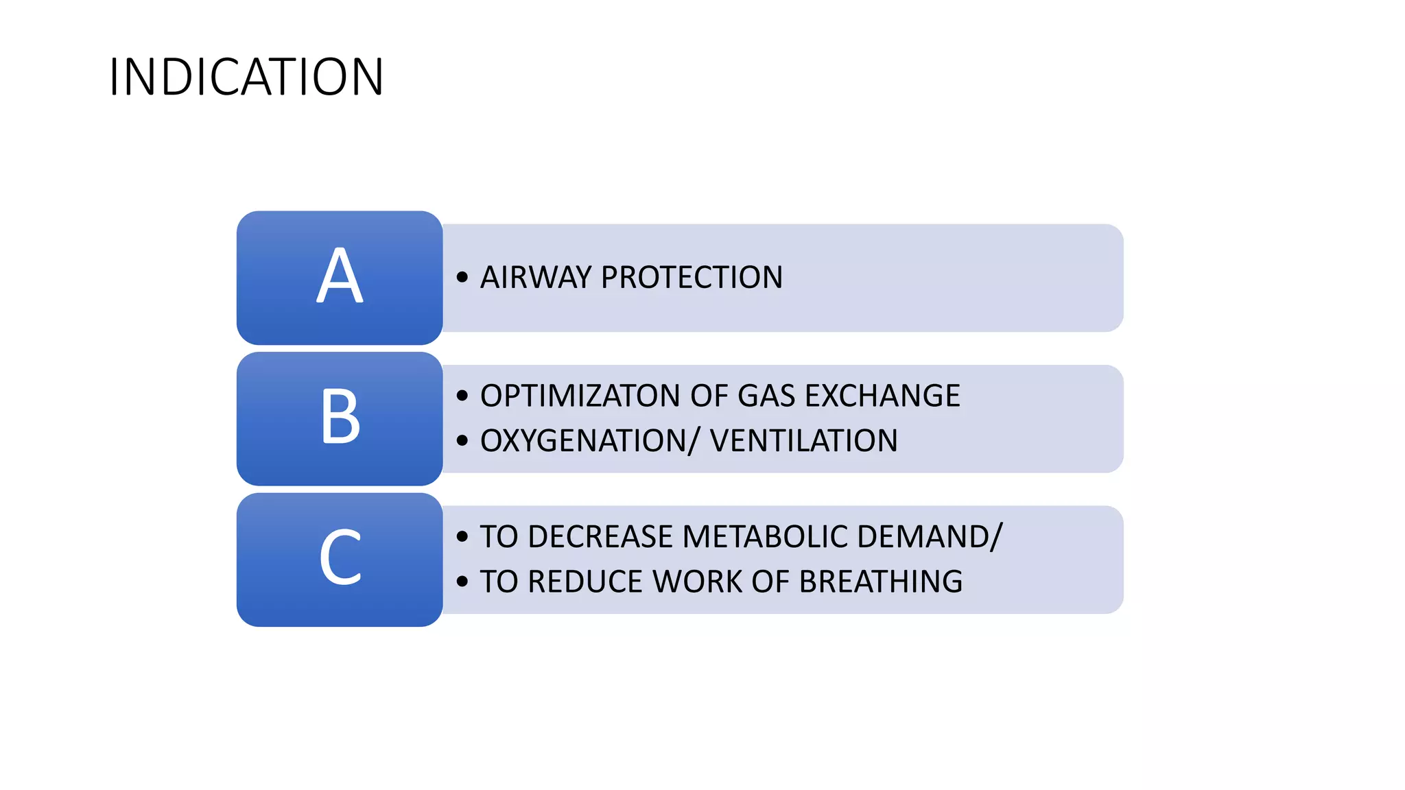
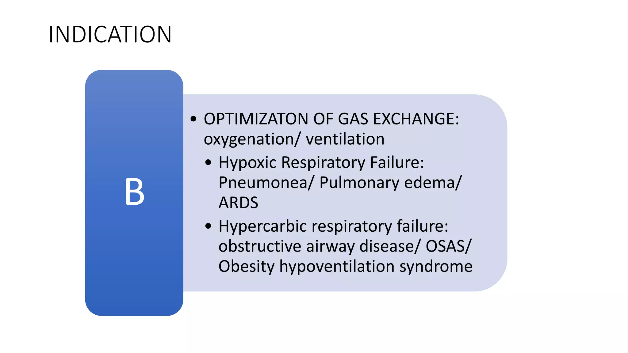
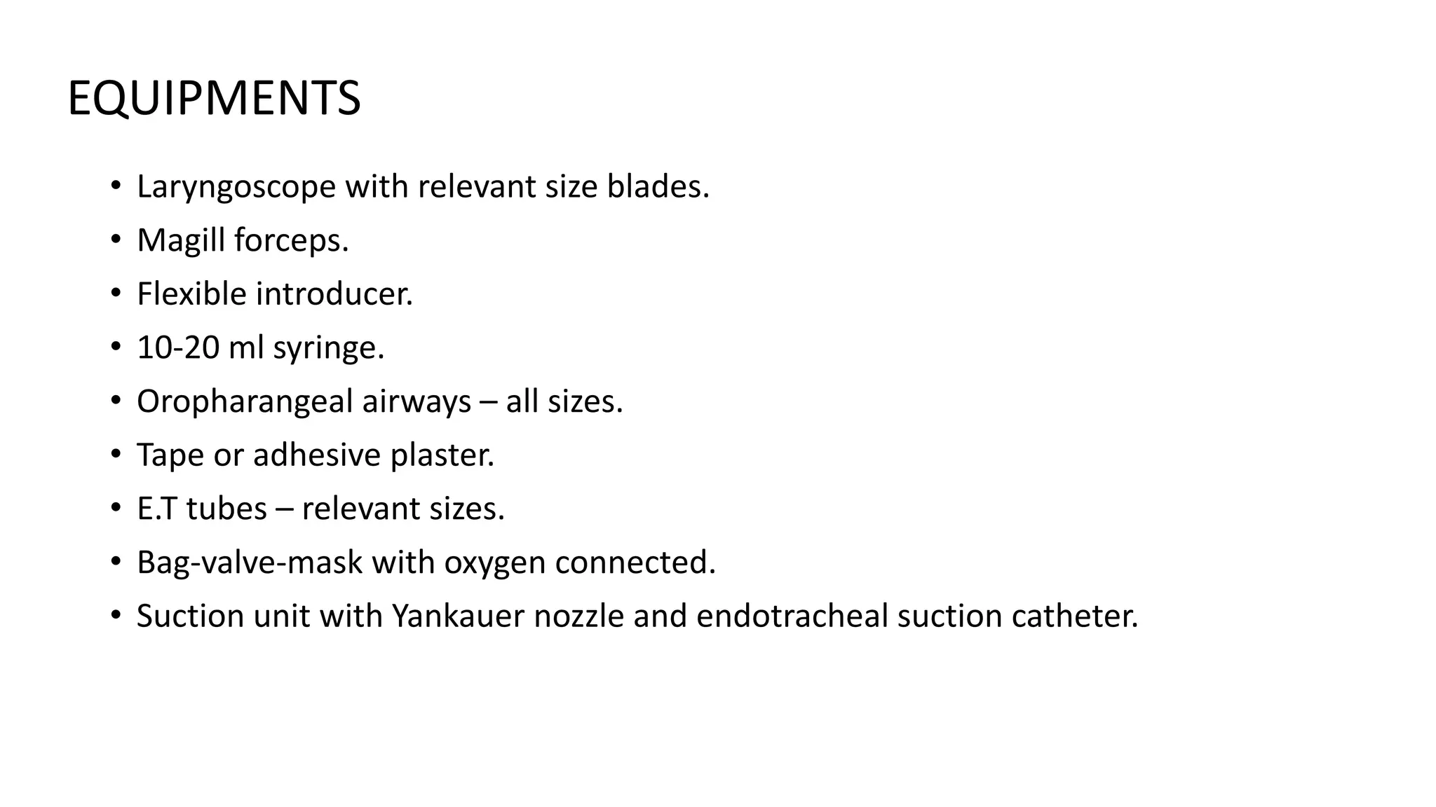
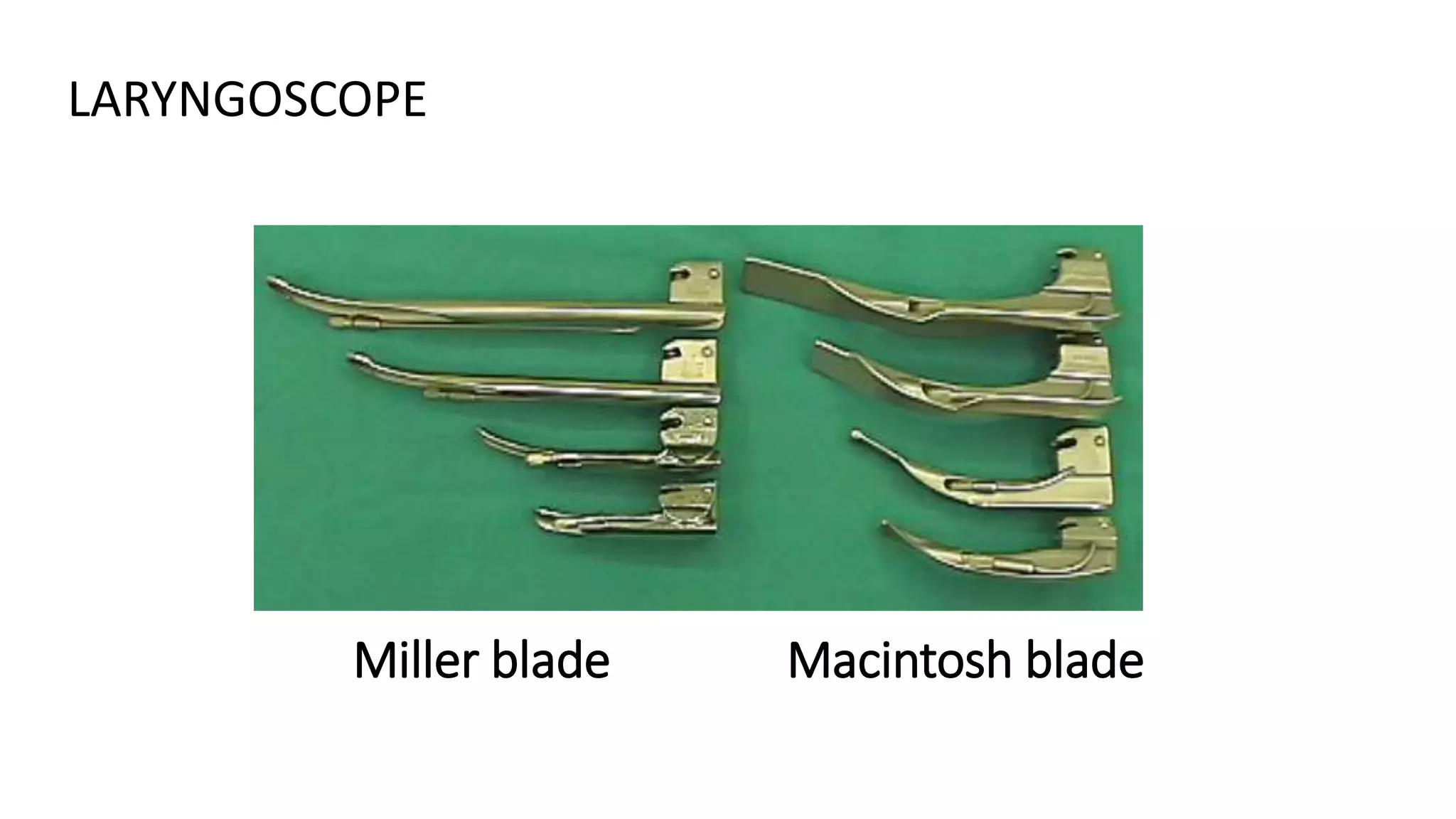
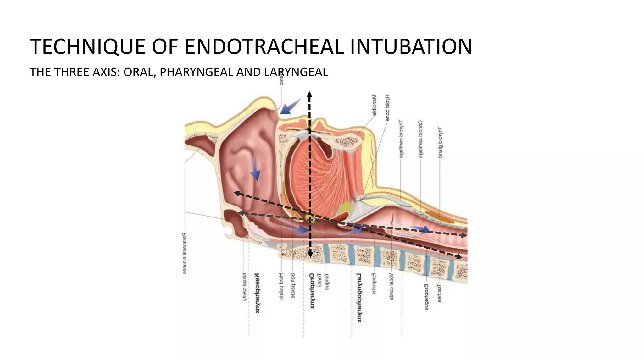
ENDOTRACHEAL INTUBATION
Dr. Ubaidur Rahaman
M.D. (Medicine), European Diploma in Intensive Care
INDICATION
• AIRWAY PROTECTIONA
• OPTIMIZATON OF GAS EXCHANGE
• OXYGENATION/ VENTILATIONB
• TO DECREASE METABOLIC DEMAND/
• TO REDUCE WORK OF BREATHINGC
INDICATION
• AIRWAY PROTECTION
• decrease LOC
• lower cranial nerve palsy
• laryngeal edema
• orofacial Injury
• Copious tracheo-bronchial secretions
A
INDICATION
• OPTIMIZATON OF GAS EXCHANGE:
oxygenation/ ventilation
• Hypoxic Respiratory Failure:
Pneumonea/ Pulmonary edema/
ARDS
• Hypercarbic respiratory failure:
obstructive airway disease/ OSAS/
Obesity hypoventilation syndrome
B
INDICATION
• TO DECREASE METABOLIC
DEMAND/ TO DECREASE WoB
• Severe septic shock/ Burn/
polytrauma
C
ANATOMY OF AIRWAY
CONGENITAL ANOMALIES
PIERRE ROBIN SYNDROME , DOWN’S SYNDROME
INFECTION IN AIRWAY
RETROPHARYNGEAL ABSCESS, EPIGLOTTITIS
TUMOR IN ORAL CAVITY OR LARYNX
ENLARGE THYROID CAUSING COMPRESSION/ DISPLACEMENT OF
TRACHEAL
CONDITION ASSOCIATED WITH DIFFICULT INTUBATION
Maxillofacial ,cervical or laryngeal trauma
Temperomandibular joint dysfunction
Burn scar at face and neck
Morbid obese or pregnancy
CONDITION ASSOCIATED WITH DIFFICULT INTUBATION
AIRWAY ASSESSMENT
INTER INCISOR DISTANCE > 3 CM
Soft palate
Uvula
AIRWAY ASSESSMENT
MALLAMPATI CLASSIFICATION
DIFFICULT INTUBATION
LARYNGOSCOPY VIEW
AIRWAY ASSESSMENT
DIFFICULT INTUBATION
THYRO MENTAL DISTACNE > 6 CM
AIRWAY ASSESSMENT
FLEXION/ EXTENSION OF NECK
AIRWAY ASSESSMENT
GRINDING
MOVEMENT OF TEMPEROMANDIBULAR JOINT (TMJ)
AIRWAY ASSESSMENT
• Laryngoscope with relevant size blades.
• Magill forceps.
• Flexible introducer.
• 10-20 ml syringe.
• Oropharangeal airways – all sizes.
• Tape or adhesive plaster.
• E.T tubes – relevant sizes.
• Bag-valve-mask with oxygen connected.
• Suction unit with Yankauer nozzle and endotracheal suction catheter.
EQUIPMENTS
Miller blade Macintosh blade
LARYNGOSCOPE
LARYNGOSCOPE
ENDOTRACHEAL TUBE
OROPHARYNGEAL/ NASOPHARYNGEAL AIRWAY
ENDOTRACHEAL TUBE: SIZE (mm internal diameter)
NEW BORN - 3 MONTHS
• 3 mm ID
3-9 MONTHS
• 3.5 mm ID
9-18 MONTHS
• ID 4.0 MMS
2- 6 YRS
• (AGE/3) + 3.5
> 6 YR
• (AGE/4) + 4.5
ADULT MALE = 8- 8.5 mm ID
ADULT FEMALE = 7-7.5 mm ID
6) Depth of endotracheal tube :
Midtrachea or below vocal cord ~ 2
cms
 Adult -> Male = 23 cms ,Female = 21
cms
 Children
Oral endotracheal tube = (Age/2) +
12 (cm)
Nasal endotracheal tube = (Age/2) +
15 (cm)
TECHNIQUE OF ENDOTRACHEAL INTUBATION
THE THREE AXIS: ORAL, PHARYNGEAL AND LARYNGEAL
NECK FLEXION
AT LOWER CERVICAL JOINT
HEAD EXTENSION
AT ATLANTO-OCCIPETAL JOINT
SNIFFING POSITION: ALIGNING THE THREE AXIS
• Position the patient supine, open the airway with a head-tilt chin-lift
maneuver.(Suspected spinal injury, attempt naso-tracheal intubation, spine in neutral
position).
• Open mouth by separating the lips and pulling on upper jaw with the
index finger.
TECHNIQUE OF ENDOTRACHEAL INTUBATION
TECHNIQUE CONT…
• Hold laryngoscope in left hand, insert scope into mouth with blade directed to right tonsil.
• Once right tonsil is reached, sweep the blade to the midline keeping the tongue on the left.
• This brings the epiglottis into view.” DO NOT LOOSE SIGHT OF IT!”
• Advance the blade until it reaches the angle between the base of the tongue and
epiglottis(velecular space).
• Lift the laryngoscope upwards and away from the nose – towards the chest. This should
bring the vocal cords into view. It may be necessary for a colleague to press on the trachea
to improve the view of the larynx.
• Place the ETT in the right hand. Keep the concavity of the tube facing
the right side of the mouth.
• Insert the tube watching it enter through the cords.
• Insert the tube just so the cuff has passed the cords and then inflate the
cuff.
• Listen for air entry at both apices and both axillae to ensure correct
placement using a stethoscope.
TECHNIQUE CONT…
• PLACEMENT UNDER VISION
• FOUR QUADRANT AUSCULATION
• CAPNOMETRY/ CAPNOGRAPHY
• VENTILATOR GRAPHS
CONFIRMATION OF PROPER TUBE PLACEMENT
RULES OF INTUBATION
• Always have a suction unit available.
• An intubation attempt should never exceed 30 seconds.
• Oxygenate the patient pre and post intubation with a bag-valve-mask and monitor SpO2 continuously.
• Have sedative/ analgesic medication available.
• Always confirm tube placement by more than one methods.
• Do not attempt intubation unless you are totally skilled, rather perform bag-valve-mask ventilation.
• Always reconfirm tube placement from time to time.
Endotracheal intubation

Endotracheal intubation

  • 1.
    ENDOTRACHEAL INTUBATION Dr. UbaidurRahaman M.D. (Medicine), European Diploma in Intensive Care
  • 2.
    INDICATION • AIRWAY PROTECTIONA •OPTIMIZATON OF GAS EXCHANGE • OXYGENATION/ VENTILATIONB • TO DECREASE METABOLIC DEMAND/ • TO REDUCE WORK OF BREATHINGC
  • 3.
    INDICATION • AIRWAY PROTECTION •decrease LOC • lower cranial nerve palsy • laryngeal edema • orofacial Injury • Copious tracheo-bronchial secretions A
  • 4.
    INDICATION • OPTIMIZATON OFGAS EXCHANGE: oxygenation/ ventilation • Hypoxic Respiratory Failure: Pneumonea/ Pulmonary edema/ ARDS • Hypercarbic respiratory failure: obstructive airway disease/ OSAS/ Obesity hypoventilation syndrome B
  • 5.
    INDICATION • TO DECREASEMETABOLIC DEMAND/ TO DECREASE WoB • Severe septic shock/ Burn/ polytrauma C
  • 6.
  • 7.
    CONGENITAL ANOMALIES PIERRE ROBINSYNDROME , DOWN’S SYNDROME INFECTION IN AIRWAY RETROPHARYNGEAL ABSCESS, EPIGLOTTITIS TUMOR IN ORAL CAVITY OR LARYNX ENLARGE THYROID CAUSING COMPRESSION/ DISPLACEMENT OF TRACHEAL CONDITION ASSOCIATED WITH DIFFICULT INTUBATION
  • 8.
    Maxillofacial ,cervical orlaryngeal trauma Temperomandibular joint dysfunction Burn scar at face and neck Morbid obese or pregnancy CONDITION ASSOCIATED WITH DIFFICULT INTUBATION
  • 9.
  • 10.
    Soft palate Uvula AIRWAY ASSESSMENT MALLAMPATICLASSIFICATION DIFFICULT INTUBATION
  • 11.
  • 12.
    THYRO MENTAL DISTACNE> 6 CM AIRWAY ASSESSMENT
  • 13.
    FLEXION/ EXTENSION OFNECK AIRWAY ASSESSMENT
  • 14.
    GRINDING MOVEMENT OF TEMPEROMANDIBULARJOINT (TMJ) AIRWAY ASSESSMENT
  • 15.
    • Laryngoscope withrelevant size blades. • Magill forceps. • Flexible introducer. • 10-20 ml syringe. • Oropharangeal airways – all sizes. • Tape or adhesive plaster. • E.T tubes – relevant sizes. • Bag-valve-mask with oxygen connected. • Suction unit with Yankauer nozzle and endotracheal suction catheter. EQUIPMENTS
  • 16.
    Miller blade Macintoshblade LARYNGOSCOPE
  • 17.
  • 18.
  • 19.
  • 20.
    ENDOTRACHEAL TUBE: SIZE(mm internal diameter) NEW BORN - 3 MONTHS • 3 mm ID 3-9 MONTHS • 3.5 mm ID 9-18 MONTHS • ID 4.0 MMS 2- 6 YRS • (AGE/3) + 3.5 > 6 YR • (AGE/4) + 4.5 ADULT MALE = 8- 8.5 mm ID ADULT FEMALE = 7-7.5 mm ID
  • 21.
    6) Depth ofendotracheal tube : Midtrachea or below vocal cord ~ 2 cms  Adult -> Male = 23 cms ,Female = 21 cms  Children Oral endotracheal tube = (Age/2) + 12 (cm) Nasal endotracheal tube = (Age/2) + 15 (cm)
  • 22.
    TECHNIQUE OF ENDOTRACHEALINTUBATION THE THREE AXIS: ORAL, PHARYNGEAL AND LARYNGEAL
  • 23.
    NECK FLEXION AT LOWERCERVICAL JOINT HEAD EXTENSION AT ATLANTO-OCCIPETAL JOINT SNIFFING POSITION: ALIGNING THE THREE AXIS
  • 25.
    • Position thepatient supine, open the airway with a head-tilt chin-lift maneuver.(Suspected spinal injury, attempt naso-tracheal intubation, spine in neutral position). • Open mouth by separating the lips and pulling on upper jaw with the index finger. TECHNIQUE OF ENDOTRACHEAL INTUBATION
  • 26.
    TECHNIQUE CONT… • Holdlaryngoscope in left hand, insert scope into mouth with blade directed to right tonsil. • Once right tonsil is reached, sweep the blade to the midline keeping the tongue on the left. • This brings the epiglottis into view.” DO NOT LOOSE SIGHT OF IT!” • Advance the blade until it reaches the angle between the base of the tongue and epiglottis(velecular space). • Lift the laryngoscope upwards and away from the nose – towards the chest. This should bring the vocal cords into view. It may be necessary for a colleague to press on the trachea to improve the view of the larynx.
  • 27.
    • Place theETT in the right hand. Keep the concavity of the tube facing the right side of the mouth. • Insert the tube watching it enter through the cords. • Insert the tube just so the cuff has passed the cords and then inflate the cuff. • Listen for air entry at both apices and both axillae to ensure correct placement using a stethoscope. TECHNIQUE CONT…
  • 28.
    • PLACEMENT UNDERVISION • FOUR QUADRANT AUSCULATION • CAPNOMETRY/ CAPNOGRAPHY • VENTILATOR GRAPHS CONFIRMATION OF PROPER TUBE PLACEMENT
  • 29.
    RULES OF INTUBATION •Always have a suction unit available. • An intubation attempt should never exceed 30 seconds. • Oxygenate the patient pre and post intubation with a bag-valve-mask and monitor SpO2 continuously. • Have sedative/ analgesic medication available. • Always confirm tube placement by more than one methods. • Do not attempt intubation unless you are totally skilled, rather perform bag-valve-mask ventilation. • Always reconfirm tube placement from time to time.