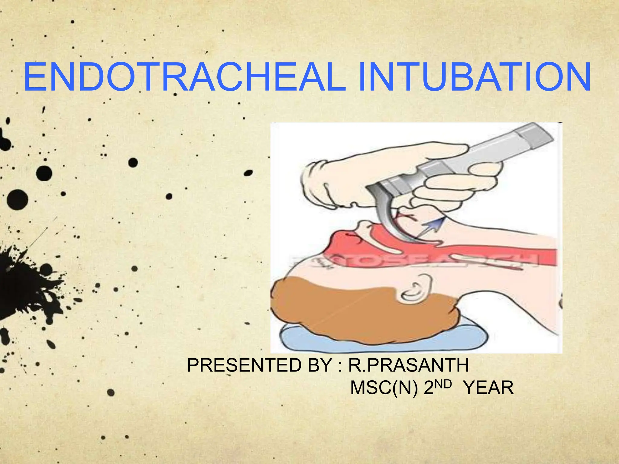
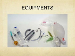
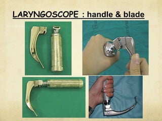
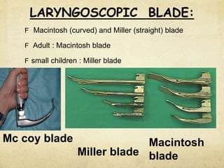
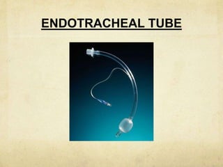
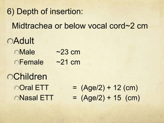
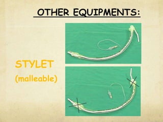
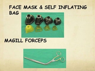
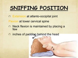
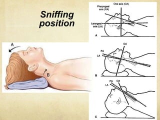
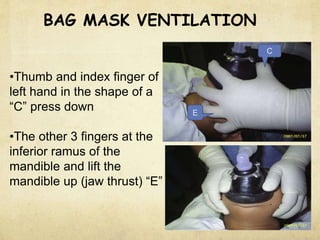
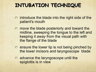
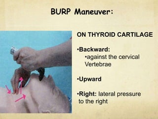
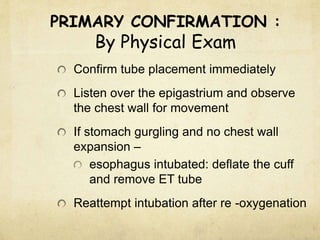
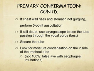
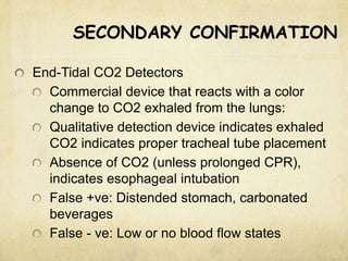
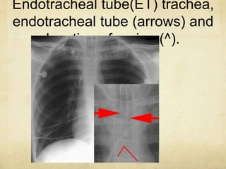
ENDOTRACHEAL INTUBATION
PRESENTED BY : R.PRASANTH
MSC(N) 2ND YEAR
DEFINITION
It is a procedure of passing of an
endotracheal tube into trachea
through the nose or mouth.
PURPOSE
It is performed to establish and
maintain a patent airway, facilitate
oxygenation and ventilation, reduce
the risk of aspiration, and assist with
the clearance of secretions.
INDICATIONS
AND
CONTRAINDICATIONS
ADVANTAGES
DISADVANTAGES
Need advanced training to properly perform
procedure
Bypasses the nares function of warming and
filtering the air
Increased incidence of trauma due to neck
manipulation when spinal cord injury is
suspected
May increase respiratory resistance
Improper placement
EQUIPMENTS
LARYNGOSCOPE : handle & blade
F Macintosh (curved) and Miller (straight) blade
F Adult : Macintosh blade
F small children : Miller blade
LARYNGOSCOPIC BLADE:
Mc coy blade
Miller blade
Macintosh
blade
ENDOTRACHEAL TUBE
TYPES OF ETTs:
1) Portex tubes:
Semirigid, with little tendency to kink.
Most commonly used.
2) Rubber tubes:
Soft, easily kinked.
3) Reinforced tubes:
- Cuffed or non cuffed. Reinforced with
wire to prevent kinking.
4) Special tubes:
Double lumen (Robertshaw
ENDOTRACHEAL TUBE: (ETT)
Male : ID 8.0 mms
Female : ID 7.5 mms
New born - 3 mths : ID 3.0 mms
3-9 months : ID 3.5 mms
9-18 months : ID 4.0 mms
2- 6 yrs : ID = (Age/3) + 3.5
> 6 yrs : ID = (Age/4) + 4.5
1) Size of ETT : internal diameter (ID)
2) MATERIAL : Red rubber or PVC
3) ETT CUFF
High volume
Low pressure cuff
Low volume
High pressure cuff
ETT CUFF
• cuff inflating system consisting of:
• valve, balloon, inflating tube & cuff.
• Uncuffed tubes used in children to minimise pressure
injury
• Purpose of cuff is:
• seal between tube & trachea
• Protect from aspiration of blood, mucus or vomitus.
4) BEVEL
5) MURPHY’S EYE
6) Depth of insertion:
Midtrachea or below vocal cord~2 cm
Adult
Male ~23 cm
Female ~21 cm
Children
Oral ETT = (Age/2) + 12 (cm)
Nasal ETT = (Age/2) + 15 (cm)
OTHER EQUIPMENTS:
STYLET
(malleable)
FACE MASK & SELF INFLATING
BAG
MAGILL FORCEPS
LOCAL ANAESTHETIC
SPRAY
 Syringe
 Lubricating jelly
 Dynaplast/ tape to strap
endotracheal tube
 Monitoring success of intubation:
 Stethoscope
 Endtidal - CO2
 Pulse oximeter
PREOXYGENATION
PROCEDURE
• ventilate with 100 % oxygen for
approximately 3 min
• Position bed / table height:
bring the patient's head to naval height
SNIFFING POSITION
Extension at atlanto-occipital joint
Flexion at lower cervical spine
Neck flexion is maintained by placing a
few
inches of padding behind the head
Sniffing
position
STEPS OF OROENDOTRACHEAL
INTUBATION
BAG MASK VENTILATION
•Thumb and index finger of
left hand in the shape of a
“C” press down
•The other 3 fingers at the
inferior ramus of the
mandible and lift the
mandible up (jaw thrust) “E”
C
E
HOLDING A LARYNGOSCOPE
Hold the
handle of
the
laryngoscop
e with your
left hand
OPEN MOUTH TECHNIQUES
Hyper-extension technique
(no touch technique)
Cross fingers techniques
INTUBATION TECHNIQUE
introduce the blade into the right side of the
patient's mouth
move the blade posteriorly and toward the
midline, sweeping the tongue to the left and
keeping it away from the visual path with
the flange of the blade
ensure the lower lip is not being pinched by
the lower incisors and laryngoscope blade
advance the laryngoscope until the
epiglottis is in view
INSERTING THE BLADE
INTUBATION TECHNIQUE
lift the laryngoscope upward and forward
insert the ETT from the right angle of mouth
with its concave curve facing downward and
to the right side of the patient
maneuver the endotracheal tube into the
larynx, midway between the cricoid cartilage
and the sternal angle
LIFTING UP A LARYNGOSCOPE:
•Pull the blade forward and upward
using firm but Steady pressure without
rotating the wrist
•Avoid leaning on the upper teeth with
EXPOSURE OF THE LARYNX:
•In most situations vocal cords should become
visible
• If not, exert gentle pressure over the cricoid
area to help bring them into view
BURP Maneuver:
ON THYROID CARTILAGE
•Backward:
•against the cervical
Vertebrae
•Upward
•Right: lateral pressure
to the right
HOW TO CONFIRM THE
CORRECT PLACEMENT OF ETT?
Primary Confirmation
Secondary Confirmation
PRIMARY CONFIRMATION :
By Physical Exam
Confirm tube placement immediately
Listen over the epigastrium and observe
the chest wall for movement
If stomach gurgling and no chest wall
expansion –
esophagus intubated: deflate the cuff
and remove ET tube
Reattempt intubation after re -oxygenation
PRIMARY CONFIRMATION:
CONTD.
If chest wall rises and stomach not gurgling,
perform 5-point auscultation
If still doubt, use laryngoscope to see the tube
passing through the vocal cords (best)
Secure the tube
Look for moisture condensation on the inside
of the tracheal tube
(not 100%: false +ve with esophageal
intubations)
SECONDARY CONFIRMATION
End-Tidal CO2 Detectors
Commercial device that reacts with a color
change to CO2 exhaled from the lungs:
Qualitative detection device indicates exhaled
CO2 indicates proper tracheal tube placement
Absence of CO2 (unless prolonged CPR),
indicates esophageal intubation
False +ve: Distended stomach, carbonated
beverages
False - ve: Low or no blood flow states
Endotracheal tube(ET) trachea,
endotracheal tube (arrows) and
location of carina (^).
AFTER CARE OF THE
PATIENT
PROCEDURE FOR
REMOVAL
THANK YOU

209415893-Endotracheal-tube-insertion-ppt.pptx

  • 1.
    ENDOTRACHEAL INTUBATION PRESENTED BY: R.PRASANTH MSC(N) 2ND YEAR
  • 2.
    DEFINITION It is aprocedure of passing of an endotracheal tube into trachea through the nose or mouth.
  • 3.
    PURPOSE It is performedto establish and maintain a patent airway, facilitate oxygenation and ventilation, reduce the risk of aspiration, and assist with the clearance of secretions.
  • 4.
  • 5.
  • 6.
    DISADVANTAGES Need advanced trainingto properly perform procedure Bypasses the nares function of warming and filtering the air Increased incidence of trauma due to neck manipulation when spinal cord injury is suspected May increase respiratory resistance Improper placement
  • 7.
  • 8.
  • 9.
    F Macintosh (curved)and Miller (straight) blade F Adult : Macintosh blade F small children : Miller blade LARYNGOSCOPIC BLADE: Mc coy blade Miller blade Macintosh blade
  • 10.
  • 11.
    TYPES OF ETTs: 1)Portex tubes: Semirigid, with little tendency to kink. Most commonly used. 2) Rubber tubes: Soft, easily kinked. 3) Reinforced tubes: - Cuffed or non cuffed. Reinforced with wire to prevent kinking. 4) Special tubes: Double lumen (Robertshaw
  • 12.
    ENDOTRACHEAL TUBE: (ETT) Male: ID 8.0 mms Female : ID 7.5 mms New born - 3 mths : ID 3.0 mms 3-9 months : ID 3.5 mms 9-18 months : ID 4.0 mms 2- 6 yrs : ID = (Age/3) + 3.5 > 6 yrs : ID = (Age/4) + 4.5 1) Size of ETT : internal diameter (ID)
  • 13.
    2) MATERIAL :Red rubber or PVC 3) ETT CUFF High volume Low pressure cuff Low volume High pressure cuff
  • 14.
    ETT CUFF • cuffinflating system consisting of: • valve, balloon, inflating tube & cuff. • Uncuffed tubes used in children to minimise pressure injury • Purpose of cuff is: • seal between tube & trachea • Protect from aspiration of blood, mucus or vomitus.
  • 15.
  • 16.
    6) Depth ofinsertion: Midtrachea or below vocal cord~2 cm Adult Male ~23 cm Female ~21 cm Children Oral ETT = (Age/2) + 12 (cm) Nasal ETT = (Age/2) + 15 (cm)
  • 17.
  • 18.
    FACE MASK &SELF INFLATING BAG MAGILL FORCEPS
  • 19.
  • 20.
     Syringe  Lubricatingjelly  Dynaplast/ tape to strap endotracheal tube  Monitoring success of intubation:  Stethoscope  Endtidal - CO2  Pulse oximeter
  • 21.
    PREOXYGENATION PROCEDURE • ventilate with100 % oxygen for approximately 3 min • Position bed / table height: bring the patient's head to naval height
  • 22.
    SNIFFING POSITION Extension atatlanto-occipital joint Flexion at lower cervical spine Neck flexion is maintained by placing a few inches of padding behind the head
  • 23.
  • 24.
  • 25.
    BAG MASK VENTILATION •Thumband index finger of left hand in the shape of a “C” press down •The other 3 fingers at the inferior ramus of the mandible and lift the mandible up (jaw thrust) “E” C E
  • 26.
    HOLDING A LARYNGOSCOPE Holdthe handle of the laryngoscop e with your left hand
  • 27.
    OPEN MOUTH TECHNIQUES Hyper-extensiontechnique (no touch technique) Cross fingers techniques
  • 28.
    INTUBATION TECHNIQUE introduce theblade into the right side of the patient's mouth move the blade posteriorly and toward the midline, sweeping the tongue to the left and keeping it away from the visual path with the flange of the blade ensure the lower lip is not being pinched by the lower incisors and laryngoscope blade advance the laryngoscope until the epiglottis is in view
  • 29.
  • 30.
    INTUBATION TECHNIQUE lift thelaryngoscope upward and forward insert the ETT from the right angle of mouth with its concave curve facing downward and to the right side of the patient maneuver the endotracheal tube into the larynx, midway between the cricoid cartilage and the sternal angle
  • 31.
    LIFTING UP ALARYNGOSCOPE: •Pull the blade forward and upward using firm but Steady pressure without rotating the wrist •Avoid leaning on the upper teeth with
  • 32.
    EXPOSURE OF THELARYNX: •In most situations vocal cords should become visible • If not, exert gentle pressure over the cricoid area to help bring them into view
  • 33.
    BURP Maneuver: ON THYROIDCARTILAGE •Backward: •against the cervical Vertebrae •Upward •Right: lateral pressure to the right
  • 34.
    HOW TO CONFIRMTHE CORRECT PLACEMENT OF ETT? Primary Confirmation Secondary Confirmation
  • 35.
    PRIMARY CONFIRMATION : ByPhysical Exam Confirm tube placement immediately Listen over the epigastrium and observe the chest wall for movement If stomach gurgling and no chest wall expansion – esophagus intubated: deflate the cuff and remove ET tube Reattempt intubation after re -oxygenation
  • 36.
    PRIMARY CONFIRMATION: CONTD. If chestwall rises and stomach not gurgling, perform 5-point auscultation If still doubt, use laryngoscope to see the tube passing through the vocal cords (best) Secure the tube Look for moisture condensation on the inside of the tracheal tube (not 100%: false +ve with esophageal intubations)
  • 37.
    SECONDARY CONFIRMATION End-Tidal CO2Detectors Commercial device that reacts with a color change to CO2 exhaled from the lungs: Qualitative detection device indicates exhaled CO2 indicates proper tracheal tube placement Absence of CO2 (unless prolonged CPR), indicates esophageal intubation False +ve: Distended stomach, carbonated beverages False - ve: Low or no blood flow states
  • 38.
    Endotracheal tube(ET) trachea, endotrachealtube (arrows) and location of carina (^).
  • 39.
    AFTER CARE OFTHE PATIENT
  • 40.
  • 41.