endotracheal intubation-Anesthesia
•
3 likes•208 views

This document discusses endotracheal tubes and intubation. It covers indications for intubation including airway protection, optimizing gas exchange, decreasing metabolic demand, and reducing work of breathing. Conditions associated with difficult intubation are described such as congenital anomalies, infections, tumors, injuries, and obesity. Proper equipment, tube sizing, intubation technique including positioning and confirmation of placement are outlined. Golden rules of intubation emphasize preparation, oxygenation, skills, confirmation, and monitoring.

Recommended
More Related Content
What's hot
What's hot (20)
Similar to endotracheal intubation-Anesthesia
Similar to endotracheal intubation-Anesthesia (20)
More from NISAR ARAIN
More from NISAR ARAIN (20)
Recently uploaded
Recently uploaded (20)
endotracheal intubation-Anesthesia
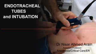
- 1. ENDOTRACHEAL TUBES and INTUBATION Dr Nisar Ahmed Arain Assistant Professor Anesthesia/Critical Care/ER
- 2. -INDICATIONS --A =Airway Protection =Optimization of gas exchange --B =Oxygenation / Ventilation =To Decrease Metabolic Demand --C =To Reduce work of breathing
- 3. -INDICATIONS =Airway protection =Decrease LOC =Lower cranial nerve palsy A =Laryngeal edema =Orofacial injury =Copious trachea-bronchial secretions
- 4. -INDICATIONS =Optimization of Gas exchange Oxygenation / Ventilation B =Hypoxic Respiratory Failure Pneumonia / Pulmonary edema/ARDS =Hyperbaric respiratory failure Obstructive airway disease/ OSAS Obesity hypoventilation syndrome
- 5. -INDICATIONS =To decrease metabolic demand/ To decrease WoB -C =Severe septic shock / Burn / polytrauma
- 7. -Conditions associated with difficult intubation -Congenital anomalies Pierre Robin Syndrome, Down’s Syndrome -Infection in airway Retropharyngeal abscess, epiglottis -Tumor in oral cavity or Larynx -Enlarge thyroid causing compression / displacement of trachea
- 8. -Conditions associated with difficult intubation --Maxillofacial cervical of laryngeal trauma --Temporomandibular joint dysfunction --Burn scar at Face and Neck --Morbidly obese or pregnancy
- 9. -Airway assesment -Inter Incisor distance > 3 cm
- 10. -Airway assesment -Mallampati classification --Difficult Intubation
- 11. -Airway assesment -Laryngoscopy View -Difficult Intubation
- 12. -Airway assesment -Thyro mental distance > 6 cm -Difficult Intubation
- 13. -Airway assesment -Flexion / Extension of Neck
- 14. -Airway Assesment -Movement of Temporomandibular joint (TMJ) -GRINDING
- 15. -EQUIPMENTS --Laryngoscope with relevant size blade --Magill's Forceps --Flexible Introducer --10 to 20 ml syringe --Oropharyngeal airways –All sizes --Tape or adhesive Plaster --E.T tubes--relevant sizes --Bag—Valve—Mask with oxygen connected --Suction unit with Yankauer nozzle and endotracheal suction catheter
- 16. -Laryngoscope
- 17. -Laryngoscope
- 19. -Oropharyngeal / Nasopharyngeal Airway
- 20. -Endotracheal Tube: Size( mm Internal Diameter) --New Born—3 Months = 3.0 mm ID --3 to 9 Months = 3.5 mm ID --9 to 18 Months = 4.0 mm ID --2 Years to 6 Years = (Age/3) + 3.5 -- >6 Years = (Age/4) + 4.5 --Adult Male = 8 to 8.5 mm ID --Adult Female =7.0 to 7.5 mm ID
- 21. -Depth of endotracheal tube : should be placed at Mid trachea or below vocal cords = 2 cms --Adult -> Male = 23 cms --Adult -> Female = 21 cms --Children a- Oral endotracheal tube = (Age/2) + 12 cms b- Nasal endotracheal tube = (Age/2) + 15 cms
- 22. -Technique of endotracheal Intubation The three Axis : Oral, Pharyngeal and Laryngeal
- 23. -Sniffing Position : Aligning the three Axis
- 25. -Technique of endotracheal Intubation --Position the patient supine, open the airway with a head- tilt-chin-lift maneuver. (suspected spinal injury, attempt Nasotracheal intubation spine in neutral position --Open mouth by separating the lips and pulling on upper jaw with the index finger
- 26. -Technique of endotracheal Intubation --Hold laryngoscope in the left hand, insert scope into the mouth with blade directed to right tonsil. --Once right tonsil is reached, sweep the blade to the midline keeping the tongue on the left. --This brings the Epiglottis into view. “DO NOT LOOSE SITE OF IT “ --Advance the blade until it reaches the angle between the base of the tongue and epiglottis (Vallecular space) --Lift the laryngoscope upwards and away from the Nose – towards the chest. This should bring the vocal cords into view. It may be necessary for a colleague to press on the trachea to improve the view of the larynx
- 27. -Technique of endotracheal Intubation cont. --Place the ETT in the right hand. Keep the concavity of the tube facing the right side of the mouth --Insert the tube watching it enter through the cords. --Insert the tube just so the cuff has passed the cords and then inflate the cuff --Listen for air entry at both apices and both axillae to ensure correct placement using stethoscope
- 28. -CONFIRMATION OF PROPER TUBE PLACEMENT --PLACEMENT UNDER VISION --FOUR QUADRANT AUSCULTATION --CAPNOMETERY / CAPNOGRAPHY --VENTILATOR GRAPHS
- 29. --GOLDEN RULES OF INTUBATION --Always have a suction unit available --An intubation attempt should never exceed 30 seconds --Oxygenate the patient Pre and Post intubation with a Bag-valve-Mask and monitor SpO2 continuously --Have sedative / analgesic medicines available --Always confirm tube placement by more then one methods --Do not attempt intubation unless you are totally skilled, rather perform Bag-Valve-Mask Ventilation --Always confirm tube placement from time to time