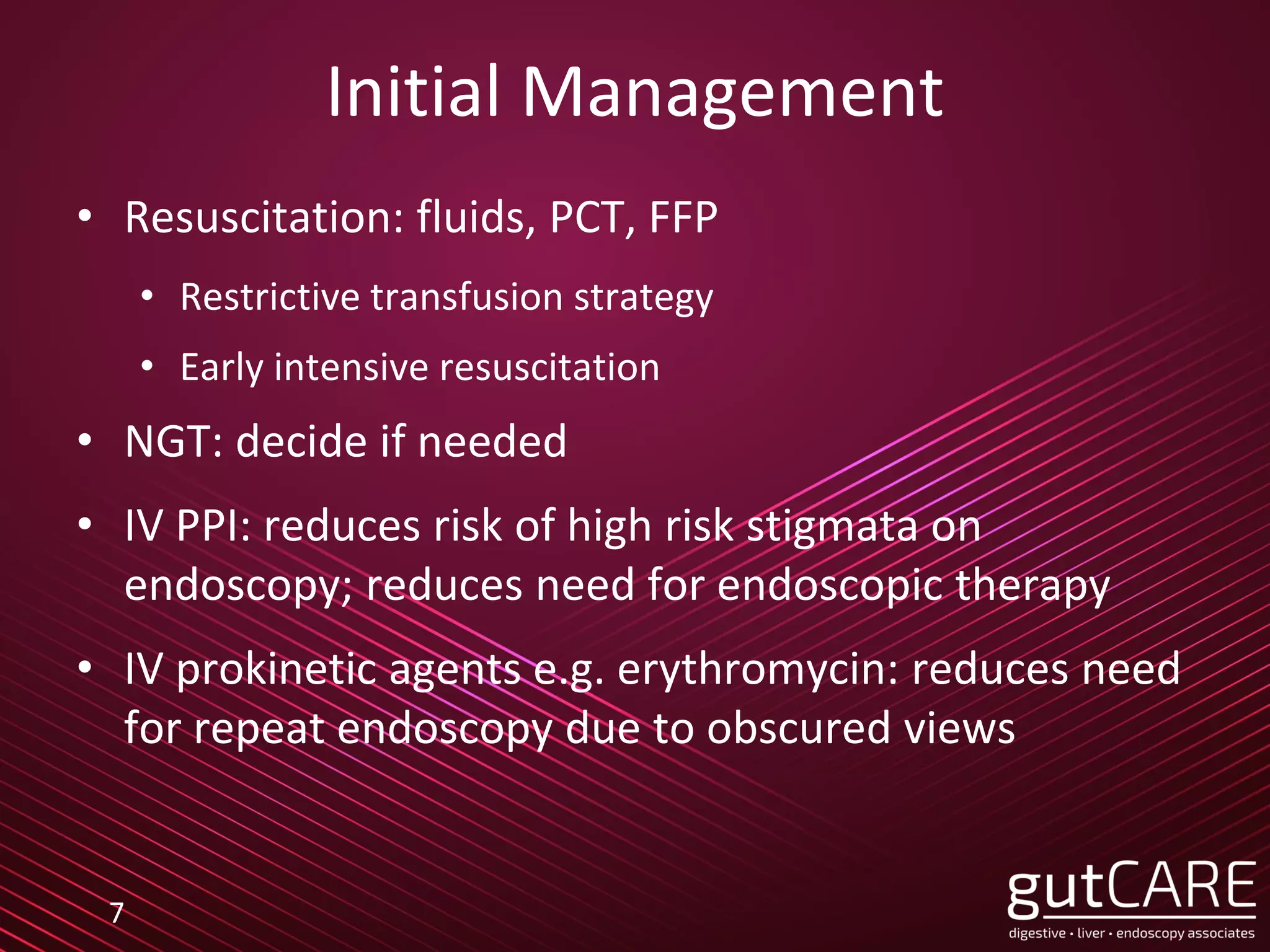
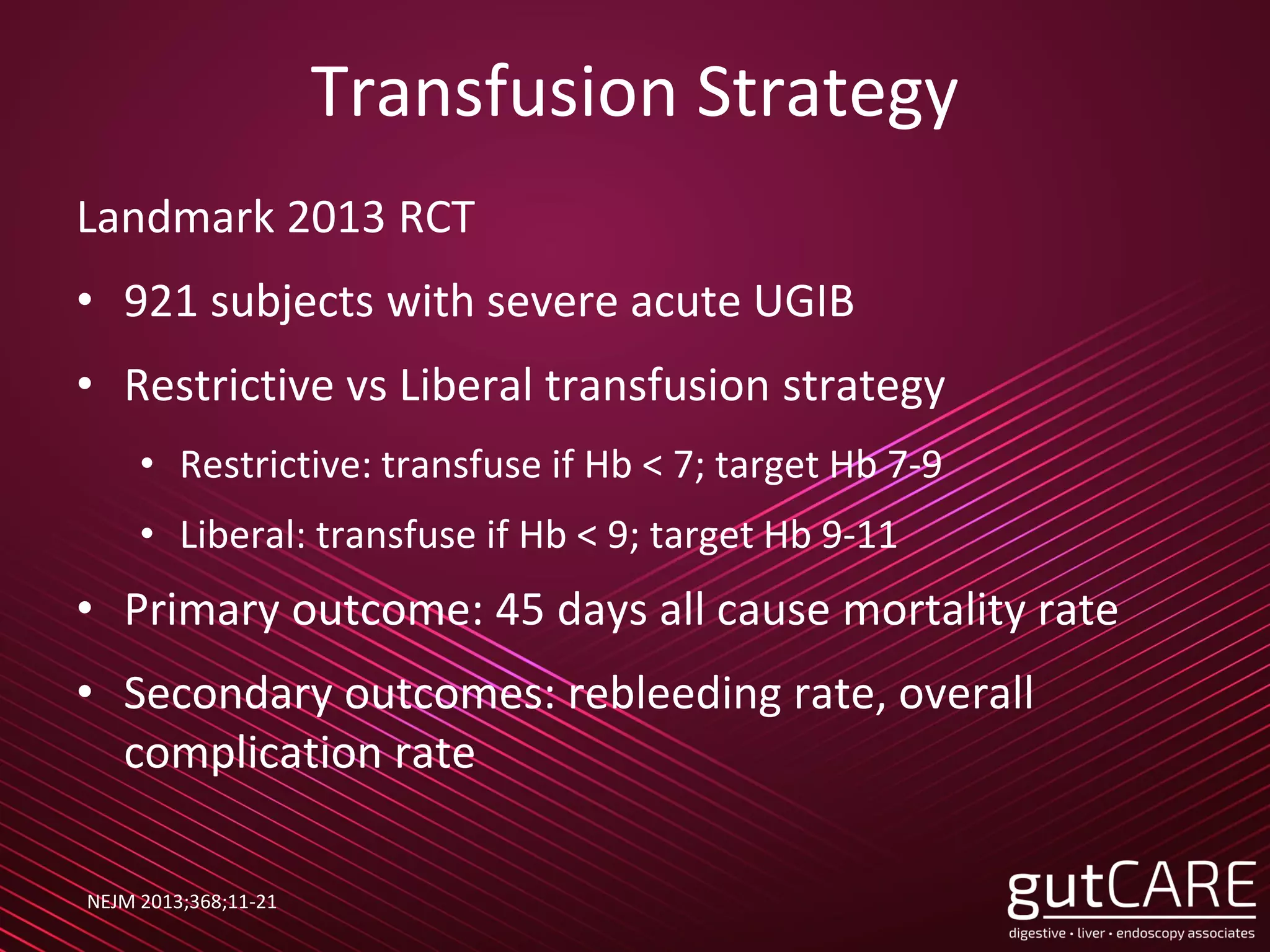
Endoscopic hemostasis is an important first line treatment modality in bleeding from the gastrointestinal tract. It is also a prerequisite skill for anyone performing therapeutic endoscopy, where bleeding is the most common intra-procedural endoscopic complication. This lecture is aimed at endoscopy nurses assisting the endoscopist, and gives an overview of endoscopic hemostasis in routine endoscopy today.
2. About gutCARE
• Founded 2014
• Singapore's only digestive specialist group practice
• Regional authorities in sub-specialty areas
• Comprehensive & niche services
From 2020:
• 6 clinics in 6 hospitals
• 7 doctors in all main subspecialty disciplines
A full gastroenterology department in private practice
3. 3
Scope
• Upper GI Bleed (UGIB)
• PUD
• Varices
• Endoscopic modalities
• Lower GI Bleed (LGIB)
• Bleeding due to
interventional endoscopy
5. Upper GI Bleed
• Definition: bleeding source proximal to ligament of
Treitz (corresponds to DJ flexure)
• Symptoms: hematemesis, malena, coffee ground
vomitus, hematochezia
• 80% of all bleeding cases
• Mortality: 3.5-10%
5
7. Initial Management
• Resuscitation: fluids, PCT, FFP
• Restrictive transfusion strategy
• Early intensive resuscitation
• NGT: decide if needed
• IV PPI: reduces risk of high risk stigmata on
endoscopy; reduces need for endoscopic therapy
• IV prokinetic agents e.g. erythromycin: reduces need
for repeat endoscopy due to obscured views
7
8. Transfusion Strategy
Landmark 2013 RCT
• 921 subjects with severe acute UGIB
• Restrictive vs Liberal transfusion strategy
• Restrictive: transfuse if Hb < 7; target Hb 7-9
• Liberal: transfuse if Hb < 9; target Hb 9-11
• Primary outcome: 45 days all cause mortality rate
• Secondary outcomes: rebleeding rate, overall
complication rate
NEJM 2013;368;11-21
9. Restrictive Strategy Superior
Restrictive Liberal P value
Mortality rate 5% 9% 0.02
Rate of further
bleeding
10% 16% 0.01
Overall
complication rate
40% 48% 0.02
NEJM 2013;368;11-21
Benefit seen primarily in
Child A/B cirrhotics
10. Early Intensive Resuscitation
2004 Study
• Consecutive series of patients with hemodynamically
significant UGIB
• 1st 36 subjects: Observation Group (no intervention)
• 2nd 36 subjects: Intensive Resuscitation Group
• Intense guidance provided; decreased time to correct
hemodynamics, Hct and coagulopathy
• Both groups same in demographics
Am J Gastroenterol 2004;99:619
12. Causes of Mortality in Patients
with Peptic Ulcer Bleeding
• Patients rarely
bleed to death
• Prospective cohort
study >10,000
cases of PUD bleed
• Mortality rate 6.2%
• 80% of deaths not
related to bleeding
Am J Gastroenterol 2010;105:84
13. International Consensus Group 2019
Pre Endoscopy Management
• Initiate resuscitation if hemodynamically unstable
• Blood transfusion if Hb < 8 and no underlying
cardiovascular disease
• Consider: Nasogastric tube, PPI treatment
• Perform early endoscopy within 24H
• Variceal bleed: do ASAP, within 12H
13
15. Peptic Ulcer Disease
• 80% of bleeds stop spontaneously
• Need for endoscopic therapy depends on stigmata of
recent hemorrhage
15
16. High Risk Stigmata
• High rebleeding risk: Needs endoscopic hemostasis
16
Spurting
Vessel
Oozing
Vessel
NBVV
17. Low Risk Stigmata
• Low rebleeding risk: No need endoscopic hemostasis
17
Flat pigmented spot Clean base
18. Adherent CLot
18
• Role of endoscopic
therapy of ulcers with
adherent clot is
controversial
• Clot removal usually
attempted
• Underlying lesion can
then be assessed,
treated if necessary
19. Endoscopic Treatment
• Aim to identify and
‘seal’ the feeding
vessel
• Options:
• Injection therapy
• Thermal therapy
• Hemoclips
• Newer modalities
19
20. Endoscopic Injection Therapy
• Reduces blood flow by temporary local tamponade
• Vasoconstricting agents (e.g. adrenaline 1:10,000 to
1:100,000) also reduces blood flow
• Not recommended as monotherapy
20
21. Thermal Therapy:
Coaptive Thermal
Coagulation
• Bipolar or heater probe:
• Coagulation through alternating electric current
• Mechanical compression interrupts blood flow and
reduces heat sink effect
• Can seal arteries up to 2mm in size
21
22. Hemostatic Clips
• Permanent mechanical tamponade
• Various clips to choose from
• Studies show equivalent results to heater probe
22
23. What to Use? When to Use?
• Studies show that other than adrenaline injection
monotherapy, no single modality was superior
• Choice will depend on type of lesion, anatomical
location and operator preference
• Favours clips: recurrent bleeding, pliable lesion, < 2
mm vessel, < 2 cm ulcer, coagulopathy
• Favours heater probe: initial hemostasis, fibrotic/
indurated base
23
24. Limitations
• Large ulcer > 2 cm
• Large vessel > 2 mm
• Difficult positions:
• Posterior wall stomach
• Upper lesser curve stomach
• Posterior wall duodenum
• Fibrotic base for hemoclips
• Anticoagulation
24
26. Hemostatic Nanopowder Spray
• 95% effective for acute hemostasis
• Advantages:
• Non contact, simple to use
• Can cover large area
• Effective for difficult locations, tumour bleeding
• Disadvantages:
• Only effective in actively bleeding or oozing lesions
• Moisture can plug catheter tip
• Loss of view after application
26
34. • Effective for refractory bleeding
• Generally safe
• Advantages
• Entraps large amount of tissue
• Band ligation technique
• Generally safe
• Disadvantages
• Withdraw scope to mount
• Endoscopic view impaired
• Suction traps fluid in cap
• Limited scope angulation
34
35. Coagulation Forceps
• Designed for endoscopic bleeding
• Flat jaws for grasping
• Rotational ability
• Usually used during EMR or ESD to ablate vessels
prophylactically or treat bleeding
• Studies show equivalent to clips and heater probe
for bleeding ulcers
35
40. Variceal Bleeding
• Suspect if background cirrhosis or portal
hypertension
• Rule of ‘thirds’
• Occurs in 1/3 of cirrhotic patients
• 1/3 of initial bleeding are fatal
• 1/3 will rebleed in 6 weeks
• Only 1/3 will survive > 1 year
40
41. Resuscitation
• Vasoconstrictor therapy: IV terlipressin or
somatostatin/ octreotide
• Antibiotics: bacterial infections occur in 2/3 of cases
• Restrictive transfusion strategy
• Platelets and FFP usually not needed
• HDU care; avoid volume overexpansion
• Endoscopy as soon as possible (<12H), may need
intubation
41
42. Varices: Endoscopic Management
• Endoscopy as soon as possible (<12H)
• May need intubation
• Oesophageal varices:
• Look for ‘red’ and ‘white’ signs
• Gastric varices:
• GOV1: Banding or glue injection
• GOV2 or IGV: glue injection or TIPS
• Ectopic varices: case by case basis
42
53. Salvage Therapy
• Definition:
• Failure to control acute variceal bleed in 24H
• Failure to prevent clinically significant rebleeding within 5
days after treatment initiation
• Options
• TIPS
• Sengstaken Blackmore Tube
• Covered metal stent
53
59. Urgent Colonoscopy
• No standardized approach; few studies to date do
not show any improvement in clinical outcomes
• Rapid bowel prep required: 4-6L PEG over 3-4H
• Colonoscopy performed within 1H of clearing stool,
blood and clots
• Should be performed in 12-24H in stable patient
• Need for bowel prep and sedation may be
prohibitive in unstable patient
59
60. Endoscopic Therapy
• Diverticular bleeding
• Difficult to identify bleeding site
• If bleeding site identified, clips preferred
• Angiectasia: APC preferred
• Radiation proctitis
• APC preferred
• RFA and thermal therapies also used
60
66. Risk Factors for Endoscopic Failure
• Hemodynamic instability on presentation:
tachycardia, hypotension, syncope
• Ongoing bleeding
• Comorbid illnesses
• Age > 60
• Initial Hct < 35%, elevated creatinine
• History of diverticulosis or angioectasia
66
67. When to go straight to Angiogram?
ACG guidelines
“Radiographic interventions should be considered in
patients with high-risk clinical features and ongoing
bleeding who have a negative upper endoscopy and do
not respond adequately to hemodynamic resuscitation
efforts and are therefore unlikely to tolerate bowel
preparation and urgent colonoscopy (strong
recommendation, very low quality evidence)”
67
71. Polypectomy
• Biggest risk factor is polyp
size
• Delayed bleeding can occur
up to 2 weeks later
• If large polyp > 2cm,
consider prophylactic loop
or clip placement
• In immediate or delayed
bleeding, all modalities can
be used except thermal
therapy in right colon
71
75. EMR/ ESD
• High risk of bleeding
• Intraprocedural 10-15%
• Delayed 5-10%
• Must differentiate intra-procedural from delayed bleeding
• Use dissection knife, hemostatic forceps or clips
• Prophylactic hemostasis of all visible vessels with
hemostatic forceps prevents delayed bleed
• Closing of scars with clips may prevent delayed bleed
75
88. Conclusion
• Endoscopy hemostasis is
the mainstay of treatment
for bleeding GIT
• Many studies and
techniques have
emerged. Endoscopy
Centres need to keep
pace to ensure best
outcomes.
88