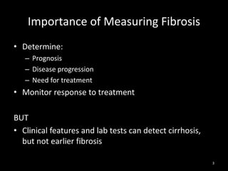
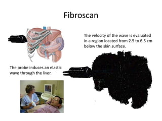
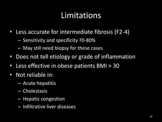
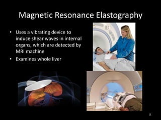
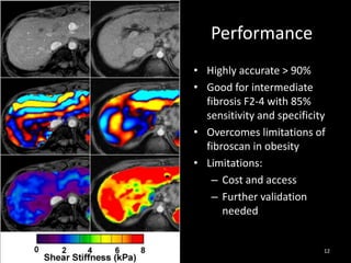
Detecting Early Liver Fibrosis
A Nutshell for Primary Care
Dr Jarrod Lee
Mt Elizabeth Novena Hospital
1
Stage 1: Portal Fibrosis

Stage 0: Normal

Stage 2: Periportal Fibrosis

Fibrosis to
Cirrhosis
Stage 4: Cirrhosis

Stage 3: Septal Fibrosis

2
Importance of Measuring Fibrosis
• Determine:
– Prognosis
– Disease progression
– Need for treatment

• Monitor response to treatment
BUT
• Clinical features and lab tests can detect cirrhosis,
but not earlier fibrosis
3
Liver Biopsy
• ‘Gold standard’ for
staging fibrosis
• Staging by accepted
classifications
• Can evaluate:
– Etiology
– Inflammation
– Steatosis

4
Problems with Liver Biopsy
• Invasive
– 30% have pain after biopsy
– 1% severe complications

• Inter and intra observer variability
• Sampling error
– Samples 1:50,000 of liver mass
– Difference between right and left lobes
• Stage of fibrosis: 33%
• Diagnosis of cirrhosis: 15%
5
How about using ultrasound?
• Systemic review of 21 studies (2010)
– Liver surface is most robust technique: high specificity (7895%), moderate sensitivity (51-73%)
– Poor observer agreement

• Singapore study (2012)
– Significant fibrosis: sensitivity 32%, specificity 85%

6
Fibroscan
• Uses mild amplitude low frequency
vibration transmitted through liver
• Velocity of shear wave correlates with
liver stiffness
– Travels faster through stiffer fibrotic
tissue
• Sampled volume 1:500
• Painless bedside test; takes < 5 min
• Good reproducibility, well validated
• 90% accurate for early fibrosis (F0-1)
and cirrhosis
7
Fibroscan
The velocity of the wave is evaluated
in a region located from 2.5 to 6.5 cm
below the skin surface.

The probe induces an elastic
wave through the liver.

25 to 65 mm

8
Liver Stiffness
Portal fibrosis
Cholestasis
Centrolobular fibrosis

Sinusoidal fibrosis

Stiffness

Steatosis

Portal blood flow

Inflammation
9
Limitations
• Less accurate for intermediate fibrosis (F2-4)
– Sensitivity and specificity 70-80%
– May still need biopsy for these cases

• Does not tell etiology or grade of inflammation
• Less effective in obese patients BMI > 30
• Not reliable in:
–
–
–
–

Acute hepatitis
Cholestasis
Hepatic congestion
Infiltrative liver diseases
10
Magnetic Resonance Elastography
• Uses a vibrating device to
induce shear waves in internal
organs, which are detected by
MRI machine
• Examines whole liver

11
Performance
• Highly accurate > 90%
• Good for intermediate
fibrosis F2-4 with 85%
sensitivity and specificity
• Overcomes limitations of
fibroscan in obesity
• Limitations:
– Cost and access
– Further validation
needed

12

Detecting Early Liver Fibrosis - A Nutshell for Primary Care

  • 1.
    Detecting Early LiverFibrosis A Nutshell for Primary Care Dr Jarrod Lee Mt Elizabeth Novena Hospital 1
  • 2.
    Stage 1: PortalFibrosis Stage 0: Normal Stage 2: Periportal Fibrosis Fibrosis to Cirrhosis Stage 4: Cirrhosis Stage 3: Septal Fibrosis 2
  • 3.
    Importance of MeasuringFibrosis • Determine: – Prognosis – Disease progression – Need for treatment • Monitor response to treatment BUT • Clinical features and lab tests can detect cirrhosis, but not earlier fibrosis 3
  • 4.
    Liver Biopsy • ‘Goldstandard’ for staging fibrosis • Staging by accepted classifications • Can evaluate: – Etiology – Inflammation – Steatosis 4
  • 5.
    Problems with LiverBiopsy • Invasive – 30% have pain after biopsy – 1% severe complications • Inter and intra observer variability • Sampling error – Samples 1:50,000 of liver mass – Difference between right and left lobes • Stage of fibrosis: 33% • Diagnosis of cirrhosis: 15% 5
  • 6.
    How about usingultrasound? • Systemic review of 21 studies (2010) – Liver surface is most robust technique: high specificity (7895%), moderate sensitivity (51-73%) – Poor observer agreement • Singapore study (2012) – Significant fibrosis: sensitivity 32%, specificity 85% 6
  • 7.
    Fibroscan • Uses mildamplitude low frequency vibration transmitted through liver • Velocity of shear wave correlates with liver stiffness – Travels faster through stiffer fibrotic tissue • Sampled volume 1:500 • Painless bedside test; takes < 5 min • Good reproducibility, well validated • 90% accurate for early fibrosis (F0-1) and cirrhosis 7
  • 8.
    Fibroscan The velocity ofthe wave is evaluated in a region located from 2.5 to 6.5 cm below the skin surface. The probe induces an elastic wave through the liver. 25 to 65 mm 8
  • 9.
    Liver Stiffness Portal fibrosis Cholestasis Centrolobularfibrosis Sinusoidal fibrosis Stiffness Steatosis Portal blood flow Inflammation 9
  • 10.
    Limitations • Less accuratefor intermediate fibrosis (F2-4) – Sensitivity and specificity 70-80% – May still need biopsy for these cases • Does not tell etiology or grade of inflammation • Less effective in obese patients BMI > 30 • Not reliable in: – – – – Acute hepatitis Cholestasis Hepatic congestion Infiltrative liver diseases 10
  • 11.
    Magnetic Resonance Elastography •Uses a vibrating device to induce shear waves in internal organs, which are detected by MRI machine • Examines whole liver 11
  • 12.
    Performance • Highly accurate> 90% • Good for intermediate fibrosis F2-4 with 85% sensitivity and specificity • Overcomes limitations of fibroscan in obesity • Limitations: – Cost and access – Further validation needed 12