This document provides guidance on the management of upper gastrointestinal bleeding (UGIB). It outlines the following key points:
1. Initial steps include assessing hemodynamic stability, IV access, monitoring, fluid resuscitation, and basic lab tests. Endoscopy within 24 hours is critical for diagnosis and treatment.
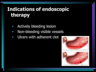
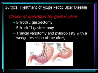
2. For non-variceal bleeding, endoscopic therapies like injection, thermal coagulation, and clipping are first-line. Refractory or high-risk bleeding may require surgery.
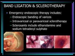
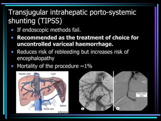
3. Variceal bleeding requires urgent endoscopic ligation or sclerotherapy. Transjugular intrahepatic portosystemic shunting or surgical shunting may be needed if initial measures
Information about Management of Appendicular Lump by Dr Dhaval Mangukiya.
Details of Appendicular Lump, Basic to Above the Basics, Incidence, Safe Approach Interval Laparoscopy, Early Surgery etc.
https://drdhavalmangukiya.com/
http://www.youtube.com/c/DrDhavalMangukiyaGastrosurgeonSurat
https://gastrosurgerysurat.blogspot.com/
Approach to Management of Upper Gastrointestinal (GI) BleedingArun Vasireddy
Upper gastrointestinal bleeding is gastrointestinal bleeding in the upper gastrointestinal tract, commonly defined as bleeding arising from the esophagus, stomach, or duodenum. Blood may be observed in vomit (hematemesis) or in altered form in the stool (melena). Depending on the severity of the blood loss, there may be symptoms of insufficient circulating blood volume and shock. As a result, upper gastrointestinal bleeding is considered a medical emergency and typically requires hospital care for urgent diagnosis and treatment. Upper gastrointestinal bleeding can be caused by peptic ulcers, gastric erosions, esophageal varices, and some rarer causes such as gastric cancer.
The initial assessment includes measurement of the blood pressure and heart rate, as well as blood tests to determine hemoglobin concentration. In significant bleeding, fluid replacement is often required, as well as blood transfusion, before the source of bleeding can be determined by endoscopy of the upper digestive tract with an esophagogastroduodenoscopy. Depending on the source, endoscopic therapy can be applied to reduce rebleeding risk. Specific medical treatments (such as proton pump inhibitors for peptic ulcer disease) or procedures (such as TIPS for variceal hemorrhage) may be used. Recurrent or refractory bleeding may lead to need for surgery, although this has become uncommon as a result of improved endoscopic and medical treatment.
by Bushra Ibnauf as part of SAMA's Visiting Faculty Program in Salam Rotana Hotel on June 24th 2011. This was in collaboration with the Sudanese Society for Gastroenterology.
Please find the power point on Carcinoma of rectum. I tried present it on understandable way and all the contents are reviewed by experts and from very reliable references.
Probably Pediatric Surgery is the only surgical specialty which opens all four body cavities to treat their little patients. This is a pictorial glimpse of the wide spectrum of Pediatric Surgery.
Sites of the highest risk are the duodenum, for adenocarcinomas, and the ileum, for carcinoids and lymphomas.
In industrialized countries, small bowel cancers are predominantly adenocarcinomas;
In developing countries, lymphomas are much more common.
The incidence of small bowel cancer rises with age and has generally been higher among males than among females.
The risk factors for small bowel cancer include
Dietary factor
Cigarette smoking,
Alcohol intake,
Medical conditions -Crohn's disease, familial adenomatous polyposis, cholecystectomy, peptic ulcer disease, and cystic fibrosis.
The protective factors may include rapid cell turnover, a general absence of bacteria, an alkaline environment, and low levels of activating enzymes of precarcinogens.
Endoscopic Hemostasis - for Endoscopy NursesJarrod Lee
Endoscopic hemostasis is an important first line treatment modality in bleeding from the gastrointestinal tract. It is also a prerequisite skill for anyone performing therapeutic endoscopy, where bleeding is the most common intra-procedural endoscopic complication. This lecture is aimed at endoscopy nurses assisting the endoscopist, and gives an overview of endoscopic hemostasis in routine endoscopy today.
Surgery Resident clinical seminar on the management of a 60yr old male with upper gastrointestinal bleeding presented to the department of surgery, Niger Delta University Teaching Hospital, Okolobiri, Bayelsa State
micro teaching on communication m.sc nursing.pdfAnurag Sharma
Microteaching is a unique model of practice teaching. It is a viable instrument for the. desired change in the teaching behavior or the behavior potential which, in specified types of real. classroom situations, tends to facilitate the achievement of specified types of objectives.
Flu Vaccine Alert in Bangalore Karnatakaaddon Scans
As flu season approaches, health officials in Bangalore, Karnataka, are urging residents to get their flu vaccinations. The seasonal flu, while common, can lead to severe health complications, particularly for vulnerable populations such as young children, the elderly, and those with underlying health conditions.
Dr. Vidisha Kumari, a leading epidemiologist in Bangalore, emphasizes the importance of getting vaccinated. "The flu vaccine is our best defense against the influenza virus. It not only protects individuals but also helps prevent the spread of the virus in our communities," he says.
This year, the flu season is expected to coincide with a potential increase in other respiratory illnesses. The Karnataka Health Department has launched an awareness campaign highlighting the significance of flu vaccinations. They have set up multiple vaccination centers across Bangalore, making it convenient for residents to receive their shots.
To encourage widespread vaccination, the government is also collaborating with local schools, workplaces, and community centers to facilitate vaccination drives. Special attention is being given to ensuring that the vaccine is accessible to all, including marginalized communities who may have limited access to healthcare.
Residents are reminded that the flu vaccine is safe and effective. Common side effects are mild and may include soreness at the injection site, mild fever, or muscle aches. These side effects are generally short-lived and far less severe than the flu itself.
Healthcare providers are also stressing the importance of continuing COVID-19 precautions. Wearing masks, practicing good hand hygiene, and maintaining social distancing are still crucial, especially in crowded places.
Protect yourself and your loved ones by getting vaccinated. Together, we can help keep Bangalore healthy and safe this flu season. For more information on vaccination centers and schedules, residents can visit the Karnataka Health Department’s official website or follow their social media pages.
Stay informed, stay safe, and get your flu shot today!
Title: Sense of Taste
Presenter: Dr. Faiza, Assistant Professor of Physiology
Qualifications:
MBBS (Best Graduate, AIMC Lahore)
FCPS Physiology
ICMT, CHPE, DHPE (STMU)
MPH (GC University, Faisalabad)
MBA (Virtual University of Pakistan)
Learning Objectives:
Describe the structure and function of taste buds.
Describe the relationship between the taste threshold and taste index of common substances.
Explain the chemical basis and signal transduction of taste perception for each type of primary taste sensation.
Recognize different abnormalities of taste perception and their causes.
Key Topics:
Significance of Taste Sensation:
Differentiation between pleasant and harmful food
Influence on behavior
Selection of food based on metabolic needs
Receptors of Taste:
Taste buds on the tongue
Influence of sense of smell, texture of food, and pain stimulation (e.g., by pepper)
Primary and Secondary Taste Sensations:
Primary taste sensations: Sweet, Sour, Salty, Bitter, Umami
Chemical basis and signal transduction mechanisms for each taste
Taste Threshold and Index:
Taste threshold values for Sweet (sucrose), Salty (NaCl), Sour (HCl), and Bitter (Quinine)
Taste index relationship: Inversely proportional to taste threshold
Taste Blindness:
Inability to taste certain substances, particularly thiourea compounds
Example: Phenylthiocarbamide
Structure and Function of Taste Buds:
Composition: Epithelial cells, Sustentacular/Supporting cells, Taste cells, Basal cells
Features: Taste pores, Taste hairs/microvilli, and Taste nerve fibers
Location of Taste Buds:
Found in papillae of the tongue (Fungiform, Circumvallate, Foliate)
Also present on the palate, tonsillar pillars, epiglottis, and proximal esophagus
Mechanism of Taste Stimulation:
Interaction of taste substances with receptors on microvilli
Signal transduction pathways for Umami, Sweet, Bitter, Sour, and Salty tastes
Taste Sensitivity and Adaptation:
Decrease in sensitivity with age
Rapid adaptation of taste sensation
Role of Saliva in Taste:
Dissolution of tastants to reach receptors
Washing away the stimulus
Taste Preferences and Aversions:
Mechanisms behind taste preference and aversion
Influence of receptors and neural pathways
Impact of Sensory Nerve Damage:
Degeneration of taste buds if the sensory nerve fiber is cut
Abnormalities of Taste Detection:
Conditions: Ageusia, Hypogeusia, Dysgeusia (parageusia)
Causes: Nerve damage, neurological disorders, infections, poor oral hygiene, adverse drug effects, deficiencies, aging, tobacco use, altered neurotransmitter levels
Neurotransmitters and Taste Threshold:
Effects of serotonin (5-HT) and norepinephrine (NE) on taste sensitivity
Supertasters:
25% of the population with heightened sensitivity to taste, especially bitterness
Increased number of fungiform papillae
These simplified slides by Dr. Sidra Arshad present an overview of the non-respiratory functions of the respiratory tract.
Learning objectives:
1. Enlist the non-respiratory functions of the respiratory tract
2. Briefly explain how these functions are carried out
3. Discuss the significance of dead space
4. Differentiate between minute ventilation and alveolar ventilation
5. Describe the cough and sneeze reflexes
Study Resources:
1. Chapter 39, Guyton and Hall Textbook of Medical Physiology, 14th edition
2. Chapter 34, Ganong’s Review of Medical Physiology, 26th edition
3. Chapter 17, Human Physiology by Lauralee Sherwood, 9th edition
4. Non-respiratory functions of the lungs https://academic.oup.com/bjaed/article/13/3/98/278874
TEST BANK for Operations Management, 14th Edition by William J. Stevenson, Ve...kevinkariuki227
TEST BANK for Operations Management, 14th Edition by William J. Stevenson, Verified Chapters 1 - 19, Complete Newest Version.pdf
Factory Supply Best Quality Pmk Oil CAS 28578–16–7 PMK Powder in Stockrebeccabio
Factory Supply Best Quality Pmk Oil CAS 28578–16–7 PMK Powder in Stock
Telegram: bmksupplier
signal: +85264872720
threema: TUD4A6YC
You can contact me on Telegram or Threema
Communicate promptly and reply
Free of customs clearance, Double Clearance 100% pass delivery to USA, Canada, Spain, Germany, Netherland, Poland, Italy, Sweden, UK, Czech Republic, Australia, Mexico, Russia, Ukraine, Kazakhstan.Door to door service
Hot Selling Organic intermediates
Report Back from SGO 2024: What’s the Latest in Cervical Cancer?bkling
Are you curious about what’s new in cervical cancer research or unsure what the findings mean? Join Dr. Emily Ko, a gynecologic oncologist at Penn Medicine, to learn about the latest updates from the Society of Gynecologic Oncology (SGO) 2024 Annual Meeting on Women’s Cancer. Dr. Ko will discuss what the research presented at the conference means for you and answer your questions about the new developments.
Title: Sense of Smell
Presenter: Dr. Faiza, Assistant Professor of Physiology
Qualifications:
MBBS (Best Graduate, AIMC Lahore)
FCPS Physiology
ICMT, CHPE, DHPE (STMU)
MPH (GC University, Faisalabad)
MBA (Virtual University of Pakistan)
Learning Objectives:
Describe the primary categories of smells and the concept of odor blindness.
Explain the structure and location of the olfactory membrane and mucosa, including the types and roles of cells involved in olfaction.
Describe the pathway and mechanisms of olfactory signal transmission from the olfactory receptors to the brain.
Illustrate the biochemical cascade triggered by odorant binding to olfactory receptors, including the role of G-proteins and second messengers in generating an action potential.
Identify different types of olfactory disorders such as anosmia, hyposmia, hyperosmia, and dysosmia, including their potential causes.
Key Topics:
Olfactory Genes:
3% of the human genome accounts for olfactory genes.
400 genes for odorant receptors.
Olfactory Membrane:
Located in the superior part of the nasal cavity.
Medially: Folds downward along the superior septum.
Laterally: Folds over the superior turbinate and upper surface of the middle turbinate.
Total surface area: 5-10 square centimeters.
Olfactory Mucosa:
Olfactory Cells: Bipolar nerve cells derived from the CNS (100 million), with 4-25 olfactory cilia per cell.
Sustentacular Cells: Produce mucus and maintain ionic and molecular environment.
Basal Cells: Replace worn-out olfactory cells with an average lifespan of 1-2 months.
Bowman’s Gland: Secretes mucus.
Stimulation of Olfactory Cells:
Odorant dissolves in mucus and attaches to receptors on olfactory cilia.
Involves a cascade effect through G-proteins and second messengers, leading to depolarization and action potential generation in the olfactory nerve.
Quality of a Good Odorant:
Small (3-20 Carbon atoms), volatile, water-soluble, and lipid-soluble.
Facilitated by odorant-binding proteins in mucus.
Membrane Potential and Action Potential:
Resting membrane potential: -55mV.
Action potential frequency in the olfactory nerve increases with odorant strength.
Adaptation Towards the Sense of Smell:
Rapid adaptation within the first second, with further slow adaptation.
Psychological adaptation greater than receptor adaptation, involving feedback inhibition from the central nervous system.
Primary Sensations of Smell:
Camphoraceous, Musky, Floral, Pepperminty, Ethereal, Pungent, Putrid.
Odor Detection Threshold:
Examples: Hydrogen sulfide (0.0005 ppm), Methyl-mercaptan (0.002 ppm).
Some toxic substances are odorless at lethal concentrations.
Characteristics of Smell:
Odor blindness for single substances due to lack of appropriate receptor protein.
Behavioral and emotional influences of smell.
Transmission of Olfactory Signals:
From olfactory cells to glomeruli in the olfactory bulb, involving lateral inhibition.
Primitive, less old, and new olfactory systems with different path
New Directions in Targeted Therapeutic Approaches for Older Adults With Mantl...i3 Health
i3 Health is pleased to make the speaker slides from this activity available for use as a non-accredited self-study or teaching resource.
This slide deck presented by Dr. Kami Maddocks, Professor-Clinical in the Division of Hematology and
Associate Division Director for Ambulatory Operations
The Ohio State University Comprehensive Cancer Center, will provide insight into new directions in targeted therapeutic approaches for older adults with mantle cell lymphoma.
STATEMENT OF NEED
Mantle cell lymphoma (MCL) is a rare, aggressive B-cell non-Hodgkin lymphoma (NHL) accounting for 5% to 7% of all lymphomas. Its prognosis ranges from indolent disease that does not require treatment for years to very aggressive disease, which is associated with poor survival (Silkenstedt et al, 2021). Typically, MCL is diagnosed at advanced stage and in older patients who cannot tolerate intensive therapy (NCCN, 2022). Although recent advances have slightly increased remission rates, recurrence and relapse remain very common, leading to a median overall survival between 3 and 6 years (LLS, 2021). Though there are several effective options, progress is still needed towards establishing an accepted frontline approach for MCL (Castellino et al, 2022). Treatment selection and management of MCL are complicated by the heterogeneity of prognosis, advanced age and comorbidities of patients, and lack of an established standard approach for treatment, making it vital that clinicians be familiar with the latest research and advances in this area. In this activity chaired by Michael Wang, MD, Professor in the Department of Lymphoma & Myeloma at MD Anderson Cancer Center, expert faculty will discuss prognostic factors informing treatment, the promising results of recent trials in new therapeutic approaches, and the implications of treatment resistance in therapeutic selection for MCL.
Target Audience
Hematology/oncology fellows, attending faculty, and other health care professionals involved in the treatment of patients with mantle cell lymphoma (MCL).
Learning Objectives
1.) Identify clinical and biological prognostic factors that can guide treatment decision making for older adults with MCL
2.) Evaluate emerging data on targeted therapeutic approaches for treatment-naive and relapsed/refractory MCL and their applicability to older adults
3.) Assess mechanisms of resistance to targeted therapies for MCL and their implications for treatment selection
Ethanol (CH3CH2OH), or beverage alcohol, is a two-carbon alcohol
that is rapidly distributed in the body and brain. Ethanol alters many
neurochemical systems and has rewarding and addictive properties. It
is the oldest recreational drug and likely contributes to more morbidity,
mortality, and public health costs than all illicit drugs combined. The
5th edition of the Diagnostic and Statistical Manual of Mental Disorders
(DSM-5) integrates alcohol abuse and alcohol dependence into a single
disorder called alcohol use disorder (AUD), with mild, moderate,
and severe subclassifications (American Psychiatric Association, 2013).
In the DSM-5, all types of substance abuse and dependence have been
combined into a single substance use disorder (SUD) on a continuum
from mild to severe. A diagnosis of AUD requires that at least two of
the 11 DSM-5 behaviors be present within a 12-month period (mild
AUD: 2–3 criteria; moderate AUD: 4–5 criteria; severe AUD: 6–11 criteria).
The four main behavioral effects of AUD are impaired control over
drinking, negative social consequences, risky use, and altered physiological
effects (tolerance, withdrawal). This chapter presents an overview
of the prevalence and harmful consequences of AUD in the U.S.,
the systemic nature of the disease, neurocircuitry and stages of AUD,
comorbidities, fetal alcohol spectrum disorders, genetic risk factors, and
pharmacotherapies for AUD.
ARTIFICIAL INTELLIGENCE IN HEALTHCARE.pdfAnujkumaranit
Artificial intelligence (AI) refers to the simulation of human intelligence processes by machines, especially computer systems. It encompasses tasks such as learning, reasoning, problem-solving, perception, and language understanding. AI technologies are revolutionizing various fields, from healthcare to finance, by enabling machines to perform tasks that typically require human intelligence.
2. • Careful history and physical examinations.
• Gain IV access with large bore cannula
• Full blood count & cross matching
• Monitoring Blood pressure, pulse, urine output
• IV colloids or crystalloid
• Blood transfusion
• ENDOSCOPY for diagnosis & treatment
• PUD- IV PPI
• Bleeding recurs: surgery
3. When the patient is first seen, a
quick examination must be
made to answer the three
following critical questions :
a) Is there evidence of airway
obstruction ?
b) Is there evidence of active
bleeding ?
c) Is there evidence of
hypovolemia ?
4. Measure pulse and blood pressure
If hemodynamically stable
• Obtain full history
• Carry out full examination
• Proceed with investigation
5. If hemodynamically unstable
• Resuscitate
• If rapid responsive, then proceed as for
stable patient
• If transient or non-responsive prompt
investigation to locate the source of
bleeding and to established ideal
treatment
6. Management:
• Intravenous access
– at least 1 large bore cannula
• Initial clinical assessment
– Circulatory status
– Evidence of liver disease
– Identify comorbidity
9. • Can start with 500 ml NaCl 0.9% over the first 15
min, followed by 500 ml colloid (eg. Gelatin) over the
next 15 min
• If BP fails to come up or falls infusion rate must be
increased accordingly
• If patient becomes stable ( BP > 100 mmHg, pulse <
100/min ) rapid infusion must be stopped, and
maintenance fluids only given.
10. Indication for blood transfusion
Shock (Pallor, cold sweaty
skin, systolic BP
<100mmHg)
Hb <10 gm% in patients
with recent or active
bleeding.
Patient with coagulopathy,
low platelet count should be
transfused with fresh frozen
plasma and platelets
respectively.
11. • Oxygen therapy
– Should be given to all patients in shock.
• IV PPI: Omeprazole
12. ENDOSCOPY
• Ideally, endoscopy should be performed
within 24 hours.
• Endoscopy can be used both in diagnosis and
therapy.
15. • Bleeding can be 2 type
– 1. non- variceal bleeding
– 2. variceal bleeding
16.
17. Endoscopic therapy
• Adrenaline(1:10,000) or sclerosant injection
• Heat probes
• application of metallic clips
• Bipolar diathermy
• Laser photocoagulation using the Nd-YAG
laser
18. • Constant probe pressure
tamponade
• Argon plasma
coagulator
• Rubber band ligation
The preference is for dual
therapy, e.g. injection of
adrenaline with thermal
coagulation.
19. Repeated endoscopy
• endoscopy and endo-therapy should be
repeated within 24 hours when initial
endoscopic treatment was considered sub-
optimal (because of difficult access, poor
visualisation,technical difficulties) or in
patients in whom rebleeding is likely to be life
threatening.
20. Medication
• Injectable proton pump inhibitor / Ranitidine
• oral PPI in high doses.
• NSAIDs should be stooped and future use
should be restricted.
21. Endoscopic hemostasis vs medical therapy
SIGNS
RISK OF RECURRENT
BLEEDING WITH MEDICAL
THERAPY ALONE
RISK OF RECURRENT
BLEEDING WITH
ENDOSCOPIC
HEMOSTASIS
Active arterial bleeding
(spurting)
85%–95% 10%–20%
Nonbleeding visible vessel 50% 5%–−10%
Nonbleeding adherent clot 35% < 5%
Ulcer oozing 10%–25% < 5%
Flat spots 7% Not indicated
Clean-based ulcer 3% Not indicated
22. Surgical Treatment of Acute Peptic Ulcer
Disease ( PUD )
Indications for Surgery
• Perforation
• Pyloric obstruction
• Continued bleeding that fails to
respond to endoscopic measures
• Recurrent bleeding
• Patients > 60 years
• Cardiovascular disease with predictive
poor response to hypotension
23. • Aim of surgery :
- Stops bleeding
- Prevent recurrent bleeding
24. Choice of operation for duodenal
ulcer
– Billroth II gastrectomy
– Truncal vagotomy and
pyloroplasty with suture ligation
of the bleeding ulcer
• Selective vagotomy
• Highly selective vagotomy
– Truncal vagotomy and
antrectomy with resection or
suture ligation of the bleeding
ulcer
25. Choice of operation for gastric ulcer
– Billroth I gastrectomy
– Billroth II gastrectomy
– Truncal vagotomy and pyloroplasty with a
wedge resection of the ulcer,
27. Complications
• The complications of UGIB are self-evident. Other
complications can arise from treatments
administered. For example:
• Endoscopy:
– Aspiration pneumonia
– Perforation
– ventricular arrhythmias during endoscopy
– Complications from coagulation, laser treatments
• Surgery:
– Ileus
– Sepsis
– Wound problems
28. Prognosis
• A score of less than 3 using the Rockall system above is associated
with an excellent prognosis
• whereas a score of 8 or above is associated with high mortality
• Mortality is about 7%.
Rockall risk scoring system
29. Prognosis is worse with the following:
• Increasing age
• Co-morbidity
• Liver disease
• Shock at presentation
• Continued bleeding after presentation
• Haematemesis
• Haematochezia
• Elevated blood urea
30. Prevention
• The most important factor to consider is
treatment for H. pylori infection. This
should be completed as an outpatient.
31.
32. • The mortality of a variceal bleed is
approximately 50%
• 70% patients will have a rebleed
• Survival is dependent on the degree of
hepatic impairment
34. BALOON TAMPONADE
• Temporary tamponade can be achieved with
Sengstaken-Blackmore tube
– Should be considered as a salvage
procedure
– Unfortunately 50% patients rebleed within
24 hours of removal of tamponade
35. BAND LIGATION & SCLEROTHERAPY
• Emergency endoscopic therapy includes:
– Endoscopic banding of varices
– Intravariceal or paravariceal sclerotherapy
– Sclerosants include ethanolamine and
sodium tetradecyl sulphate
36. Transjugular intrahepatic porto-systemic
shunting (TIPSS)
• If endoscopic methods fail.
• Recommended as the treatment of choice for
uncontrolled variceal haemorrhage.
• Reduces risk of rebleeding but increases risk of
encephalopathy
• Mortality of the procedure ~1%
37. Porto-systemic shunt operation
• Only done if
– Unsuccessful endoscopic treatment
– Good liver function
• Can lead to:
– Post operative liver failure
– Hepatic encephalopathy
• Emergency shunting associated with 20% operative
mortality.
38. PROGNOSIS
• Recurrence within 2 year
– 7% for small varies
– 30% for large varies
• Poor liver function – 45%
• Mortality – 15%
39. PREVENTION
Primary prevention
Bleeding from varices more likely if poor hepatic
function or large varices
• Primary prevention of bleeding is possible
with β blockers
– Reduces risk of haemorrhage by 40-50%
• Band ligation may also be considered
Sclerotherapy or shunting is ineffective
40. Secondary prevention
• 70% of patients with an variceal
haemorrhage will rebleed
• The following have been shown to be
effective in the prevention of rebleeding
– Beta-blockers possibly combined with
isosorbide mononitrate
– Endoscopic ligation
– Sclerotherapy
– TIPSS
– Surgical shunting