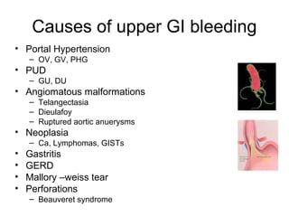
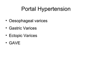
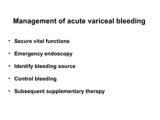
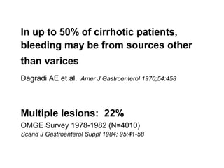
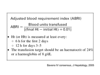
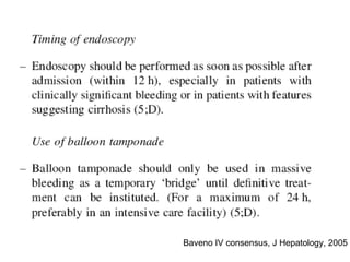
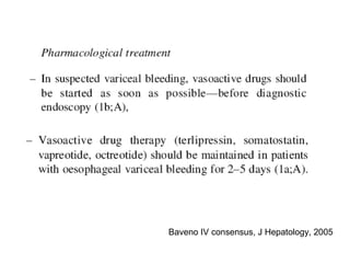
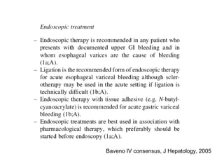
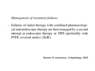
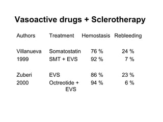
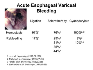
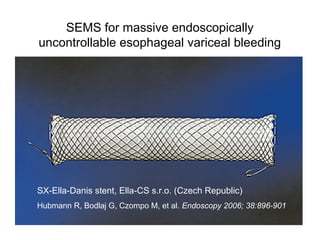
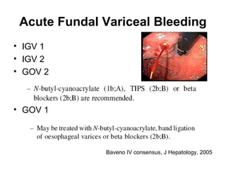
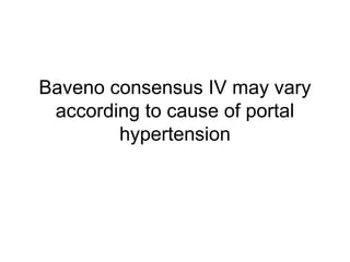
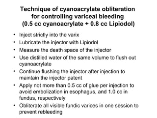
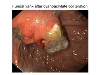
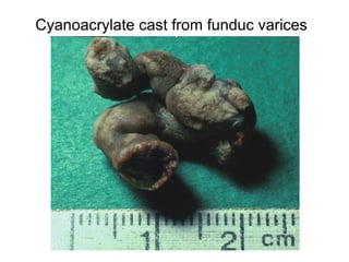
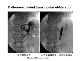
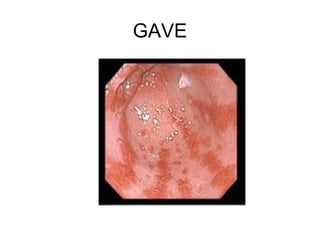
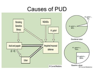
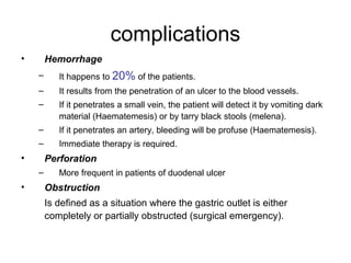
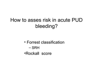
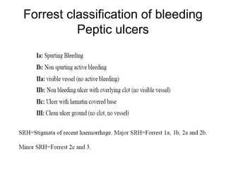
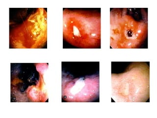
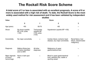
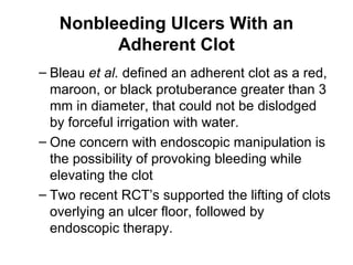
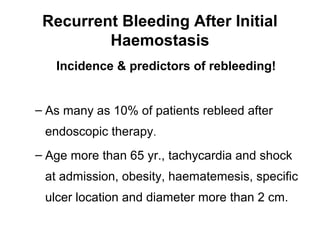
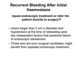
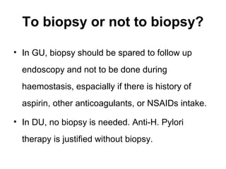
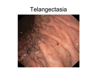
Upper GI Bleeding
Hossam GHONEIM, MD
17.6.2010
Causes of upper GI bleeding
• Portal Hypertension
– OV, GV, PHG
• PUD
– GU, DU
• Angiomatous malformations
– Telangectasia
– Dieulafoy
– Ruptured aortic anuerysms
• Neoplasia
– Ca, Lymphomas, GISTs
• Gastritis
• GERD
• Mallory –weiss tear
• Perforations
– Beauveret syndrome
History Taking
Portal Hypertension
• Oesophageal varices
• Gastric Varices
• Ectopic Varices
• GAVE
Management of acute variceal bleeding
• Secure vital functions
• Emergency endoscopy
• Identify bleeding source
• Control bleeding
• Subsequent supplementary therapy
In up to 50% of cirrhotic patients,
bleeding may be from sources other
than varices
Dagradi AE et al. Amer J Gastroenterol 1970;54:458
Multiple lesions: 22%
OMGE Survey 1978-1982 (N=4010)
Scand J Gastroenterol Suppl 1984; 95:41-58
Baveno IV consensus, J Hepatology, 2005
Vasoactive drugs + Sclerotherapy
Authors Treatment Hemostasis Rebleeding
Villanueva Somatostatin 76 % 24 %
1999 SMT + EVS 92 % 7 %
Zuberi EVS 86 % 23 %
2000 Octreotide + 94 % 6 %
EVS
Acute Esophageal Variceal
Bleeding
Ligation Sclerotherapy Cyanoacrylate
Hemostasis 97%1
76%1
100%2,3,4
Rebleeding 17%1
25%2
31%4
35%1
44%3
9%2
10%3,4
1 Lo et al. Hepatology 1997;25:1101
2 Thakeb et al. Endoscopy 1995;27:358
3 Feretis et al. Endoscopy 1995;27:355
4 Soehendra et al. Endoscopy 1987;19:221
SEMS for massive endoscopically
uncontrollable esophageal variceal bleeding
SX-Ella-Danis stent, Ella-CS s.r.o. (Czech Republic)
Hubmann R, Bodlaj G, Czompo M, et al. Endoscopy 2006; 38:896-901
Acute Fundal Variceal Bleeding
• IGV 1
• IGV 2
• GOV 2
• GOV 1
Baveno IV consensus, J Hepatology, 2005
Baveno consensus IV may vary
according to cause of portal
hypertension
Technique of cyanoacrylate obliteration
for controlling variceal bleeding
(0.5 cc cyanoacrylate + 0.8 cc Lipiodol)
• Inject strictly into the varix
• Lubricate the injector with Lipiodol
• Measure the death space of the injector
• Use distilled water of the same volume to flush out
cyanoacrylate
• Continue flushing the injector after injection to
maintain the injector patent
• Apply not more than 0.5 cc of glue per injection to
avoid embolization in esophagus, and 1.0 cc in
fundus, respectively
• Obliterate all visible fundic varices in one session to
prevent rebleeding
Fundal varix after cyanoacrylate obliteration
Cyanoacrylate cast from funduc varices
Alternative treatment modality
for bleeding fundic varices
Balloon-occluded Retrograde
Transjugular Obliteration (B-RTO )
using 5% Ethanolamine Oleate
Chikamori F et al. Dig Surg 2000;17:23-8
Balloon-occluded transjugular obliteration
FV
Balloon
GAVE
PUD
Causes of PUD
complications
• Hemorrhage
– It happens to 20% of the patients.
– It results from the penetration of an ulcer to the blood vessels.
– If it penetrates a small vein, the patient will detect it by vomiting dark
material (Haematemesis) or by tarry black stools (melena).
– If it penetrates an artery, bleeding will be profuse (Haematemesis).
– Immediate therapy is required.
• Perforation
– More frequent in patients of duodenal ulcer
• Obstruction
Is defined as a situation where the gastric outlet is either
completely or partially obstructed (surgical emergency).
How to asses risk in acute PUD
bleeding?
• Forrest classification
– SRH
•Rockall score
Forrest classification of bleeding
Peptic ulcers
The Rockall Risk Score Scheme
A total score of 3 or less is associated with an excellent prognosis. A score of 8 or
more is associated with a high risk of death. To date, the Rockall Score is the most
widely used method for risk assessment and it has been validated by independent
studies
Value Score
0 1 2 3
Age (years) <60 60-79 >80 --
Shock No shock (systolic
BP ≥100, pulse
<100)
Tachycardia
(systolic BP
≥100, pulse
≥100)
Hypotension (systolic BP <100) --
Comorbidity No major comorbidity -- Cardiac failure, ischemic heart
disease, any major comorbidity
Renal failure, liver
failure, disseminated
malignancy
Diagnosis Mallory-Weiss tear,
no lesion identified
and no SRH
All other
diagnoses
Malignancy of upper
gastrointestinal tract
--
Major stigmata
of recent
hemorrhage
None or dark spot
only
-- Blood in upper gastrointestinal
tract, adherent clot, visible or
spurting vessel
--
What have we got for
management?
•Medical
•Endoscopic
•Surgical
Do not forget
• Resuscitation
– Blood pressure
– Plasma expanders
– Blood transfusion
– Procoagulants
• Gastric lavage
• Airway protection if needed
Medical treatment
IV PPI
• Continuous Infusion Intravenous high dose omeprazole (80
mg bolus followed by 8 mg/hr), reduces the risk of rebleeding and
need for endoscopic re-treatment and surgery.
• Use of H2 receptor antagonists is not supported by clinical trials.
• Oral omeprazole in the dose of 40 mg twice a day has been
reported to decrease the risk of rebleeding in patients with stigmata
of recent bleed without endoscopic therapy by Khuroo et al.
Endoscopic
Injection therapy
• The principal mechanism of action by which diluted
epinephrine solutions work is a tamponade effect
induced by the volume of solution injected. It is,
therefore, logical that in a recent study, a large volume
(35-45 ml) epinephrine injection appeared to be more
effective than a standard volume (15-25 ml) injection.
• Other solutions, such as polidocanol, saline and even
dextrose, can produce the same effect.
Butyl-2-cyanoacrylate versus hypertonic saline-
epinephrine injection
• Lee et al reported similar initial haemostasis rates with
both butyl-2-cyanoacrylate and hypertonic saline-
epinephrine injection therapy in actively bleeding peptic
ulcers
• The glue injection group had a significantly lower
rebleeding rate (14% vs. 42%).
• Glue embolization and perforation are the two feared
complications with glue injection
Cipolleta L, Bianco MA, Marmo R, et al. Endoclips versus heater probe in preventing early recurrent bleeding from peptic ulcer: A prospective and
randomized trial. Gastrointest Endosc 2001; 53 : 147-51.
Endoscopic
Thermal devices
• Thermal devices can be divided into
– Contact (heater probe, monopolar and
bipolar electrocoagulation)
– Noncontact types (laser treatment, argon
plasma coagulation APC)
• No single method of endoscopic thermal
coagulation therapy is superior to the
others.
Endoscopic
Haemoclips
• Hemoclips have gained popularity in the past
few years.
• The deployment of hemoclips on fibrotic ulcer
floors can be difficult, particularly when they are
used tangentially (posterior wall of duodenal
bulb) or with the endoscope in a retroflexed
position (high body or fundal ulcers).
Endoscopic combination Therapy
• Benefit of combination therapy has been evaluated in many trials &
confirmed by meta-analysis.
• In a systematic review a total of 16 randomized studies involving 1,673
patients were analyzed.
• Combination reduced the rate of recurrent bleeding from 18.4%
to 10.6% (OR 0.53, 95% CI 0.40-0.69) and that of emergency
surgery from 11.3% to 7.6% (OR 0.64, 95% CI 0.46-0.90). The
mortality rate fell from 5.1% to 2.6% (OR 0.51, 95% CI 0.31-0.84).
FAQ
Nonbleeding Ulcers With an
Adherent Clot
– Bleau et al. defined an adherent clot as a red,
maroon, or black protuberance greater than 3
mm in diameter, that could not be dislodged
by forceful irrigation with water.
– One concern with endoscopic manipulation is
the possibility of provoking bleeding while
elevating the clot
– Two recent RCT’s supported the lifting of clots
overlying an ulcer floor, followed by
endoscopic therapy.
Recurrent Bleeding After Initial
Haemostasis
Incidence & predictors of rebleeding!
– As many as 10% of patients rebleed after
endoscopic therapy.
– Age more than 65 yr., tachycardia and shock
at admission, obesity, haematemesis, specific
ulcer location and diameter more than 2 cm.
Recurrent Bleeding After Initial
Haemostasis
repeat endoscopic treatment or refer the
patient directly to surgery?!
– Ulcers larger than 2 cm in diameter and
hypotension at the time of rebleeding were
two independent factors that predicted failure
of endoscopic retreatment
– Those who are poor surgical candidates might
benefit from repeated endoscopic treatment.
Eradication of Helicobacter Pylori
• There is ample evidence to prove that, for
bleeding related to Helicobacter pylori infection,
when patient is not using aspirin or NSAIDs,
curing the infection obviates the risk of ulcer
recurrence and ulcer rebleeding.
• Therapy to be started after adequate
haemostasis.
To biopsy or not to biopsy?
• In GU, biopsy should be spared to follow up
endoscopy and not to be done during
haemostasis, espacially if there is history of
aspirin, other anticoagulants, or NSAIDs intake.
• In DU, no biopsy is needed. Anti-H. Pylori
therapy is justified without biopsy.
Second-Look Endoscopy
In a meta-analysis of four studies comparing systematic second-
look endoscopy and re-treatment versus expectant treatment,
Marmo et al. showed that the risk of recurrent bleeding with the
former approach was reduced by 6.2%, but risk reductions for
surgery and mortality were insignificant.
The authors concluded that appropriate selection of patients for
second-look endoscopy is crucial.
When to discharge patient from hospital?
(stop IV PPI)
• Depending on risk assessment
• After second-look endoscopy, if needed
• Usually within 0NE week.
Angiomatous Malformations
Telangectasia
Dieulafoy lesion

Upper gi bleeding

  • 1.
    Upper GI Bleeding HossamGHONEIM, MD 17.6.2010
  • 2.
    Causes of upperGI bleeding • Portal Hypertension – OV, GV, PHG • PUD – GU, DU • Angiomatous malformations – Telangectasia – Dieulafoy – Ruptured aortic anuerysms • Neoplasia – Ca, Lymphomas, GISTs • Gastritis • GERD • Mallory –weiss tear • Perforations – Beauveret syndrome
  • 3.
  • 4.
    Portal Hypertension • Oesophagealvarices • Gastric Varices • Ectopic Varices • GAVE
  • 5.
    Management of acutevariceal bleeding • Secure vital functions • Emergency endoscopy • Identify bleeding source • Control bleeding • Subsequent supplementary therapy
  • 6.
    In up to50% of cirrhotic patients, bleeding may be from sources other than varices Dagradi AE et al. Amer J Gastroenterol 1970;54:458 Multiple lesions: 22% OMGE Survey 1978-1982 (N=4010) Scand J Gastroenterol Suppl 1984; 95:41-58
  • 7.
    Baveno IV consensus,J Hepatology, 2005
  • 8.
    Baveno IV consensus,J Hepatology, 2005
  • 9.
    Baveno IV consensus,J Hepatology, 2005
  • 10.
    Baveno IV consensus,J Hepatology, 2005
  • 11.
    Baveno IV consensus,J Hepatology, 2005
  • 12.
    Vasoactive drugs +Sclerotherapy Authors Treatment Hemostasis Rebleeding Villanueva Somatostatin 76 % 24 % 1999 SMT + EVS 92 % 7 % Zuberi EVS 86 % 23 % 2000 Octreotide + 94 % 6 % EVS
  • 13.
    Acute Esophageal Variceal Bleeding LigationSclerotherapy Cyanoacrylate Hemostasis 97%1 76%1 100%2,3,4 Rebleeding 17%1 25%2 31%4 35%1 44%3 9%2 10%3,4 1 Lo et al. Hepatology 1997;25:1101 2 Thakeb et al. Endoscopy 1995;27:358 3 Feretis et al. Endoscopy 1995;27:355 4 Soehendra et al. Endoscopy 1987;19:221
  • 14.
    SEMS for massiveendoscopically uncontrollable esophageal variceal bleeding SX-Ella-Danis stent, Ella-CS s.r.o. (Czech Republic) Hubmann R, Bodlaj G, Czompo M, et al. Endoscopy 2006; 38:896-901
  • 15.
    Acute Fundal VaricealBleeding • IGV 1 • IGV 2 • GOV 2 • GOV 1 Baveno IV consensus, J Hepatology, 2005
  • 16.
    Baveno consensus IVmay vary according to cause of portal hypertension
  • 17.
    Technique of cyanoacrylateobliteration for controlling variceal bleeding (0.5 cc cyanoacrylate + 0.8 cc Lipiodol) • Inject strictly into the varix • Lubricate the injector with Lipiodol • Measure the death space of the injector • Use distilled water of the same volume to flush out cyanoacrylate • Continue flushing the injector after injection to maintain the injector patent • Apply not more than 0.5 cc of glue per injection to avoid embolization in esophagus, and 1.0 cc in fundus, respectively • Obliterate all visible fundic varices in one session to prevent rebleeding
  • 18.
    Fundal varix aftercyanoacrylate obliteration
  • 19.
  • 20.
    Alternative treatment modality forbleeding fundic varices Balloon-occluded Retrograde Transjugular Obliteration (B-RTO ) using 5% Ethanolamine Oleate Chikamori F et al. Dig Surg 2000;17:23-8
  • 21.
  • 22.
  • 23.
  • 24.
  • 25.
    complications • Hemorrhage – Ithappens to 20% of the patients. – It results from the penetration of an ulcer to the blood vessels. – If it penetrates a small vein, the patient will detect it by vomiting dark material (Haematemesis) or by tarry black stools (melena). – If it penetrates an artery, bleeding will be profuse (Haematemesis). – Immediate therapy is required. • Perforation – More frequent in patients of duodenal ulcer • Obstruction Is defined as a situation where the gastric outlet is either completely or partially obstructed (surgical emergency).
  • 26.
    How to assesrisk in acute PUD bleeding? • Forrest classification – SRH •Rockall score
  • 27.
    Forrest classification ofbleeding Peptic ulcers
  • 29.
    The Rockall RiskScore Scheme A total score of 3 or less is associated with an excellent prognosis. A score of 8 or more is associated with a high risk of death. To date, the Rockall Score is the most widely used method for risk assessment and it has been validated by independent studies Value Score 0 1 2 3 Age (years) <60 60-79 >80 -- Shock No shock (systolic BP ≥100, pulse <100) Tachycardia (systolic BP ≥100, pulse ≥100) Hypotension (systolic BP <100) -- Comorbidity No major comorbidity -- Cardiac failure, ischemic heart disease, any major comorbidity Renal failure, liver failure, disseminated malignancy Diagnosis Mallory-Weiss tear, no lesion identified and no SRH All other diagnoses Malignancy of upper gastrointestinal tract -- Major stigmata of recent hemorrhage None or dark spot only -- Blood in upper gastrointestinal tract, adherent clot, visible or spurting vessel --
  • 30.
    What have wegot for management? •Medical •Endoscopic •Surgical
  • 31.
    Do not forget •Resuscitation – Blood pressure – Plasma expanders – Blood transfusion – Procoagulants • Gastric lavage • Airway protection if needed
  • 32.
    Medical treatment IV PPI •Continuous Infusion Intravenous high dose omeprazole (80 mg bolus followed by 8 mg/hr), reduces the risk of rebleeding and need for endoscopic re-treatment and surgery. • Use of H2 receptor antagonists is not supported by clinical trials. • Oral omeprazole in the dose of 40 mg twice a day has been reported to decrease the risk of rebleeding in patients with stigmata of recent bleed without endoscopic therapy by Khuroo et al.
  • 33.
    Endoscopic Injection therapy • Theprincipal mechanism of action by which diluted epinephrine solutions work is a tamponade effect induced by the volume of solution injected. It is, therefore, logical that in a recent study, a large volume (35-45 ml) epinephrine injection appeared to be more effective than a standard volume (15-25 ml) injection. • Other solutions, such as polidocanol, saline and even dextrose, can produce the same effect.
  • 34.
    Butyl-2-cyanoacrylate versus hypertonicsaline- epinephrine injection • Lee et al reported similar initial haemostasis rates with both butyl-2-cyanoacrylate and hypertonic saline- epinephrine injection therapy in actively bleeding peptic ulcers • The glue injection group had a significantly lower rebleeding rate (14% vs. 42%). • Glue embolization and perforation are the two feared complications with glue injection Cipolleta L, Bianco MA, Marmo R, et al. Endoclips versus heater probe in preventing early recurrent bleeding from peptic ulcer: A prospective and randomized trial. Gastrointest Endosc 2001; 53 : 147-51.
  • 35.
    Endoscopic Thermal devices • Thermaldevices can be divided into – Contact (heater probe, monopolar and bipolar electrocoagulation) – Noncontact types (laser treatment, argon plasma coagulation APC) • No single method of endoscopic thermal coagulation therapy is superior to the others.
  • 36.
    Endoscopic Haemoclips • Hemoclips havegained popularity in the past few years. • The deployment of hemoclips on fibrotic ulcer floors can be difficult, particularly when they are used tangentially (posterior wall of duodenal bulb) or with the endoscope in a retroflexed position (high body or fundal ulcers).
  • 37.
    Endoscopic combination Therapy •Benefit of combination therapy has been evaluated in many trials & confirmed by meta-analysis. • In a systematic review a total of 16 randomized studies involving 1,673 patients were analyzed. • Combination reduced the rate of recurrent bleeding from 18.4% to 10.6% (OR 0.53, 95% CI 0.40-0.69) and that of emergency surgery from 11.3% to 7.6% (OR 0.64, 95% CI 0.46-0.90). The mortality rate fell from 5.1% to 2.6% (OR 0.51, 95% CI 0.31-0.84).
  • 38.
  • 39.
    Nonbleeding Ulcers Withan Adherent Clot – Bleau et al. defined an adherent clot as a red, maroon, or black protuberance greater than 3 mm in diameter, that could not be dislodged by forceful irrigation with water. – One concern with endoscopic manipulation is the possibility of provoking bleeding while elevating the clot – Two recent RCT’s supported the lifting of clots overlying an ulcer floor, followed by endoscopic therapy.
  • 40.
    Recurrent Bleeding AfterInitial Haemostasis Incidence & predictors of rebleeding! – As many as 10% of patients rebleed after endoscopic therapy. – Age more than 65 yr., tachycardia and shock at admission, obesity, haematemesis, specific ulcer location and diameter more than 2 cm.
  • 41.
    Recurrent Bleeding AfterInitial Haemostasis repeat endoscopic treatment or refer the patient directly to surgery?! – Ulcers larger than 2 cm in diameter and hypotension at the time of rebleeding were two independent factors that predicted failure of endoscopic retreatment – Those who are poor surgical candidates might benefit from repeated endoscopic treatment.
  • 42.
    Eradication of HelicobacterPylori • There is ample evidence to prove that, for bleeding related to Helicobacter pylori infection, when patient is not using aspirin or NSAIDs, curing the infection obviates the risk of ulcer recurrence and ulcer rebleeding. • Therapy to be started after adequate haemostasis.
  • 43.
    To biopsy ornot to biopsy? • In GU, biopsy should be spared to follow up endoscopy and not to be done during haemostasis, espacially if there is history of aspirin, other anticoagulants, or NSAIDs intake. • In DU, no biopsy is needed. Anti-H. Pylori therapy is justified without biopsy.
  • 44.
    Second-Look Endoscopy In ameta-analysis of four studies comparing systematic second- look endoscopy and re-treatment versus expectant treatment, Marmo et al. showed that the risk of recurrent bleeding with the former approach was reduced by 6.2%, but risk reductions for surgery and mortality were insignificant. The authors concluded that appropriate selection of patients for second-look endoscopy is crucial.
  • 45.
    When to dischargepatient from hospital? (stop IV PPI) • Depending on risk assessment • After second-look endoscopy, if needed • Usually within 0NE week.
  • 46.
  • 47.
  • 48.