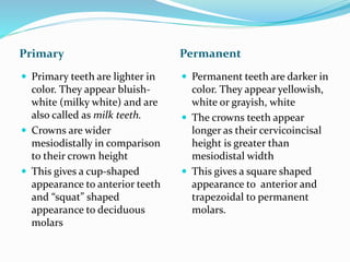
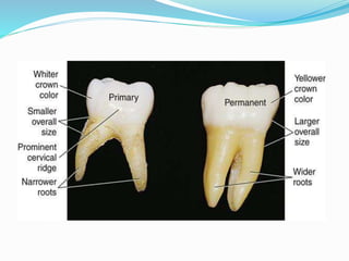
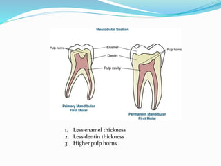
Department of Pedodontics
By: Akshdeep singh
Roll no: 07
Submitted To:
Dr. Virinder Goyal, Prof. & HOD
Dr. Kanika Gupta Verma, Prof.
Dr. Suruchi Juneja Sukhija, Reader
Dr. Sakshi Bamba, Sr. Lecturer
Introduction
 Humans possess 2 type of dentition:
 Deciduous dentition
No of tooth: 20
Eruption time: 8-29 months
Eruption Sequence: AB DC E
A B D CE
 Permanent dentition
No of tooth: 32
Eruption time: 6-21 years
 Eruption Sequence:
Maxillary teeth:6 1 2 4 3 5 7 or 6 1 2 4 5 3 7
Mandibular teeth: 6 1 2 3 4 5 7
Crown
Primary Permanent
 Shorter
 Constricted in cervical
portion i.e. are narrower at
their necks.
 Cervical ridges on buccal
aspect are more prominent
 Bigger
 Cervical constriction is not
well marked
 Cervical ridges on permanent
crowns are flatter
Primary Permanent
 Primary teeth are lighter in
color. They appear bluish-
white (milky white) and are
also called as milk teeth.
 Crowns are wider
mesiodistally in comparison
to their crown height
 This gives a cup-shaped
appearance to anterior teeth
and “squat” shaped
appearance to deciduous
molars
 Permanent teeth are darker in
color. They appear yellowish,
white or grayish, white
 The crowns teeth appear
longer as their cervicoincisal
height is greater than
mesiodistal width
 This gives a square shaped
appearance to anterior and
trapezoidal to permanent
molars.
Clinical Significance
 As deciduous dentition is lighter in colour when
compared to permanent dentition. Thus while
restoring primary teeth lighter shades are used.
Incisors
Primary Permanent
 Primary incisors do not
exhibit mamelones.
 They are noticeably wider
mesio-distally than they are
long cervico-incisally.
 Newly erupted permanent
incisors exhibit mamelons.
 They are longer cervico-
incisally than they are wider
mesio-distally.
mamelones
Canines
Primary Permanent
 Primary canines tend to be
more conical in shape and
cusp tip is more pointed and
sharp
 Permanent canines are less
conical i.e. their cusps tips are
less pointed
Premolars
Primary Permanent
 No premolars in deciduous
dentition.
 There are two premolars in
each quadrant
Molars
Primary Permanent
 There are only 2 molars in each
quadrant.
 Crown of 2nd molar is larger than
the crown of 1st molar.
 Molars are more bulbous, bell-
shaped and with marked
cervical constriction.
 Buccal and lingual surfaces of
molars, especially that of 1st
molars converge sharply
occlusally, thus forming narrow
occlusal table in buccolingual
dimension
 There are 3 molars in each
quadrant.
 1st molar is larger than 2nd
and 3rd molars.
 Permanent molars have less
constriction of neck
 There is less convergence of
buccal and lingual surfaces of
molars towards occlusal
surface. Thus have broader
occlusal table
(A) Prominent cervical ridge;
(B) Flatter cervical ridge
(A)Primary molars have
narrow occlusal table
(B) Permanent molars
have wider occlusal
table
Clinical Significance
 Cervical ridges are more pronounced especially on
buccal aspect of 1st primary molars.
 These cervical bulges have to be reproduced during
restoration/ crown prosthesis.
 Sharp cervical constriction has to be kept in mind and
special care should be taken while forming gingival
floor during class II cavity preparation.
 During class II cavity preparation for amalgam buccal
and lingual extensions/walls of class II cavity should
be located in selfcleansing areas.
 Occlusal plane is
relatively flat.
 Supplemental grooves
are more
 Contact areas between
molars are broader,
flatter and situated
gingivally.
 Occlusal plane have
more curved contour
 Supplemental grooves
are less
 Contact areas between
molars are narrower and
situated occlusally
More supplementary grooves in
primary molar
Less supplementary grooves in
permanent molar
Clinical Significance
 Deciduous molars are functionally adapted to
withstand less occlusal load.
 Occlusal cavity preparations should be kept narrow in
buccolingual plane.
 Primary molars are more caries prone due to easy food
lodgment because of more supplementary grooves . Pit
and fissure sealants are advisable to prevent caries
 Maxillary 1st molar has 3
cusps (resembles a
premolar)
 Maxillary 2nd molar has 4
cusps + one accessory
cusp (resembles
permanent upper 1st
molar)
 Maxillary 1st molar has 4
cusps + 1 accessory cusp.
 Maxillary 2nd molar has 4
cusps
Maxillary deciduous
1st molar
Maxillary
deciduous 2ndmolar
Permanent
maxillary 1st molar Permanent
maxillary 2ndmolar
Primary Permanent
 Mandibular 1st molar has 4
cusps (does not resemble any
permanent tooth)
 Mandibular 2nd molar has 5
cusps (resembles permanent
lower 1st molar)
 Mandibular 1st molar has 5
cusps
 Mandibular 2nd molar has 4
cups.
Mandibular
deciduous 1st molar
Mandibular
deciduous 2nd molar
Permanent
mandibular 1st molar
Permanent mandibular
2nd molar
Enamel
Primary Permanent
 Enamel is thinner and is
about 1 mm thick but of
uniform thickness
 Enamel rods at the cervical
third of primary crowns are
directed occlusally instead of
gingivally as seen in
permanent teeth
 Enamel is 2–3 mm thick and
is not uniform in thickness.
 Enamel rods at the cervix are
directed apically
Enamel rods are directed occlusally at
the cervical 3rd
Enamel rods at cervical 3rd directed
apically
1. Less enamel thickness
2. Less dentin thickness
3. Higher pulp horns
Clinical Significance
 Less pressure/force is required during cavity
preparation of primary teeth. Depth of the cavity
preparation is less.
 Etching time in primary teeth is prolonged to 90–120
seconds whereas Usual etching time for permanent
teeth is 30 seconds due to difference between their
mineralisation.
Dentin
Primary Permanent
 Dentin thickness is half that
of permanent teeth.
 Dentinal tubules are less
regular.
 Greater thickness of dentin
over pulpal roof.
 Dentinal tubules are more
regular.
Less regular dentinal tubules in
deciduous dentition
More regular dentinal tubules in
permanent dentition
Clinical Significance
 Depth of occlusal cavity preparation in primary molars
should be kept shallow (There is less thickness of
protective dentin over pulp).
Histological features
Primary Permanent
 Incremental lines of Retzius
are less common in enamel.
 Primary enamel is less
mineralized and more
organic content is present.
 Interglobular dentin is
absent.
 Lines of Retzius are more
common in enamel.
 Enamel is highly mineralized.
 Interglobular dentin is
present beneath the
wellcalcified mantle layer of
dentin.
Common for both enamel and
dentin
Primary Permanent
 Both enamel and dentin are
less mineralized and less
dense.
 Neonatal lines are present in
all primary teeth both in
enamel and dentin.
 Enamel and dentin are more
mineralized.
 Neonatal lines are seen only
in 1st molar (since
mineralization begins at
birth)
Clinical Significance
 This difference can be easily appreciated clinically by
the resistance offered during cavity cutting.
Pulp
Primary Permanent
 Pulp chambers are
proportionately larger when
compared to crown size
 Pulpal outline follows DEJ
more closely
 Pulp horns are higher and
closer to outer surface
 Root canals are more ribbon-
like, follows a thin, tortuous
and branching path.
 Pulp chamber is smaller in
relation to crown size
 Pulp outline follows DEJ less
closely
 Pulp horns are comparatively
lower and away from outer
surface
 Root canals of permanent
teeth are well-defined and
less branching
Pulp chamber size is smaller in permanent
teeth when compared to deciduous teeth
Primary Permanent
 Floor of the pulp chamber is
more porous. Accessory
canals in pulp chambers
directly lead to inter-
radicular furcation areas
 Floor of the pulp chamber do
not have many accessory
canals. Accessory canal are
present in apical third area.
Primary Permanent
 Apical portion of the canal is
less constricted and apical
foramen is wider.
 Thus primary teeth have
abundant blood supply and
exhibit a more typical
inflammatory response.
Thus, poor localization of
infection and inflammation.
 Apical portion of the canal is
constricted and apical
foramen is smaller/narrower.
 Reduced blood supply follows
healing by calcific scarring.
Thus, infection and
inflammation are
comparatively well-localized.
Primary Permanent
 Primary pulp is less densely
innervated. Nerve fibers
terminate near odontoblastic
zone as free nerve endings.
 Permanent pulp is densely
innervated. Nerve fibers
terminate among
odontoblasts and even pass
beyond predentin.
Clinical Significance
 Depth of cavity preparation in primary teeth should be
kept shallow as pulp horns are near DEJ. Care should
be taken not to expose the pulp.
 Multiple ramification of primary pulp make complete
debridement (almost) impossible.
 Inflammation/infection from pulp can easily reach
periodontium and vice versa in case of primary molars.
 Enamel of underlying permanent successor teeth may
become hypoplastic due to spread of inflammation.
This can result in ‘turner’s hypoplasia’ of permanent
tooth.
 Primary pulp is less densely innervated, thus primary
teeth are less sensitive to operating procedure
compared to permanent teeth.
Cementum
Primary Permanent
 In primary teeth the
cementum is thin and made-
up of only primary cementum
 Cementum is thick. Both
primary and secondary
cementum present.
Clinical Significance
 As cementum is only made of primary cementum in
deciduous dentition anchorage of primary teeth is
comparatively less firm and easily resorbed, and can be
easily extracted as compared to permanent teeth.
Root
Primary Permanent
 Primary roots are more
delicate.
 Roots are proportionately
longer and more slender in
comparison to crown size
 Roots are narrower
mesiodistally
 Furcation of molar roots is
placed more cervically thus
root trunk is much smaller.
 Permanent roots are stronger
and provide good anchorage
in jaw bone.
 They are shorter and bulkier
in comparison to their crown
 Roots are broader
mesiodistally
 Furcation in molars is placed
more apically and thus root
trunk is larger
Primary Permanent
 Roots of primary molars flare
out markedly from cervical
area to their tips.
 Primary roots undergo
physiologic resorption and
the primary teeth are shed
naturally.
 Marked flaring of roots is
absent.
 Physiologic resorption is
absent.
Marked flaring of roots in primary teeth
Clinical Significance
 Roots of primary molars are flared out to
accommodate permanent tooth buds between their
roots
Peridontium
Primary Permanent
 Primary teeth are less firmly
attached and easily resorbed,
and can be easily extracted
 Primary teeth have abundant
blood supply and exhibit a
more typical inflammatory
response. Thus, poor
localization of infection and
inflammation.
 Permanent teeth are firmly
anchored in alveolar bone
and are not easily resorbed.
 Reduced blood supply
follows healing by calcific
scarring. Thus, infection and
inflammation are
comparatively well-localized.
Conclusion
 Both primary and permanent dentition are unique in
their anatomy, histology and physiological function
and their pattern of eruption.
 Due to uniqueness in their anatomy, while doing any
procedure anatomic differences should be taken care
of.
Reference
 Textbook Of Dental Anatomy, Physiology And
Occlusion; Rashmi G S (Phulari)
 Wheeler’s Dental Anatomy, Physiology And
Occlusion; Stanley J. Nelson
 Finn Clinical Pedodontics; Sidney B. Finn
Differences between primary and permanent dentition

Differences between primary and permanent dentition

  • 1.
    Department of Pedodontics By:Akshdeep singh Roll no: 07 Submitted To: Dr. Virinder Goyal, Prof. & HOD Dr. Kanika Gupta Verma, Prof. Dr. Suruchi Juneja Sukhija, Reader Dr. Sakshi Bamba, Sr. Lecturer
  • 3.
    Introduction  Humans possess2 type of dentition:  Deciduous dentition No of tooth: 20 Eruption time: 8-29 months Eruption Sequence: AB DC E A B D CE  Permanent dentition No of tooth: 32 Eruption time: 6-21 years  Eruption Sequence: Maxillary teeth:6 1 2 4 3 5 7 or 6 1 2 4 5 3 7 Mandibular teeth: 6 1 2 3 4 5 7
  • 4.
    Crown Primary Permanent  Shorter Constricted in cervical portion i.e. are narrower at their necks.  Cervical ridges on buccal aspect are more prominent  Bigger  Cervical constriction is not well marked  Cervical ridges on permanent crowns are flatter
  • 5.
    Primary Permanent  Primaryteeth are lighter in color. They appear bluish- white (milky white) and are also called as milk teeth.  Crowns are wider mesiodistally in comparison to their crown height  This gives a cup-shaped appearance to anterior teeth and “squat” shaped appearance to deciduous molars  Permanent teeth are darker in color. They appear yellowish, white or grayish, white  The crowns teeth appear longer as their cervicoincisal height is greater than mesiodistal width  This gives a square shaped appearance to anterior and trapezoidal to permanent molars.
  • 7.
    Clinical Significance  Asdeciduous dentition is lighter in colour when compared to permanent dentition. Thus while restoring primary teeth lighter shades are used.
  • 8.
    Incisors Primary Permanent  Primaryincisors do not exhibit mamelones.  They are noticeably wider mesio-distally than they are long cervico-incisally.  Newly erupted permanent incisors exhibit mamelons.  They are longer cervico- incisally than they are wider mesio-distally.
  • 9.
  • 10.
    Canines Primary Permanent  Primarycanines tend to be more conical in shape and cusp tip is more pointed and sharp  Permanent canines are less conical i.e. their cusps tips are less pointed
  • 11.
    Premolars Primary Permanent  Nopremolars in deciduous dentition.  There are two premolars in each quadrant
  • 12.
    Molars Primary Permanent  Thereare only 2 molars in each quadrant.  Crown of 2nd molar is larger than the crown of 1st molar.  Molars are more bulbous, bell- shaped and with marked cervical constriction.  Buccal and lingual surfaces of molars, especially that of 1st molars converge sharply occlusally, thus forming narrow occlusal table in buccolingual dimension  There are 3 molars in each quadrant.  1st molar is larger than 2nd and 3rd molars.  Permanent molars have less constriction of neck  There is less convergence of buccal and lingual surfaces of molars towards occlusal surface. Thus have broader occlusal table
  • 13.
    (A) Prominent cervicalridge; (B) Flatter cervical ridge (A)Primary molars have narrow occlusal table (B) Permanent molars have wider occlusal table
  • 14.
    Clinical Significance  Cervicalridges are more pronounced especially on buccal aspect of 1st primary molars.  These cervical bulges have to be reproduced during restoration/ crown prosthesis.  Sharp cervical constriction has to be kept in mind and special care should be taken while forming gingival floor during class II cavity preparation.  During class II cavity preparation for amalgam buccal and lingual extensions/walls of class II cavity should be located in selfcleansing areas.
  • 15.
     Occlusal planeis relatively flat.  Supplemental grooves are more  Contact areas between molars are broader, flatter and situated gingivally.  Occlusal plane have more curved contour  Supplemental grooves are less  Contact areas between molars are narrower and situated occlusally
  • 16.
    More supplementary groovesin primary molar Less supplementary grooves in permanent molar
  • 17.
    Clinical Significance  Deciduousmolars are functionally adapted to withstand less occlusal load.  Occlusal cavity preparations should be kept narrow in buccolingual plane.  Primary molars are more caries prone due to easy food lodgment because of more supplementary grooves . Pit and fissure sealants are advisable to prevent caries
  • 18.
     Maxillary 1stmolar has 3 cusps (resembles a premolar)  Maxillary 2nd molar has 4 cusps + one accessory cusp (resembles permanent upper 1st molar)  Maxillary 1st molar has 4 cusps + 1 accessory cusp.  Maxillary 2nd molar has 4 cusps
  • 19.
    Maxillary deciduous 1st molar Maxillary deciduous2ndmolar Permanent maxillary 1st molar Permanent maxillary 2ndmolar
  • 20.
    Primary Permanent  Mandibular1st molar has 4 cusps (does not resemble any permanent tooth)  Mandibular 2nd molar has 5 cusps (resembles permanent lower 1st molar)  Mandibular 1st molar has 5 cusps  Mandibular 2nd molar has 4 cups.
  • 21.
    Mandibular deciduous 1st molar Mandibular deciduous2nd molar Permanent mandibular 1st molar Permanent mandibular 2nd molar
  • 22.
    Enamel Primary Permanent  Enamelis thinner and is about 1 mm thick but of uniform thickness  Enamel rods at the cervical third of primary crowns are directed occlusally instead of gingivally as seen in permanent teeth  Enamel is 2–3 mm thick and is not uniform in thickness.  Enamel rods at the cervix are directed apically
  • 23.
    Enamel rods aredirected occlusally at the cervical 3rd Enamel rods at cervical 3rd directed apically
  • 24.
    1. Less enamelthickness 2. Less dentin thickness 3. Higher pulp horns
  • 25.
    Clinical Significance  Lesspressure/force is required during cavity preparation of primary teeth. Depth of the cavity preparation is less.  Etching time in primary teeth is prolonged to 90–120 seconds whereas Usual etching time for permanent teeth is 30 seconds due to difference between their mineralisation.
  • 26.
    Dentin Primary Permanent  Dentinthickness is half that of permanent teeth.  Dentinal tubules are less regular.  Greater thickness of dentin over pulpal roof.  Dentinal tubules are more regular.
  • 28.
    Less regular dentinaltubules in deciduous dentition More regular dentinal tubules in permanent dentition
  • 29.
    Clinical Significance  Depthof occlusal cavity preparation in primary molars should be kept shallow (There is less thickness of protective dentin over pulp).
  • 30.
    Histological features Primary Permanent Incremental lines of Retzius are less common in enamel.  Primary enamel is less mineralized and more organic content is present.  Interglobular dentin is absent.  Lines of Retzius are more common in enamel.  Enamel is highly mineralized.  Interglobular dentin is present beneath the wellcalcified mantle layer of dentin.
  • 31.
    Common for bothenamel and dentin Primary Permanent  Both enamel and dentin are less mineralized and less dense.  Neonatal lines are present in all primary teeth both in enamel and dentin.  Enamel and dentin are more mineralized.  Neonatal lines are seen only in 1st molar (since mineralization begins at birth)
  • 32.
    Clinical Significance  Thisdifference can be easily appreciated clinically by the resistance offered during cavity cutting.
  • 33.
    Pulp Primary Permanent  Pulpchambers are proportionately larger when compared to crown size  Pulpal outline follows DEJ more closely  Pulp horns are higher and closer to outer surface  Root canals are more ribbon- like, follows a thin, tortuous and branching path.  Pulp chamber is smaller in relation to crown size  Pulp outline follows DEJ less closely  Pulp horns are comparatively lower and away from outer surface  Root canals of permanent teeth are well-defined and less branching
  • 34.
    Pulp chamber sizeis smaller in permanent teeth when compared to deciduous teeth
  • 35.
    Primary Permanent  Floorof the pulp chamber is more porous. Accessory canals in pulp chambers directly lead to inter- radicular furcation areas  Floor of the pulp chamber do not have many accessory canals. Accessory canal are present in apical third area.
  • 36.
    Primary Permanent  Apicalportion of the canal is less constricted and apical foramen is wider.  Thus primary teeth have abundant blood supply and exhibit a more typical inflammatory response. Thus, poor localization of infection and inflammation.  Apical portion of the canal is constricted and apical foramen is smaller/narrower.  Reduced blood supply follows healing by calcific scarring. Thus, infection and inflammation are comparatively well-localized.
  • 37.
    Primary Permanent  Primarypulp is less densely innervated. Nerve fibers terminate near odontoblastic zone as free nerve endings.  Permanent pulp is densely innervated. Nerve fibers terminate among odontoblasts and even pass beyond predentin.
  • 38.
    Clinical Significance  Depthof cavity preparation in primary teeth should be kept shallow as pulp horns are near DEJ. Care should be taken not to expose the pulp.  Multiple ramification of primary pulp make complete debridement (almost) impossible.  Inflammation/infection from pulp can easily reach periodontium and vice versa in case of primary molars.  Enamel of underlying permanent successor teeth may become hypoplastic due to spread of inflammation. This can result in ‘turner’s hypoplasia’ of permanent tooth.
  • 39.
     Primary pulpis less densely innervated, thus primary teeth are less sensitive to operating procedure compared to permanent teeth.
  • 40.
    Cementum Primary Permanent  Inprimary teeth the cementum is thin and made- up of only primary cementum  Cementum is thick. Both primary and secondary cementum present.
  • 41.
    Clinical Significance  Ascementum is only made of primary cementum in deciduous dentition anchorage of primary teeth is comparatively less firm and easily resorbed, and can be easily extracted as compared to permanent teeth.
  • 42.
    Root Primary Permanent  Primaryroots are more delicate.  Roots are proportionately longer and more slender in comparison to crown size  Roots are narrower mesiodistally  Furcation of molar roots is placed more cervically thus root trunk is much smaller.  Permanent roots are stronger and provide good anchorage in jaw bone.  They are shorter and bulkier in comparison to their crown  Roots are broader mesiodistally  Furcation in molars is placed more apically and thus root trunk is larger
  • 43.
    Primary Permanent  Rootsof primary molars flare out markedly from cervical area to their tips.  Primary roots undergo physiologic resorption and the primary teeth are shed naturally.  Marked flaring of roots is absent.  Physiologic resorption is absent.
  • 44.
    Marked flaring ofroots in primary teeth
  • 45.
    Clinical Significance  Rootsof primary molars are flared out to accommodate permanent tooth buds between their roots
  • 46.
    Peridontium Primary Permanent  Primaryteeth are less firmly attached and easily resorbed, and can be easily extracted  Primary teeth have abundant blood supply and exhibit a more typical inflammatory response. Thus, poor localization of infection and inflammation.  Permanent teeth are firmly anchored in alveolar bone and are not easily resorbed.  Reduced blood supply follows healing by calcific scarring. Thus, infection and inflammation are comparatively well-localized.
  • 47.
    Conclusion  Both primaryand permanent dentition are unique in their anatomy, histology and physiological function and their pattern of eruption.  Due to uniqueness in their anatomy, while doing any procedure anatomic differences should be taken care of.
  • 48.
    Reference  Textbook OfDental Anatomy, Physiology And Occlusion; Rashmi G S (Phulari)  Wheeler’s Dental Anatomy, Physiology And Occlusion; Stanley J. Nelson  Finn Clinical Pedodontics; Sidney B. Finn