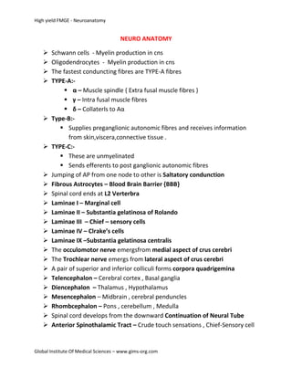
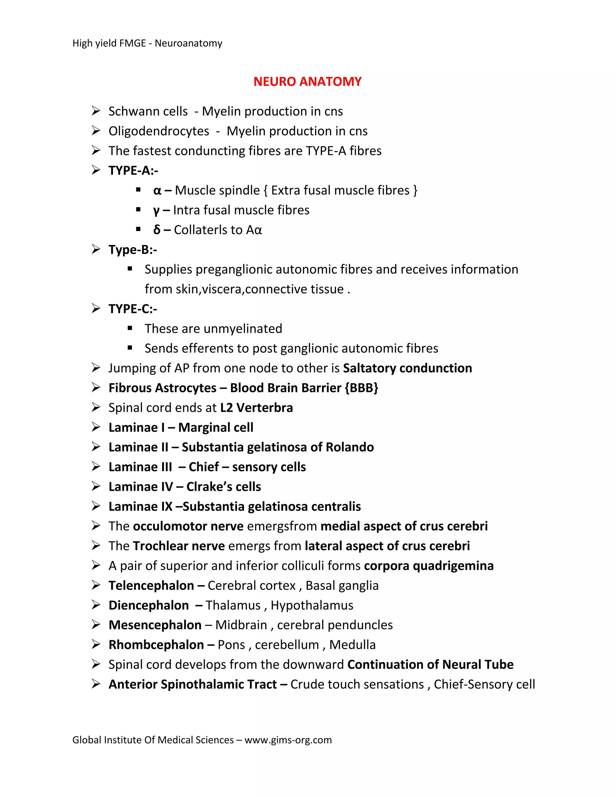
High yield FMGE - Neuroanatomy


                                      NEURO ANATOMY

      Schwann cells - Myelin production in cns
      Oligodendrocytes - Myelin production in cns
      The fastest conduncting fibres are TYPE-A fibres
      TYPE-A:-
             α – Muscle spindle { Extra fusal muscle fibres }
             γ – Intra fusal muscle fibres
             δ – Collaterls to Aα
      Type-B:-
           Supplies preganglionic autonomic fibres and receives information
              from skin,viscera,connective tissue .
      TYPE-C:-
           These are unmyelinated
           Sends efferents to post ganglionic autonomic fibres
      Jumping of AP from one node to other is Saltatory condunction
      Fibrous Astrocytes – Blood Brain Barrier {BBB}
      Spinal cord ends at L2 Verterbra
      Laminae I – Marginal cell
      Laminae II – Substantia gelatinosa of Rolando
      Laminae III – Chief – sensory cells
      Laminae IV – Clrake’s cells
      Laminae IX –Substantia gelatinosa centralis
      The occulomotor nerve emergsfrom medial aspect of crus cerebri
      The Trochlear nerve emergs from lateral aspect of crus cerebri
      A pair of superior and inferior colliculi forms corpora quadrigemina
      Telencephalon – Cerebral cortex , Basal ganglia
      Diencephalon – Thalamus , Hypothalamus
      Mesencephalon – Midbrain , cerebral penduncles
      Rhombcephalon – Pons , cerebellum , Medulla
      Spinal cord develops from the downward Continuation of Neural Tube
      Anterior Spinothalamic Tract – Crude touch sensations , Chief-Sensory cell


Global Institute Of Medical Sciences – www.gims-org.com
High yield FMGE - Neuroanatomy


    Lateral Spincothalamic Tract – Substantia geltinosa , Pain and
     Temperature
    Renshaw cell –Laminae VII
    Laminae -IX – α OR γ Motor neurons
    Medulla oblongata –3cm length , 2cm width
    Anterior surface of pons is marked in the midline by shallow groove which
     contains Basilar Artery
    Cerebello pontine angle has Facial Nerve , Vestibulo cochlear Nerve and
     Glossopharyngeal Nerve emergs from Medial To Lateral
    Spinal nucleus of trigeminus presents on the Lateral aspect of medulla ,
     More medially nucleus gracilis and nucleus cuneatus
    Medial lemniscus is formed by fibres arising from nucleus gracilis and
      nucleus cuneatus
    Cerebrum and cerebellum are separated by Tentrioum cerebella
    Cerebellar nuclei – Dentate , Emboli form , Globose ,Fastigeal
    Dentate is the largest form of cerebellar nuclei
    Anterior spinal artery – Anterior 2/3 of spinal cord
    Posterior spinal artery – Posterior 1/3 of spinal cord
    Greater Anterior segmental medullary artery – Adam Kiewilz
           Directly arising from aorta
           Major source of blood to lower third of spinal cord
           Spinal shock is more common in Lower Third Of Spinal cord
     Anterior Longitudinal artery and Intra medullary arteries are end arteries
       of spinal cord
     Spinal shock is seen Most common in Thoracic and Lumbar Area
     Lumbar spinal shock - Most common Abdominal Aortic Aneurysm
     Synaptic cleft - 20 - 40 nm Wide
     Subconscious Kinesthetic sensations from upperlimb – Cuneocerebellar
       Tract
     Toxin induced neuronal degeneratiob – Injury to dorsal columns
     Lateral spinothalamic and anterior spinothalamic ascends in the form
       Of spinal Lemniscus
      Dorsal spino cerebellar tract – Clarke’s column
Global Institute Of Medical Sciences – www.gims-org.com
High yield FMGE - Neuroanatomy


        Ventral spino cerebellat tract – Marginal cell
        Spino cerebellar tract carries Subconscious kinesthetic sensations
        Syringomyeliea – Dorsal Column Injury – Sensory Loss
        ARAS – Conscious And Awarness
        Upper motor neuron { UMN } – Spastic Paralysis
        Lower motor neuron { LMN } - Flaccid Paralysis
        Babinski sign positive – UMN Lesions
        CSF – 150 Ml
        Globus Pallidus + putamen = Lentiform Nucleus
        Lentiform Nucleus + Caudate Nucleus = Corpus Striatum
        Corpus striatum is important for Intelligence and Longterm Memory
         and Development of IQ
        Flow of CSF from Lateral 2/3 rd ventricle – Foramen of Monro
        CSF from 3rd – 4th ventricle – Aqueduct of sylvius
        Area 4 – Primary Motor Area
        Area 6 – Pre Motor Area { Association Area }
        Area 8– Frontal eye field { Conjugate Movements of eye ball }
        Area 9,10,11 – Controls personality and intellect
        Area 3,2,1 – Primary sensory area
        Damage to somato sensory area causes deficit in fine touch ,position
         and vibration sense ( Dorsal columns are more commonly involved )
        Area 17,18,19 – Visual Cortex
             Area 17 – Primary Visual Area
             Area 18 – Visual Association Area
             Area 19 – Identification and Analyzing
        Area 41 – Primary Auditory Area
        Area 42 – Auditory Association Area
        Area 22 – Wernicke’s Area
        Area 44,45 – Broca’s Area
        The posterior limb of internal capsule contains Cortico spinal tract
        Blood supply of internal capsule – Middle cerebral artery
        Specific Gravity of CSF – 1.005
        Concentration of Nacl is high in CSF when compared to plasma
Global Institute Of Medical Sciences – www.gims-org.com
High yield FMGE - Neuroanatomy


         Production of CSF – 0.3 ml / min
         Thalamus is supplied by Posterior cerebral Artery (75%) , Middle
          Cerebral Artery (25%)
         Thalamus - Striate Vein - Venous Drianage of thalamus
         Lateral Hypothalamic Area considered as feeding ccentre
         Hypoglossal nucleus – Innervates Styloglossus and Genioglossus
         Arterial Supply Of Medulla
              Anterior spinal artery – Branch of veretebral artery
              Posterior spinal artery – Branch of veretebral artery
              Posterior inferior cerebellar artery –Branch of verterbral artery
          Substantia Nigra :-
                 Dorsal part – Pars Compacta
                 Ventral part –Pars Reticularis
          Pars Compacta contains Dopaminergic and Cholinergic Neurons
          Pars Reticularis contains Dopaminergic Neurons
          Trochlear Nerve is the only cranial nerve arises from Dorsal aspect of
           brain { Posteriorly }
          Lesion in red nucleus causes Benedict’s syndrome
          Paranaud’s syndrome – Tumor at superior collicular which
           compresses pineal body
          Blood supply of pons – Superior Cerebellar Artery , Posterior –
           Inferior Cerebellar Artery
          Archicerebellum – Lingula + Floculla
               Function : Equilibrium – Tone and Posture of trunk muscles
          Paleo cerebellum – Tone and Posture of Anti gravity muscles
          Neopallial cortex – Skilled Movements
          Climbing Fibers , Mossy Fibers , Granular Fibers – { Excitatory
            Neurotransmitters are Aspartate , Glutamate }
          Purkinjie cell – Neurotransmitter – GABA
          BBB is absent in Posterior Pituitary , Median Eminence , Pineal
            Gland
          Putamen – Anterior Cerebral Artery
          Anterior Chorodial Artery – Optic Tract , Optic Chiasma
Global Institute Of Medical Sciences – www.gims-org.com
High yield FMGE - Neuroanatomy


            Globus pallidus – Straite Arteries – Branch of middle cerebral artery
            Tail of caudate nucleus and amygdaloid body are perforating
             branches of middle cerebral artery
            Lesions in corpus striatum – Parkinsonism
            The cranial nerve which does not have parasympathetic component
             –Abducens Nerve


Global Institute Of Medical Sciences – www.gims-org.com

Neuro anatomy High yield - fmge

  • 1.
    High yield FMGE- Neuroanatomy NEURO ANATOMY  Schwann cells - Myelin production in cns  Oligodendrocytes - Myelin production in cns  The fastest conduncting fibres are TYPE-A fibres  TYPE-A:-  α – Muscle spindle { Extra fusal muscle fibres }  γ – Intra fusal muscle fibres  δ – Collaterls to Aα  Type-B:-  Supplies preganglionic autonomic fibres and receives information from skin,viscera,connective tissue .  TYPE-C:-  These are unmyelinated  Sends efferents to post ganglionic autonomic fibres  Jumping of AP from one node to other is Saltatory condunction  Fibrous Astrocytes – Blood Brain Barrier {BBB}  Spinal cord ends at L2 Verterbra  Laminae I – Marginal cell  Laminae II – Substantia gelatinosa of Rolando  Laminae III – Chief – sensory cells  Laminae IV – Clrake’s cells  Laminae IX –Substantia gelatinosa centralis  The occulomotor nerve emergsfrom medial aspect of crus cerebri  The Trochlear nerve emergs from lateral aspect of crus cerebri  A pair of superior and inferior colliculi forms corpora quadrigemina  Telencephalon – Cerebral cortex , Basal ganglia  Diencephalon – Thalamus , Hypothalamus  Mesencephalon – Midbrain , cerebral penduncles  Rhombcephalon – Pons , cerebellum , Medulla  Spinal cord develops from the downward Continuation of Neural Tube  Anterior Spinothalamic Tract – Crude touch sensations , Chief-Sensory cell Global Institute Of Medical Sciences – www.gims-org.com
  • 2.
    High yield FMGE- Neuroanatomy  Lateral Spincothalamic Tract – Substantia geltinosa , Pain and Temperature  Renshaw cell –Laminae VII  Laminae -IX – α OR γ Motor neurons  Medulla oblongata –3cm length , 2cm width  Anterior surface of pons is marked in the midline by shallow groove which contains Basilar Artery  Cerebello pontine angle has Facial Nerve , Vestibulo cochlear Nerve and Glossopharyngeal Nerve emergs from Medial To Lateral  Spinal nucleus of trigeminus presents on the Lateral aspect of medulla , More medially nucleus gracilis and nucleus cuneatus  Medial lemniscus is formed by fibres arising from nucleus gracilis and nucleus cuneatus  Cerebrum and cerebellum are separated by Tentrioum cerebella  Cerebellar nuclei – Dentate , Emboli form , Globose ,Fastigeal  Dentate is the largest form of cerebellar nuclei  Anterior spinal artery – Anterior 2/3 of spinal cord  Posterior spinal artery – Posterior 1/3 of spinal cord  Greater Anterior segmental medullary artery – Adam Kiewilz  Directly arising from aorta  Major source of blood to lower third of spinal cord  Spinal shock is more common in Lower Third Of Spinal cord  Anterior Longitudinal artery and Intra medullary arteries are end arteries of spinal cord  Spinal shock is seen Most common in Thoracic and Lumbar Area  Lumbar spinal shock - Most common Abdominal Aortic Aneurysm  Synaptic cleft - 20 - 40 nm Wide  Subconscious Kinesthetic sensations from upperlimb – Cuneocerebellar Tract  Toxin induced neuronal degeneratiob – Injury to dorsal columns  Lateral spinothalamic and anterior spinothalamic ascends in the form Of spinal Lemniscus  Dorsal spino cerebellar tract – Clarke’s column Global Institute Of Medical Sciences – www.gims-org.com
  • 3.
    High yield FMGE- Neuroanatomy  Ventral spino cerebellat tract – Marginal cell  Spino cerebellar tract carries Subconscious kinesthetic sensations  Syringomyeliea – Dorsal Column Injury – Sensory Loss  ARAS – Conscious And Awarness  Upper motor neuron { UMN } – Spastic Paralysis  Lower motor neuron { LMN } - Flaccid Paralysis  Babinski sign positive – UMN Lesions  CSF – 150 Ml  Globus Pallidus + putamen = Lentiform Nucleus  Lentiform Nucleus + Caudate Nucleus = Corpus Striatum  Corpus striatum is important for Intelligence and Longterm Memory and Development of IQ  Flow of CSF from Lateral 2/3 rd ventricle – Foramen of Monro  CSF from 3rd – 4th ventricle – Aqueduct of sylvius  Area 4 – Primary Motor Area  Area 6 – Pre Motor Area { Association Area }  Area 8– Frontal eye field { Conjugate Movements of eye ball }  Area 9,10,11 – Controls personality and intellect  Area 3,2,1 – Primary sensory area  Damage to somato sensory area causes deficit in fine touch ,position and vibration sense ( Dorsal columns are more commonly involved )  Area 17,18,19 – Visual Cortex  Area 17 – Primary Visual Area  Area 18 – Visual Association Area  Area 19 – Identification and Analyzing  Area 41 – Primary Auditory Area  Area 42 – Auditory Association Area  Area 22 – Wernicke’s Area  Area 44,45 – Broca’s Area  The posterior limb of internal capsule contains Cortico spinal tract  Blood supply of internal capsule – Middle cerebral artery  Specific Gravity of CSF – 1.005  Concentration of Nacl is high in CSF when compared to plasma Global Institute Of Medical Sciences – www.gims-org.com
  • 4.
    High yield FMGE- Neuroanatomy  Production of CSF – 0.3 ml / min  Thalamus is supplied by Posterior cerebral Artery (75%) , Middle Cerebral Artery (25%)  Thalamus - Striate Vein - Venous Drianage of thalamus  Lateral Hypothalamic Area considered as feeding ccentre  Hypoglossal nucleus – Innervates Styloglossus and Genioglossus  Arterial Supply Of Medulla  Anterior spinal artery – Branch of veretebral artery  Posterior spinal artery – Branch of veretebral artery  Posterior inferior cerebellar artery –Branch of verterbral artery  Substantia Nigra :-  Dorsal part – Pars Compacta  Ventral part –Pars Reticularis  Pars Compacta contains Dopaminergic and Cholinergic Neurons  Pars Reticularis contains Dopaminergic Neurons  Trochlear Nerve is the only cranial nerve arises from Dorsal aspect of brain { Posteriorly }  Lesion in red nucleus causes Benedict’s syndrome  Paranaud’s syndrome – Tumor at superior collicular which compresses pineal body  Blood supply of pons – Superior Cerebellar Artery , Posterior – Inferior Cerebellar Artery  Archicerebellum – Lingula + Floculla  Function : Equilibrium – Tone and Posture of trunk muscles  Paleo cerebellum – Tone and Posture of Anti gravity muscles  Neopallial cortex – Skilled Movements  Climbing Fibers , Mossy Fibers , Granular Fibers – { Excitatory Neurotransmitters are Aspartate , Glutamate }  Purkinjie cell – Neurotransmitter – GABA  BBB is absent in Posterior Pituitary , Median Eminence , Pineal Gland  Putamen – Anterior Cerebral Artery  Anterior Chorodial Artery – Optic Tract , Optic Chiasma Global Institute Of Medical Sciences – www.gims-org.com
  • 5.
    High yield FMGE- Neuroanatomy  Globus pallidus – Straite Arteries – Branch of middle cerebral artery  Tail of caudate nucleus and amygdaloid body are perforating branches of middle cerebral artery  Lesions in corpus striatum – Parkinsonism  The cranial nerve which does not have parasympathetic component –Abducens Nerve Global Institute Of Medical Sciences – www.gims-org.com