
Download as PDF, PPTX


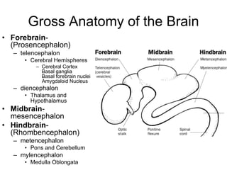
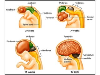


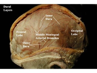


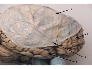
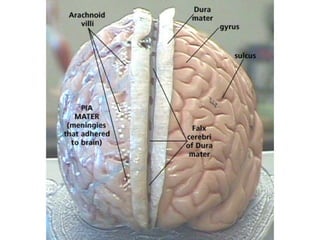
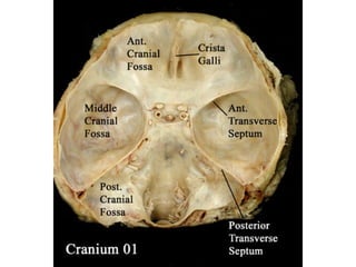


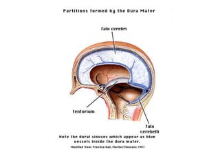

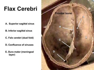


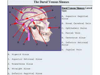
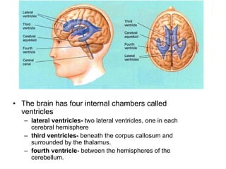


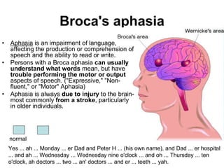


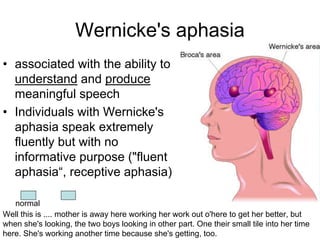


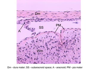
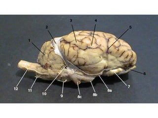

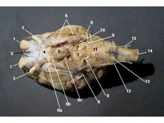

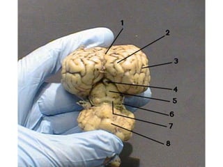


This document details the anatomy and embryonic development of the brain, explaining its major parts including the cerebrum, cerebellum, and brainstem, alongside their functions and structural features. It also describes the histology of the cerebral cortex, the meninges, and the production and circulation of cerebrospinal fluid in the brain's ventricles. Additionally, it highlights disorders such as Broca's and Wernicke's aphasia associated with brain injury, emphasizing the importance of various brain regions in motor and sensory functions.


