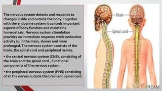
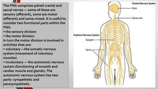
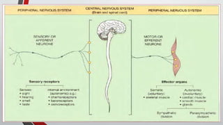
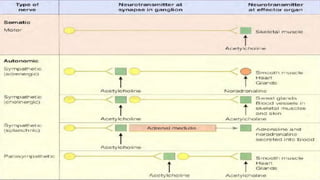
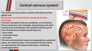
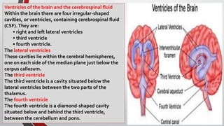
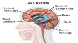
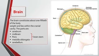
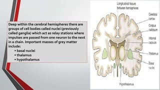
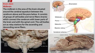
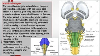
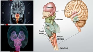
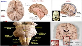
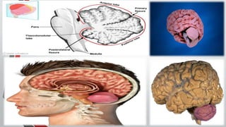
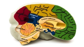
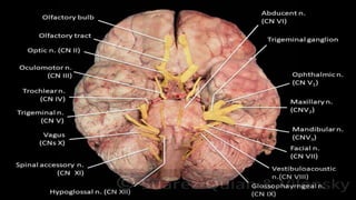
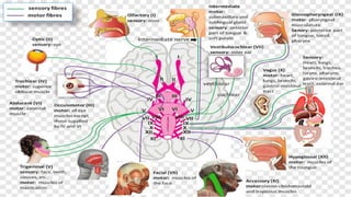
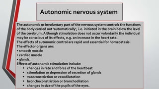
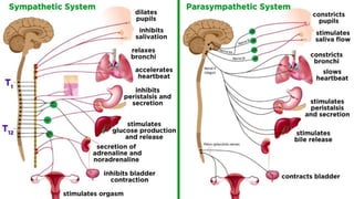
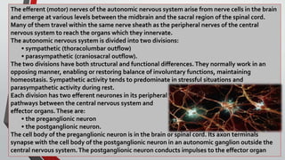
This lecture help the students such as medical ,nursing , and any health care provider to understand the basic information about anatomy of nervous system
2. The nervous system detects and responds to
changes inside and outside the body.Together
with the endocrine system it controls important
aspects of body function and maintains
homeostasis. Nervous system stimulation
provides an immediate response while endocrine
activity is, in the main, slower and more
prolonged.The nervous system consists of the
brain, the spinal cord and peripheral nerves.
• the central nervous system (CNS), consisting of
the brain and the spinal cord , Functional
components of the nervous system.
• the peripheral nervous system (PNS) consisting
of all the nerves outside the brain and spinal cord.
3. The PNS comprises paired cranial and
sacral nerves — some of these are
sensory (afferent), some are motor
(efferent) and some mixed. It is useful to
consider two functional parts within the
PNS:
• the sensory division
• the motor division.
In turn the motor division is involved in
activities that are:
• voluntary —the somatic nervous
system (movement of voluntary
muscles)
• involuntary — the autonomic nervous
system (functioning of smooth and
cardiac muscle and glands).The
autonomic nervous system has two
parts: sympathetic and
parasympathetic.
4.
5. NEURONES
The nervous system consists of a vast number
of cells called neurons supported by a special
type of connective tissue, neuroglia. Each
neuron consists of a cell body and its
processes, one axon and many dendrites.
Neurons are commonly referred to simply as
nerve cells. Bundles of axons bound together
are called nerves.
neurons can synthesize chemical energy
(ATP) only from glucose.
6. Cell bodies
Nerve cells vary considerably in size and shape, but
they are all too small to be seen by the naked eye.
Cell bodies form the grey matter of the nervous
system and are found at the periphery of the brain and
in the Centre of the
spinal cord. Groups of cell bodies are called nuclei in
the central nervous system and ganglia in the
peripheral nervous system.
Axons and dendrites
Axons and dendrites are extensions of cell bodies and
form the white matter of the nervous system. Axons
are found deep in the brain and in groups, called
tracts, at the periphery of the spinal cord. They are
referred to as nerves or nerve fibers outside the brain
and spinal cord.
7. The transmission of a nerve impulse from its origin to its destination,
whether it is sensory or motor. The point at which the nerve impulse
passes from one to another is the synapse. breaks up into minute
branches which terminate in small swellings called synaptic knobs.The
space between them is the synaptic cleft. synaptic vesicles, containing
a chemical, the neurotransmitter, which is released into synaptic clefts.
Neurotransmitters are synthesized by nerve cells, actively transported
along the axons and stored in the synaptic vesicles.
8.
9. Central nervous system
The central nervous system consists of the brain and the
spinal cord.
Membranes covering the brain and spinal cord (the
meninges):
The brain and spinal cord are completely surrounded by
three membranes, the meninges, lying between the skull
and the brain and between the vertebrae and the spinal
cord. Named from outside inwards they are:
• dura mater
• arachnoid mater
• pia mater.
The dura and arachnoid maters are separated by a potential
space, the subdural space.The arachnoid and pia maters
are separated by the subarachnoid space, containing
cerebrospinal fluid.
10. Ventricles of the brain and the cerebrospinal fluid
Within the brain there are four irregular-shaped
cavities, or ventricles, containing cerebrospinal fluid
(CSF).They are:
• right and left lateral ventricles
• third ventricle
• fourth ventricle.
The lateral ventricles
These cavities lie within the cerebral hemispheres,
one on each side of the median plane just below the
corpus callosum.
The third ventricle
The third ventricle is a cavity situated below the
lateral ventricles between the two parts of the
thalamus.
The fourth ventricle
The fourth ventricle is a diamond-shaped cavity
situated below and behind the third ventricle,
between the cerebellum and pons.
11.
12. Cerebrospinal fluid is secreted into each ventricle of the
brain by choroid plexuses.These are vascular areas were
there is a proliferation of blood vessels surrounded by
ependymal cells in the lining of ventricle walls. CSF
passes back into the blood through tiny diverticula of
arachnoid mater, called arachnoid villi (arachnoid
granulations), that project into the venous sinuses.
13. Brain
The brain constitutes about one-fiftieth
of the body
weight and lies within the cranial
cavity.The parts are:
 cerebrum
 midbrain
 Pons brain stem
 medulla oblongata
 cerebellum.
14. Cerebrum
This is the largest part of the brain, and it occupies the anterior and middle cranial fossae. It is
divided by a deep cleft, the longitudinal cerebral fissure, into right and left cerebral hemispheres,
each containing one of the lateral ventricles. Deep within the brain the hemispheres are connected
by a mass of white matter (nerve fibers) called the corpus callosum.The falx cerebri is formed by the
dura mater. It separates the two hemispheres and penetrates to the depth of the corpus callosum.
The superficial (peripheral) part of the cerebrum is composed of nerve cell bodies or grey matter,
forming the cerebral cortex, and the deeper layers consist of nerve fibers or white matter.
The cerebral cortex shows many infoldings or furrows of varying depth.The exposed areas of the
folds are the gyri or convolutions, and these are separated by sulci or fissures.These convolutions
greatly increase the surface area of the cerebrum.
For descriptive purposes, each hemisphere of the cerebrum is divided into lobes which take the
names of the bones of the cranium under which they lie:
• frontal
• parietal
• temporal
• occipital.
The boundaries of the lobes are marked by deep sulci (fissures).These are the central, lateral and
parieto-occipital sulci.
15.
16. Deep within the cerebral hemispheres there are
groups of cell bodies called nuclei (previously
called ganglia) which act as relay stations where
impulses are passed from one neuron to the next
in a chain. Important masses of grey matter
include:
• basal nuclei
• thalamus
• hypothalamus
17. Brain stem
Midbrain
The midbrain is the area of the brain situated
around the cerebral aqueduct between the
cerebrum above and the pons below. It consists
of groups of cell bodies and nerve fibers (tracts)
which connect the cerebrum with lower parts of
the brain and with the spinal cord.The cell bodies
act as relay stations for the ascending and
descending nerve fibers.
18. Pons
The pons is situated in front of the
cerebellum, below the midbrain and
above the medulla oblongata. It
consists mainly of nerve fibers which
form a bridge between the two
hemispheres of the cerebellum, and of
fibers passing between the higher levels
of the brain and the spinal cord.There
are groups of cells within the pons which
act as relay stations and some of these
are associated with the cranial nerves.
The anatomical structure of the pons
differs from that of the cerebrum in that
the cell bodies (grey matter) lie deeply,
and the nerve fibers are on the surface.
19. Medulla oblongata
The medulla oblongata extends from the pons
above and is continuous with the spinal cord
below. It is about 2.5 cm long. Its anterior and
posterior surfaces are marked by central fissures.
The outer aspect is composed of white matter
which passes between the brain and the spinal
cord, and grey matter lies centrally. Some cells
constitute relay stations for sensory nerves
passing from the spinal cord to the cerebrum.
The vital centers, consisting of groups of cells
associated with autonomic reflex activity, lie in
its deeper structure. These are the:
• cardiac centre
• respiratory centre
• vasomotor centre
• reflex centres of vomiting,
coughing, sneezing and
swallowing.
20.
21.
22. The medulla oblongata has several special features:
• Decussation (crossing) of the pyramids. In the medulla motor nerves descending from the
motor area in the cerebrum to the spinal cord in the pyramidal (corticospinal) tracts cross from
one side to the other.
This means that the left hemisphere of the cerebrum controls the right half of the body, and
vice versa.These tracts are the main pathway for impulses to skeletal (voluntary) muscles.
• Sensory decussation. Some of the sensory nerves ascending to the cerebrum from the spinal
cord cross from one side to the other in the medulla. Others decussate at lower levels, i.e. in
the spinal cord.
•The cardiovascular centre controls the rate and force of cardiac contraction. Sympathetic and
parasympathetic nerve fibres originating in the medulla pass to the heart. Sympathetic
stimulation increases the rate and force of the heartbeat and parasympathetic stimulation has
the opposite effect.
•The respiratory centre controls the rate and depth of respiration. From this centre, nerve
impulses pass to the phrenic and intercostal nerves which stimulate contraction of the
diaphragm and intercostal muscles, thus initiating inspiration. The respiratory centre is
stimulated by excess carbon dioxide and, to a lesser extent, by deficiency of oxygen in its
blood supply and by nerve impulses from the chemoreceptors in the carotid bodies.
23. •The vasomotor centre controls the diameter of the blood vessels, especially the small arteries
and arterioles which have a large proportion of smooth muscle fibres in their walls.Vasomotor
impulses reach the blood vessels through the autonomic nervous system. Stimulation may
cause either constriction or dilatation of blood vessels depending on the site.The sources of
stimulation of the vasomotor centre are the arterial baroreceptors, body temperature and
emotions such as sexual excitement and anger. Pain usually causes vasoconstriction although
severe pain may cause vasodilatation, a fall in blood pressure and fainting.
• Reflex centres. When irritating substances are present in the stomach or respiratory tract,
nerve impulses pass to the medulla oblongata, stimulating the reflex centres which initiate the
reflex actions of vomiting, coughing and sneezing to expel the irritant.
24. Reticular formation
The reticular formation is a collection of
neurones in the core of the brain stem,
surrounded by neural pathways which conduct
ascending and descending nerve impulses
between the brain and the spinal cord. It has a
vast number of synaptic links with other parts of
the brain and is therefore constantly receiving
'information’ being transmitted in ascending and
descending tracts.
Cerebellum
The cerebellum is situated behind the pons and
immediately below the posterior portion of the
cerebrum. It has two hemispheres, separated by
a narrow median strip called the vermis. Grey
matter forms the surface of the cerebellum, and
the white matter lies deeply.
25.
26.
27. Spinal cord
The spinal cord is the elongated, almost cylindrical part of the
central nervous system, which is suspended in the vertebral
canal surrounded by the meninges and cerebrospinal fluid. It is
continuous above with the medulla oblongata and extends from
the upper border of the atlas to the lower border of the 1st
lumbar vertebra . It is approximately 45 cm long in an adult
Caucasian male,and is about the thickness of the little finger.
When a specimen of cerebrospinal fluid is required, it is taken
from a point below the end of the cord, i.e. below the level of the
2nd lumbar vertebra.This procedure is called lumbar puncture.
Except for the cranial nerves, the spinal cord is the nervous
tissue link between the brain and the rest of the body
28. brain to the various organs and tissues descend
through the spinal cord. At the appropriate level they
leave the cord and pass to the structure they supply.
Similarly, sensory nerves from organs and tissues
enter and pass upwards in the spinal cord to the brain.
Some activities of the spinal cord are independent of
the brain, i.e. spinal reflexes.To facilitate these there
are extensive neurone connections between sensory
and motor neurones at the same or different levels in
the cord.
The spinal cord is incompletely divided into two equal
parts, anteriorly by a short, shallow median fissure
and posteriorly by a deep narrow septum, the
posterior median septum.
A cross-section of the spinal cord shows that it is
composed of grey matter in the centre surrounded by
white matter supported by neuroglia.
29. PERIPHERAL NERVOUS SYSTEM
This part of the nervous system consists of:
• 31 pairs of spinal nerves
• 12 pairs of cranial nerves
• the autonomic part of the nervous system.
Most of the nerves of the peripheral nervous
system are composed of sensory nerve fibres
conveying afferent impulses from sensory
end organs to the brain, and motor nerve
fibres conveying efferent impulses from the
brain through the spinal cord to the effector
organs, e.g. skeletal muscles, smooth muscle
and glands.
30. Spinal nerves
There are 31 pairs of spinal nerves that leave the
vertebral canal by passing through the
intervertebral foramina formed by adjacent
vertebrae.They are named and grouped
according to the vertebrae with which they are
associated:
• 8 cervical
• 12 thoracic
• 5 lumbar
• 5 sacral
• 1 coccygeal.
Although there are only seven cervical vertebrae,
there are eight nerves because the first pair leave
the vertebral canal between the occipital bone
and the atlas and the eighth pair leave below the
last cervical vertebra.
31. The spinal nerves arise from both sides of the spinal cord
and emerge through the intervertebral foramina.
Each nerve is formed by the union of a motor and a
sensory nerve root and is, therefore, a mixed nerve.
Each spinal nerve has a contribution from the
sympathetic part of the autonomic nervous system in
the form of a preganglionic fiber.
The anterior nerve root consists of motor nerve fibers
which are the axons of the nerve cells in the anterior
column of grey matter in the spinal cord and, in the
thoracic and lumbar regions, sympathetic nerve fibers
which are the axons of cells in the lateral columns of
grey matter.
The posterior nerve root consists of sensory nerve fibers.
Just outside the spinal cord there is a spinal ganglion
(posterior root ganglion), consisting of a little cluster of
cell bodies. Sensory nerve fibers pass through these
ganglia before entering the spinal cord.
32. Cranial nerves
There are 12 pairs of cranial nerves originating
from nuclei in the inferior surface of the brain,
some sensory, some motor and some mixed.
Their names and numbers are:
I. Olfactory: sensory
II. Optic: sensory
III. Oculomotor: motor
IV.Trochlear: motor
V.Trigeminal: mixed
VI. Abducent: motor
VII. Facial: mixed
VIII.Vestibulocochlear (auditory): sensory
IX. Glossopharyngeal: mixed
X.Vagus: mixed
XL Accessory: motor
XII. Hypoglossal: motor.
33.
34.
35. Autonomic nervous system
The autonomic or involuntary part of the nervous system controls the functions
of the body carried out 'automatically', i.e. initiated in the brain below the level
of the cerebrum. Although stimulation does not occur voluntarily the individual
may be conscious of its effects, e.g. an increase in the heart rate.
The effects of autonomic control are rapid and essential for homeostasis.
The effector organs are:
• smooth muscle
• cardiac muscle
• glands.
Effects of autonomic stimulation include:
 changes in rate and force of the heartbeat
 stimulation or depression of secretion of glands
 vasoconstriction or vasodilatation
 bronchoconstriction or bronchodilation
 changes in size of the pupils of the eyes.
36.
37. The efferent (motor) nerves of the autonomic nervous system arise from nerve cells in the brain
and emerge at various levels between the midbrain and the sacral region of the spinal cord.
Many of them travel within the same nerve sheath as the peripheral nerves of the central
nervous system to reach the organs which they innervate.
The autonomic nervous system is divided into two divisions:
• sympathetic (thoracolumbar outflow)
• parasympathetic (craniosacral outflow).
The two divisions have both structural and functional differences.They normally work in an
opposing manner, enabling or restoring balance of involuntary functions, maintaining
homeostasis. Sympathetic activity tends to predominate in stressful situations and
parasympathetic activity during rest.
Each division has two efferent neurones in its peripheral
pathways between the central nervous system and
effector organs.These are:
• the preganglionic neuron
• the postganglionic neuron.
The cell body of the preganglionic neuron is in the brain or spinal cord. Its axon terminals
synapse with the cell body of the postganglionic neuron in an autonomic ganglion outside the
central nervous system.The postganglionic neuron conducts impulses to the effector organ
38. Sympathetic nervous system
Neurones convey impulses from their origin in
the hypothalamus, reticular formation and
medulla oblongata to effector organs and
tissues.The first neurone has its cell body in the
brain and its fibre extends into the spinal cord.
In the preganglionic neurone, Acetylcholine is
the neurotransmitter. While the postganglionic
neurone, Noradrenaline is usually the
neurotransmitter.
Parasympathetic nervous system
Two neurones (preganglionic and postganglionic)
are involved in the transmission of impulses from
their source to the effector organ.The
neurotransmitter at both synapses is
acetylcholine.