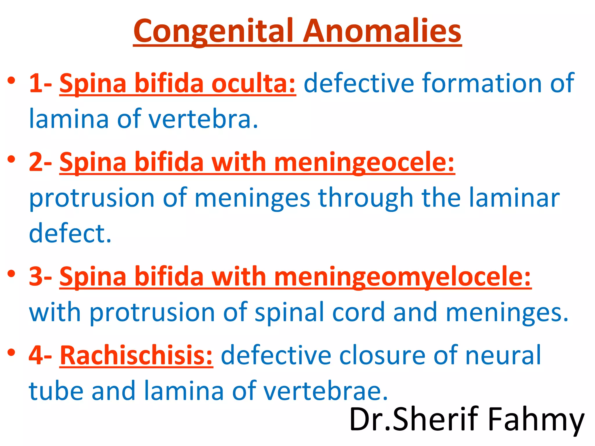
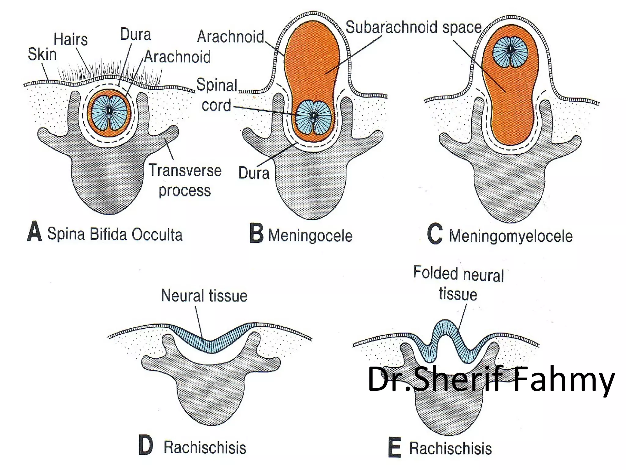
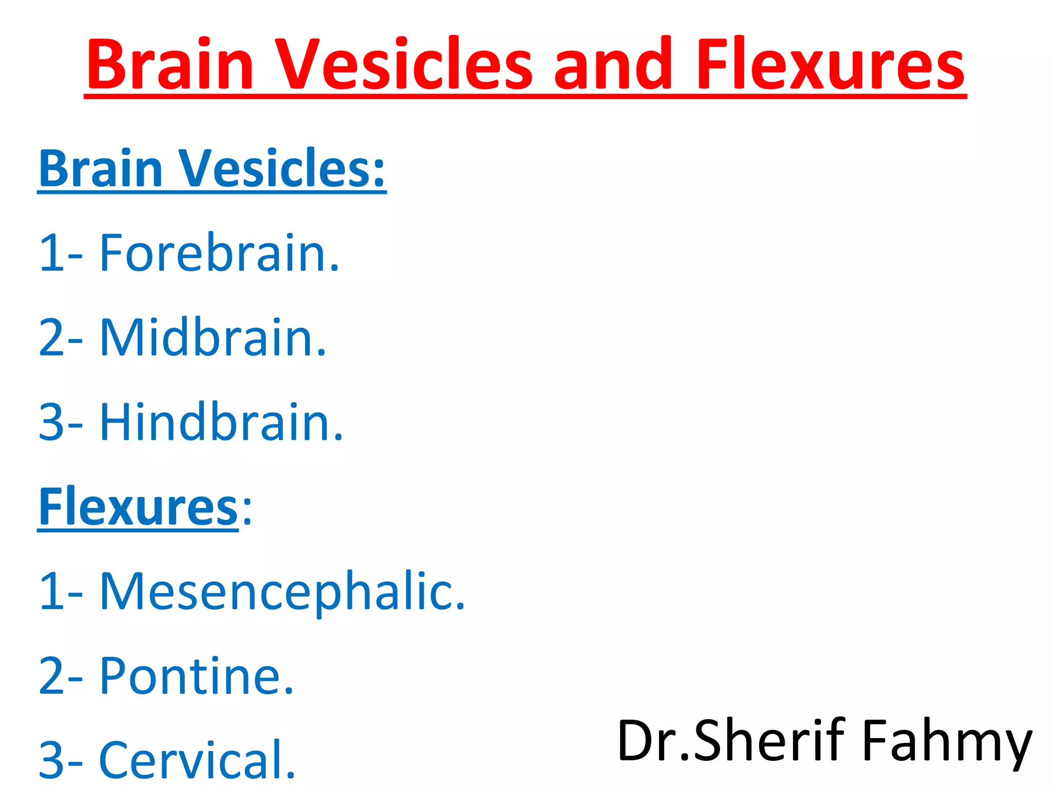
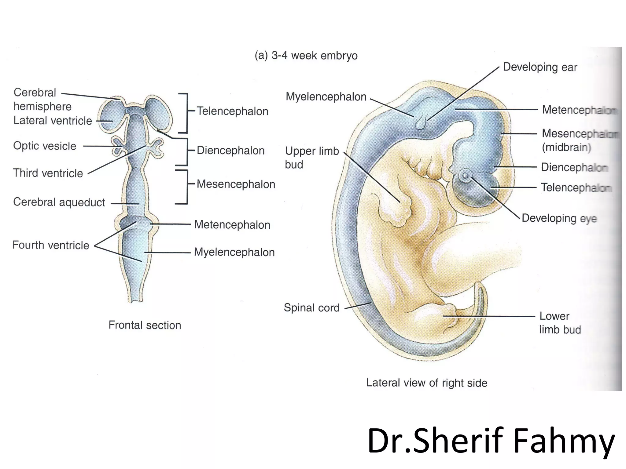
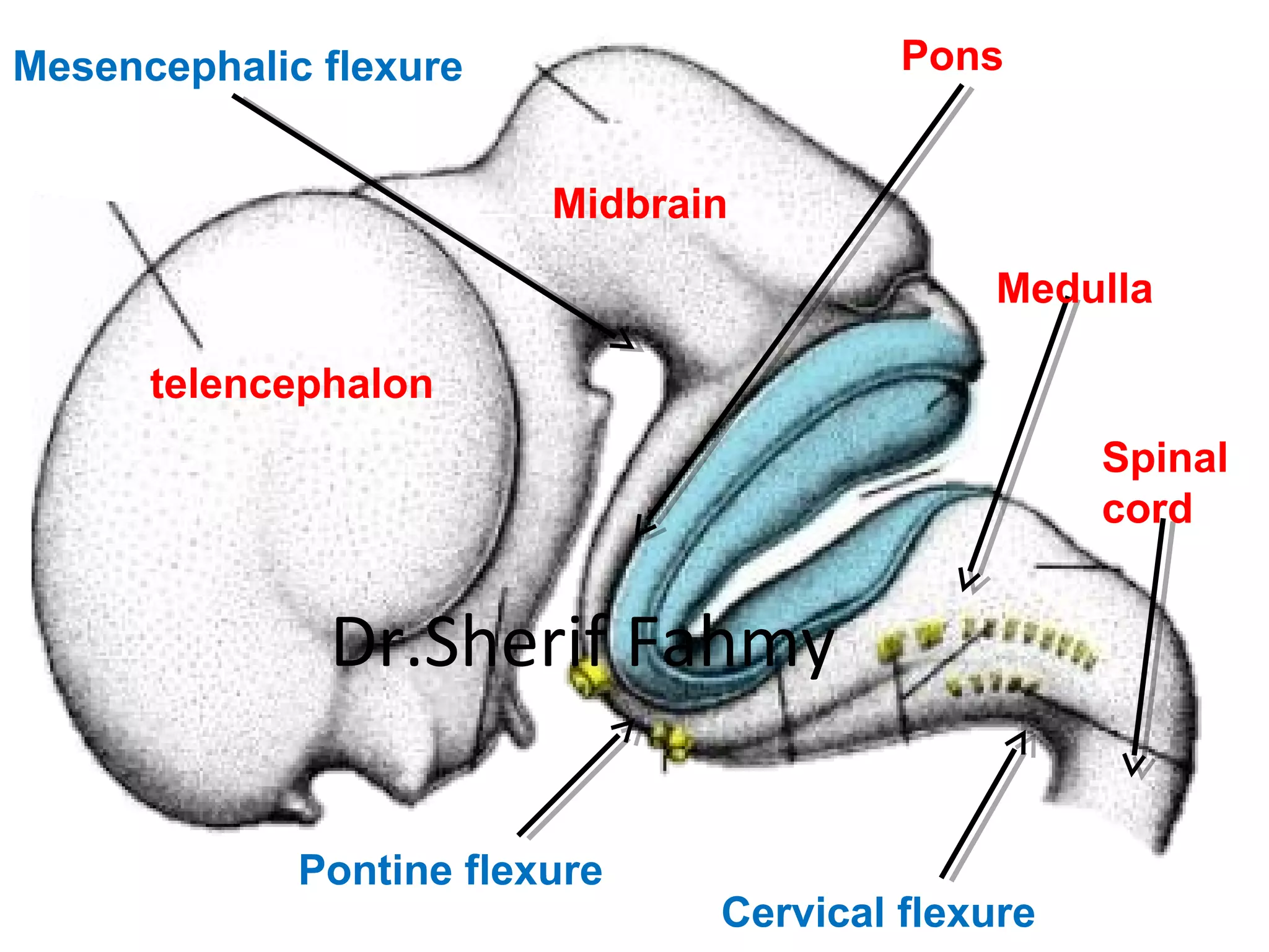
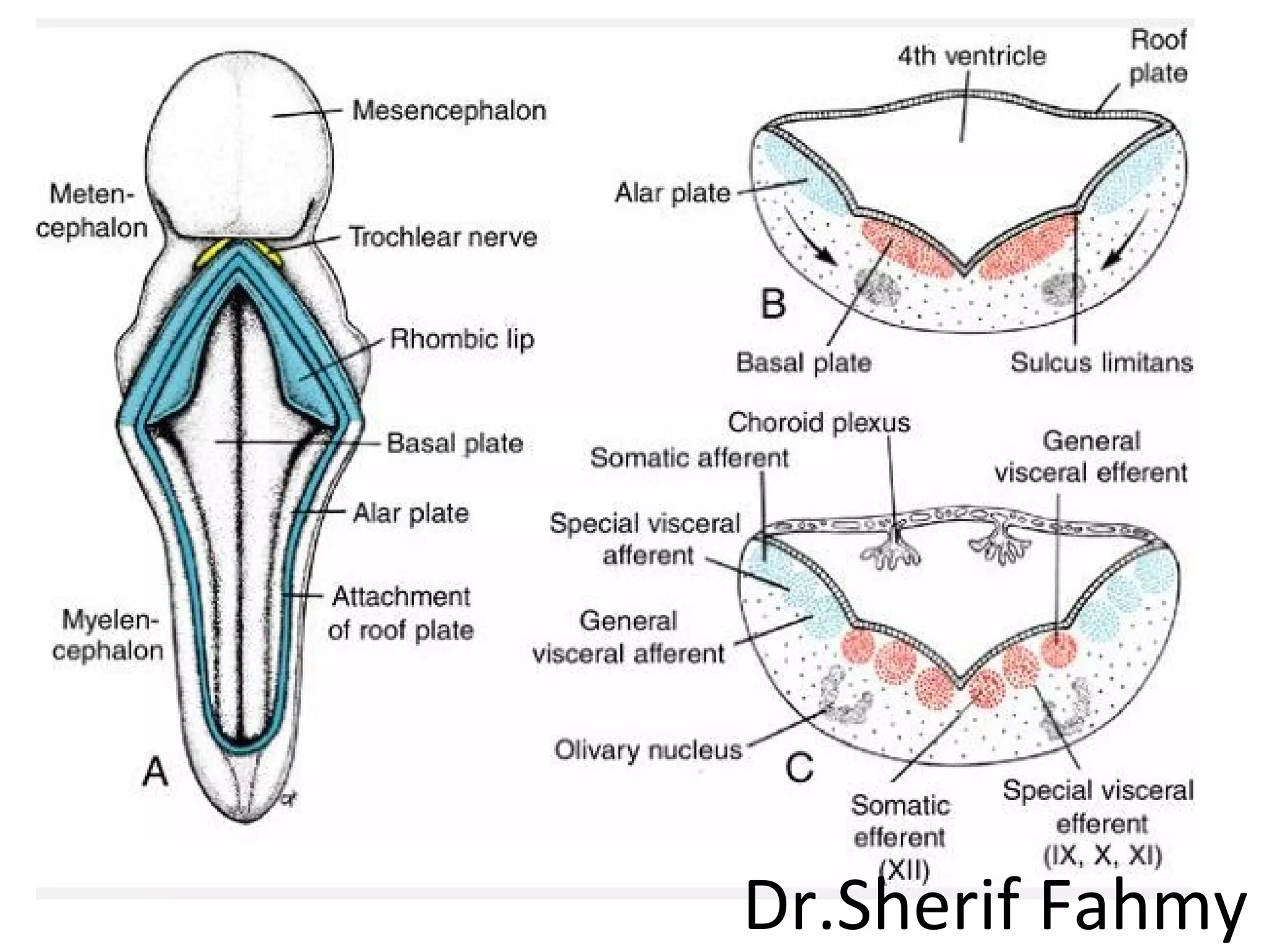
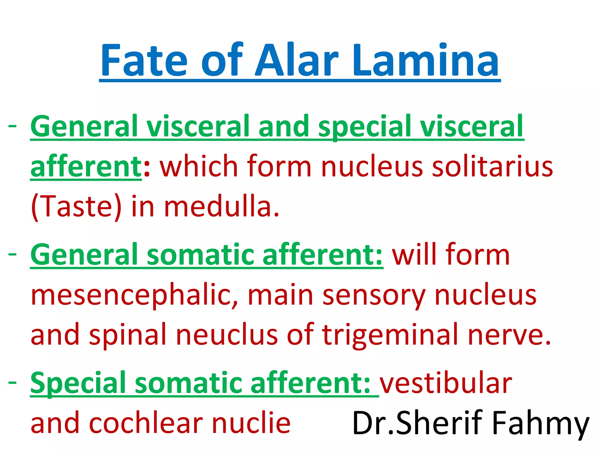
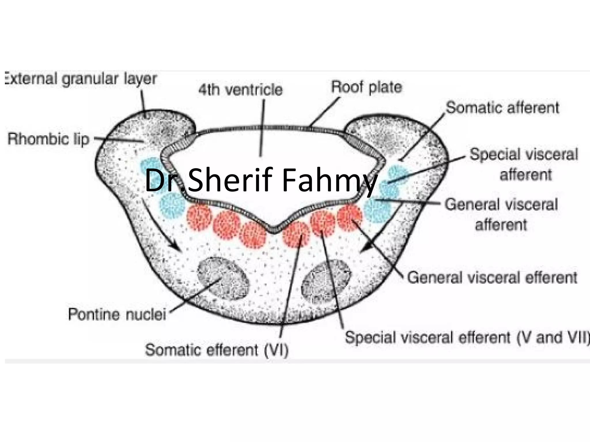
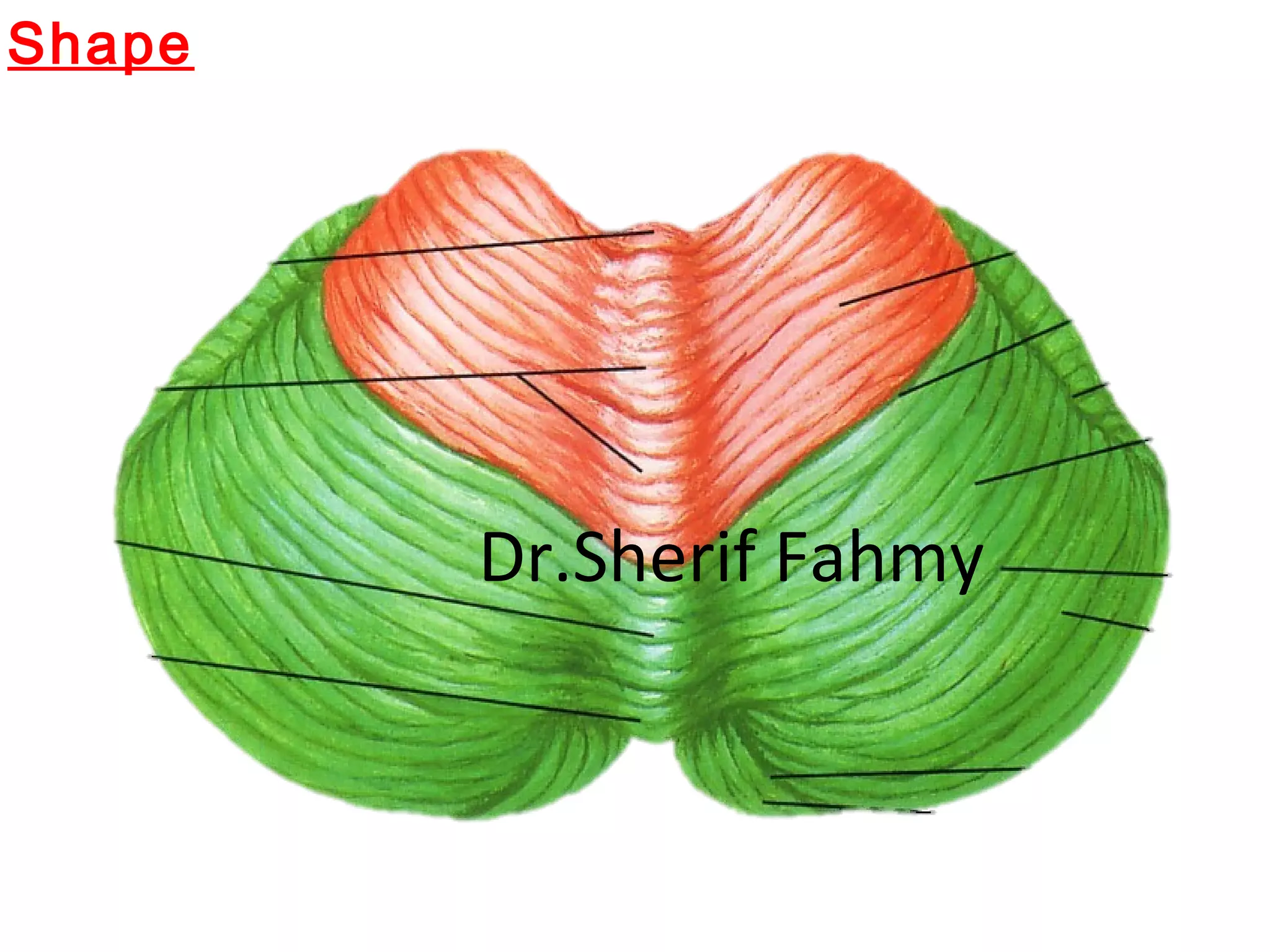
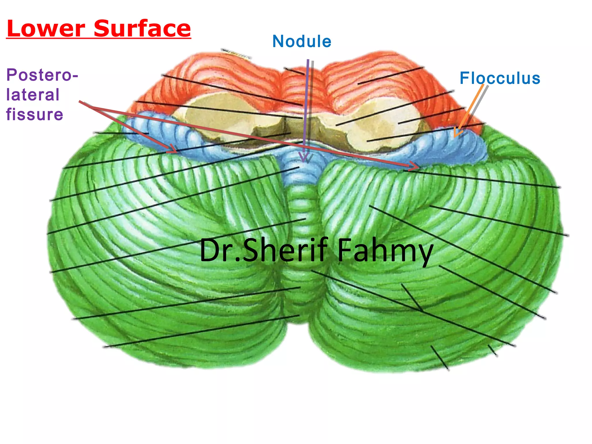
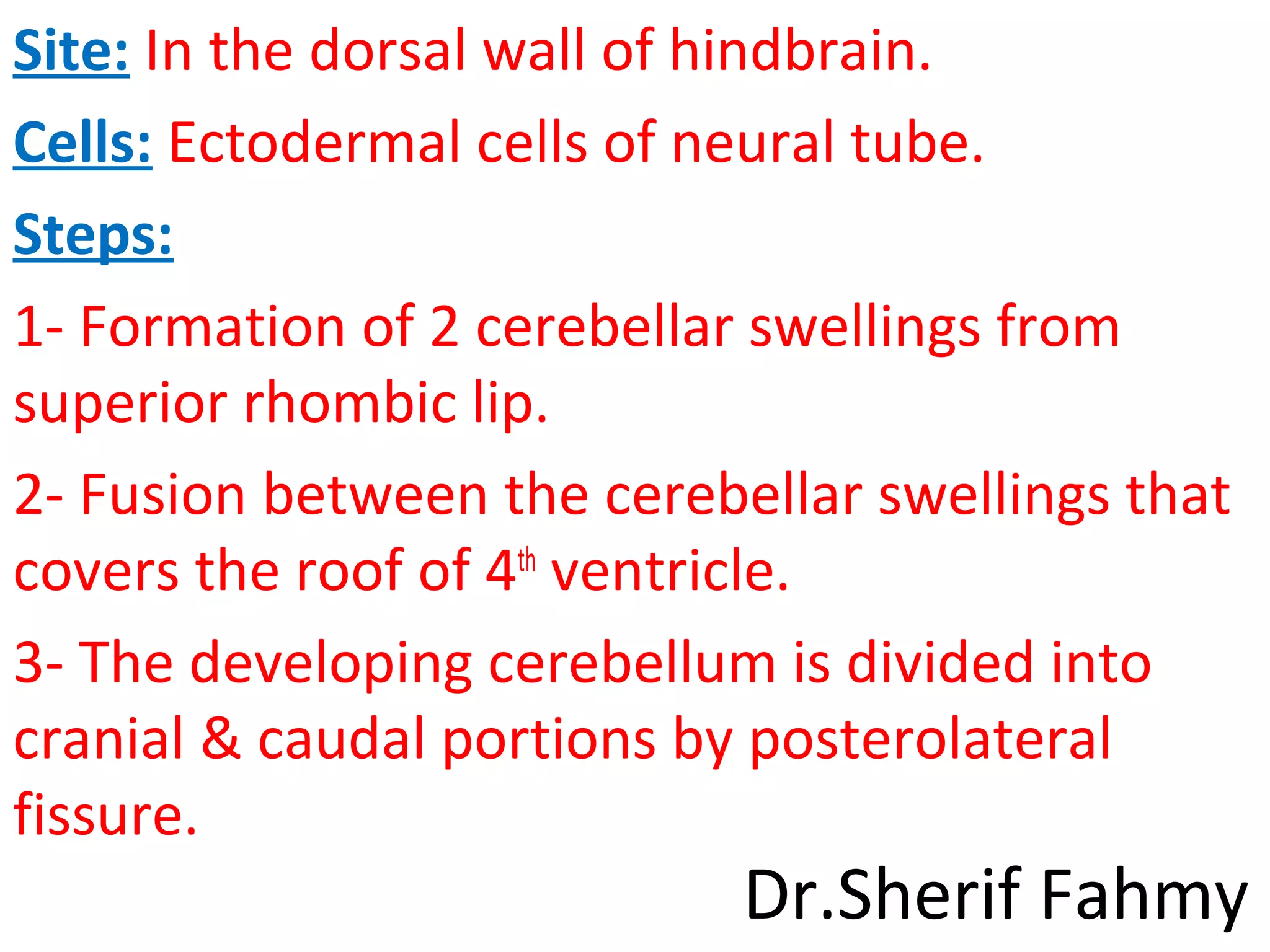
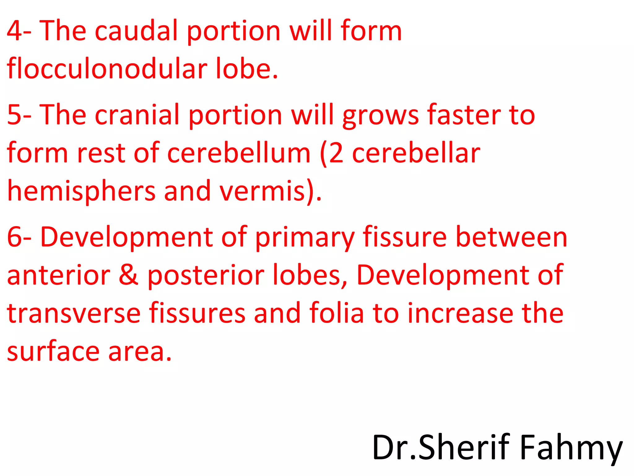
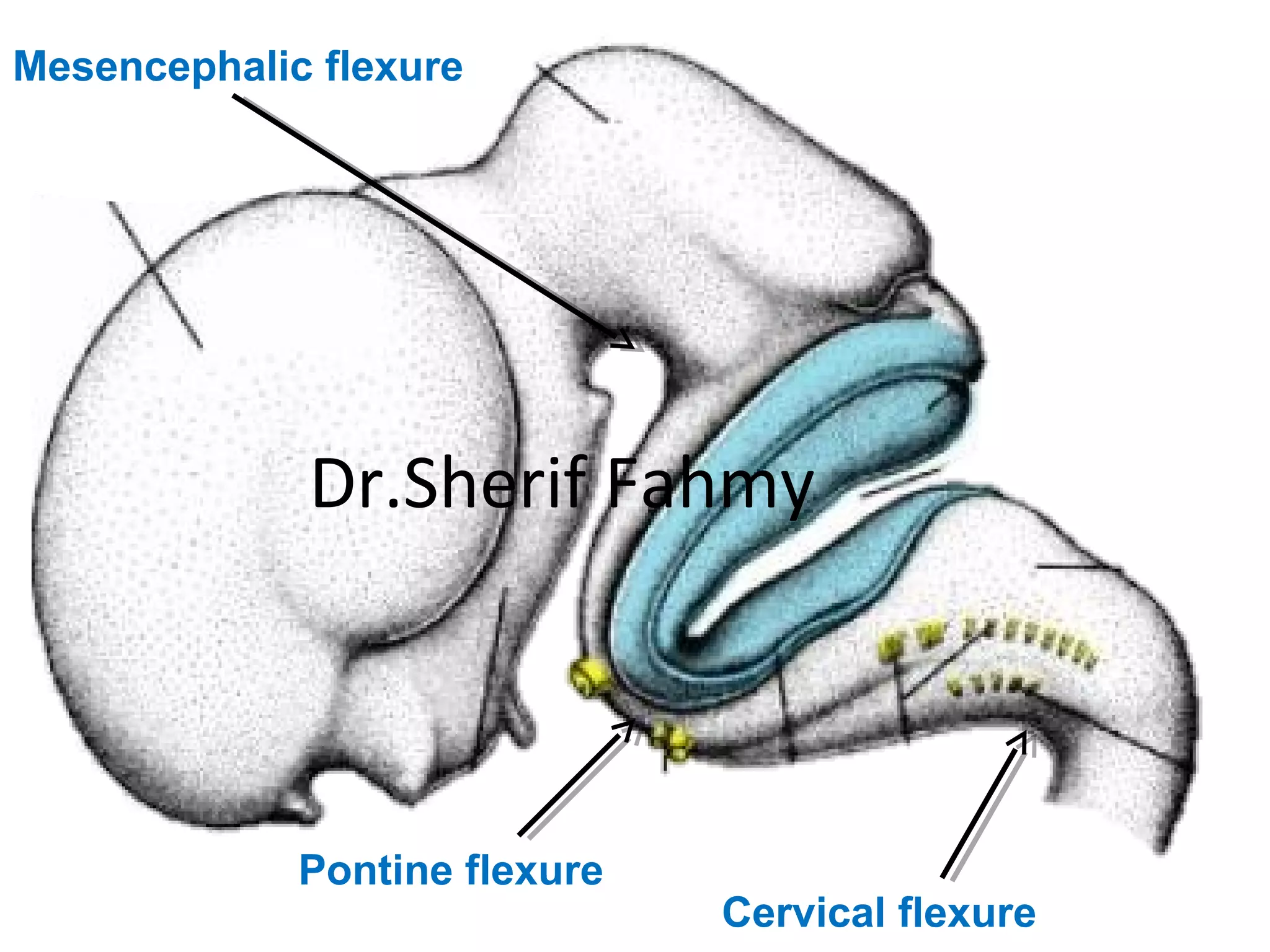
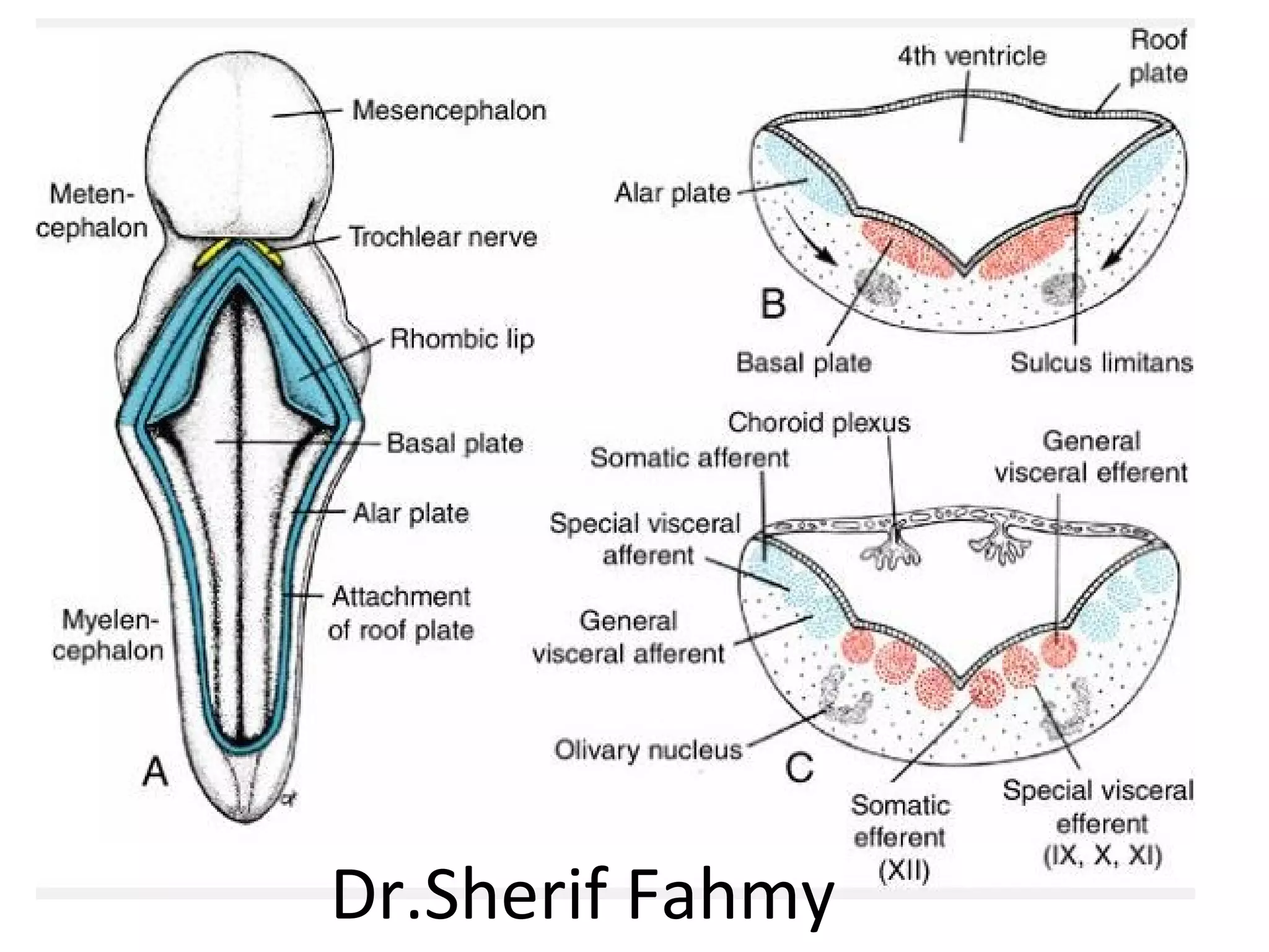
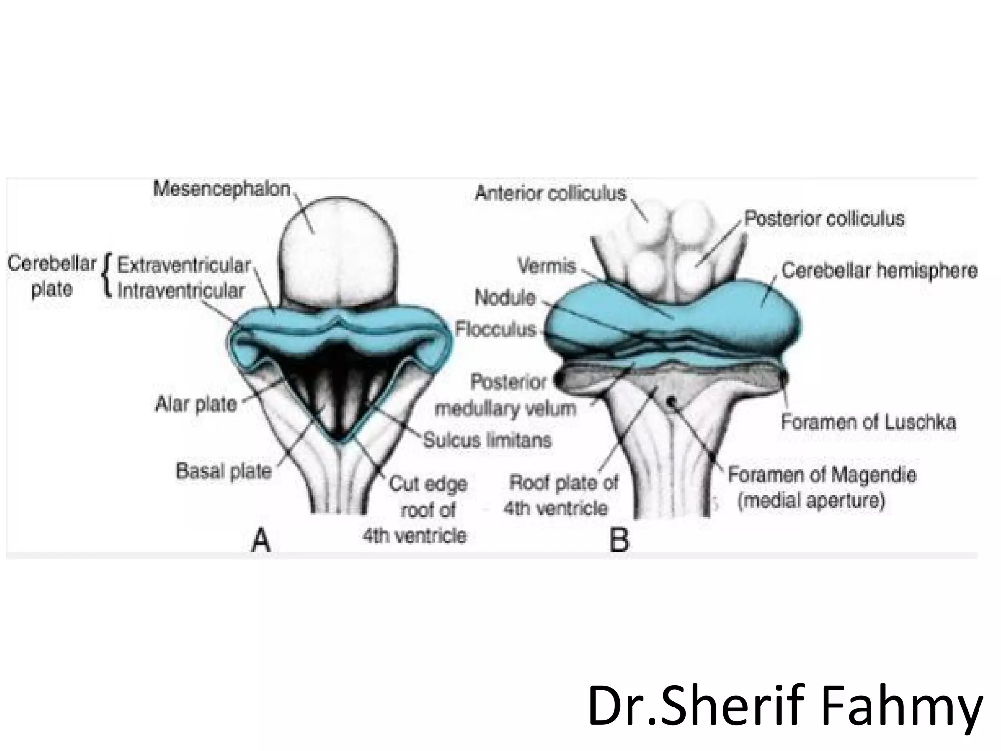
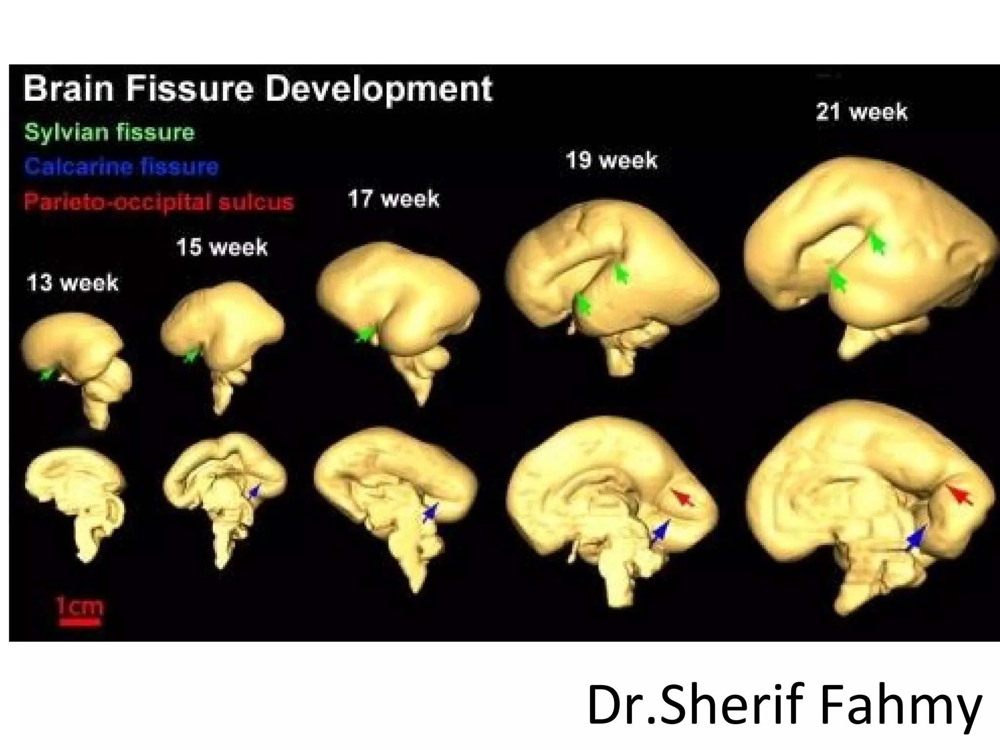
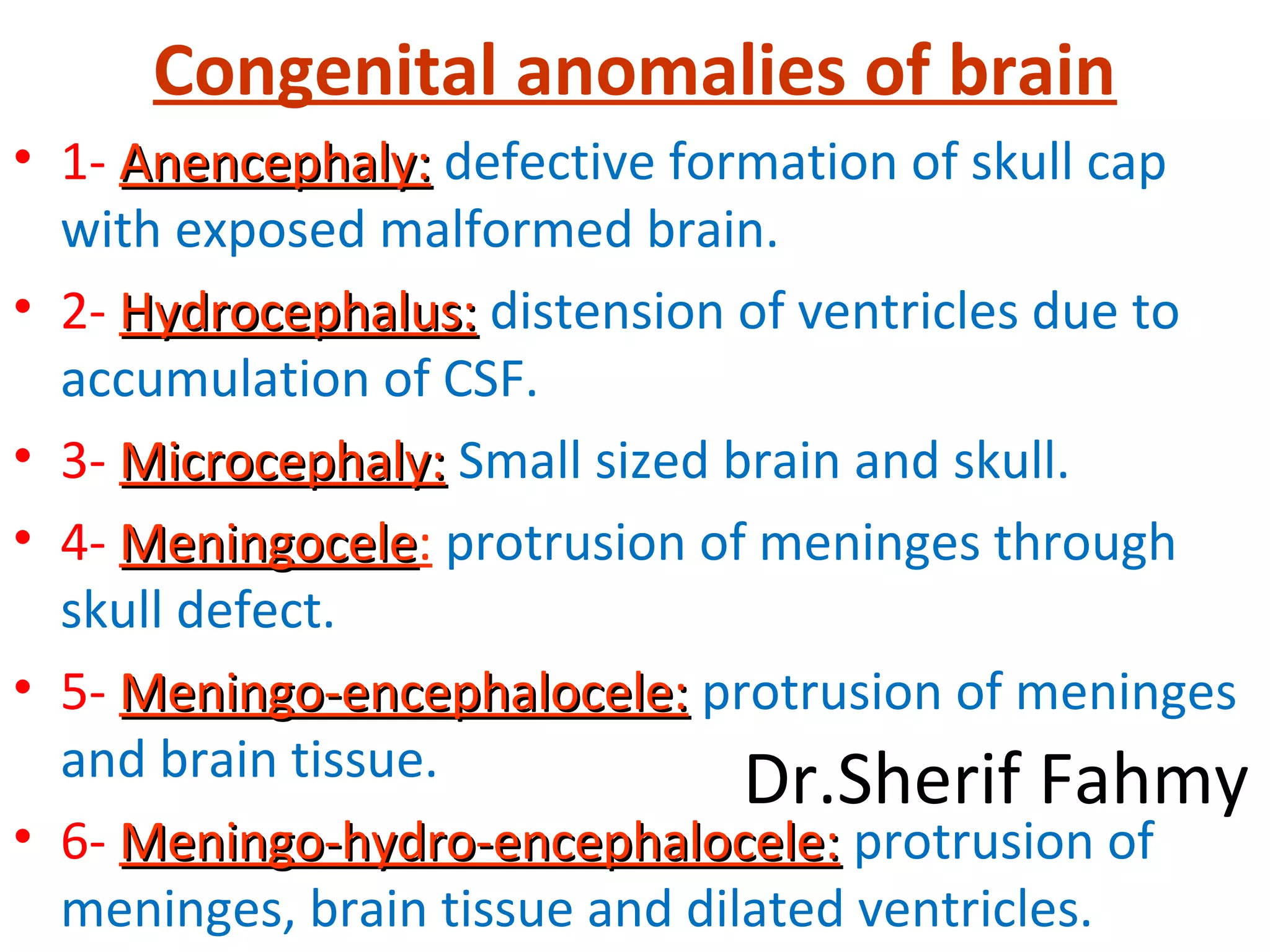
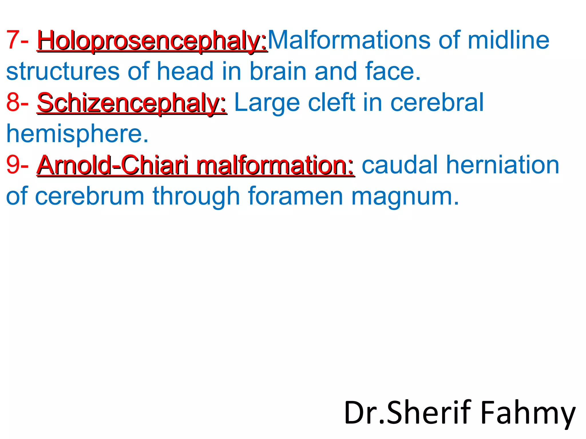
DEVELOPMENT
OF NERVOUS
SYSTEM
Dr.Sherif Fahmy
NEURAL TUBE
• Neural plate: is formed from thickened
ectoderm between primitive node and bucco-
pharyngeal membrane.
• Neural tube: fusion of neural folds at the
middle of the plate then extends cranio-
caudaly till the closure of anterior neuropore
(at 23th day) and posterior neuropore (at 25th
day).
Dr.Sherif Fahmy
Dr.Sherif Fahmy
Dr.Sherif Fahmy
Dr.Sherif Fahmy
SPINAL CORD
• It is developed from caudal part of neural tube.
Firstly the neural tube is formed of a single layer
which proliferates to form middle mantle layer with
lining ependymal layer. Marginal layer is nerve
axons that lie outside the mantle.
• Mantle layer will be differentiated into ventral basal
lamina a dorsal alar lamina. This demarcation
appears on the inner surface of neural as sulcus
limitans.
• Basal lamina forms anterior horn cells and alar
lamina forms posterior horn cells.
Dr.Sherif Fahmy
Dr.Sherif Fahmy
Level of lower end of spinal cord
• Firstly, spinal cord occupies the
whole length of vertebral canal.
• At birth: It lies at level of L3 vertebra.
• In adults: It terminates at disc
between L1/L2 verterbra.
Dr.Sherif Fahmy
Dr.Sherif Fahmy
Dr.Sherif Fahmy
Congenital Anomalies
• 1- Spina bifida oculta: defective formation of
lamina of vertebra.
• 2- Spina bifida with meningeocele:
protrusion of meninges through the laminar
defect.
• 3- Spina bifida with meningeomyelocele:
with protrusion of spinal cord and meninges.
• 4- Rachischisis: defective closure of neural
tube and lamina of vertebrae.
Dr.Sherif Fahmy
Dr.Sherif Fahmy
Development of Brain
(Page 175)
Dr.Sherif Fahmy
Brain Vesicles and Flexures
Brain Vesicles:
1- Forebrain.
2- Midbrain.
3- Hindbrain.
Flexures:
1- Mesencephalic.
2- Pontine.
3- Cervical. Dr.Sherif Fahmy
Dr.Sherif Fahmy
Cervical flexure
Pontine flexure
Mesencephalic flexure
Spinal
cord
Medulla
Pons
Midbrain
telencephalon
Dr.Sherif Fahmy
Development of Brain
Stem
Dr.Sherif Fahmy
Sagittal Section
Dr.Sherif Fahmy
Dr.Sherif Fahmy
Dr.Sherif Fahmy
Upper Part of Medulla and Pons
-Cavity is widened to form 4th
ventricle.
-Roof plate will form roof of 4th
ventricle.
-Alar lamina becomes lateral to basal
lamina.
-Dorsal edge of alar lamina of pons will
form superior rhombic lip.
-Dorsal edge of alar lamina of medulla
will form inferior rhombic lip.
Dr.Sherif Fahmy
Lower part of Medulla and Midbrain
-The cavity remains as central canal.
-Alar lamina is lateral to basal lamina.
Then the alar and basal laminae will
give rise to the following nuclei.
Dr.Sherif Fahmy
Fate of Basal Lamina
General somatic efferent (GSE): Form motor nuclei of
3rd
, 4th
, 6th
and 12th
cranial nerves.
Special visceral efferent (SVE): Forms motor nuclei of
trigeminal, facial, 9th
, 10th
and 11th
.
General Visceral efferent: Edinger-westphal n. of
oculomotor, superior salivary of facial n., inferior
salivary nuscleus of glossopharyngeal n and dorsal
nucleus of vagus
Dr.Sherif Fahmy
Fate of Alar Lamina
- General visceral and special visceral
afferent: which form nucleus solitarius
(Taste) in medulla.
- General somatic afferent: will form
mesencephalic, main sensory nucleus
and spinal neuclus of trigeminal nerve.
- Special somatic afferent: vestibular
and cochlear nuclie Dr.Sherif Fahmy
Dr.Sherif Fahmy
Development of
Cerebellum
Dr.Sherif Fahmy
Shape
Dr.Sherif Fahmy
Flocculus
Nodule
Lower Surface
Postero-
lateral
fissure
Dr.Sherif Fahmy
Site: In the dorsal wall of hindbrain.
Cells: Ectodermal cells of neural tube.
Steps:
1- Formation of 2 cerebellar swellings from
superior rhombic lip.
2- Fusion between the cerebellar swellings that
covers the roof of 4th
ventricle.
3- The developing cerebellum is divided into
cranial & caudal portions by posterolateral
fissure.
Dr.Sherif Fahmy
4- The caudal portion will form
flocculonodular lobe.
5- The cranial portion will grows faster to
form rest of cerebellum (2 cerebellar
hemisphers and vermis).
6- Development of primary fissure between
anterior & posterior lobes, Development of
transverse fissures and folia to increase the
surface area.
Dr.Sherif Fahmy
Cervical flexure
Pontine flexure
Mesencephalic flexure
Dr.Sherif Fahmy
Dr.Sherif Fahmy
Dr.Sherif Fahmy
Development of
Forebrain
(Prosencephalon)
(Page 181)
Dr.Sherif Fahmy
A- Development of Diencephalon
Site: In the cranial portion of neural tube.
Cells: Ectodermal cells.
Steps:
1- The roof plate is thin and invaded by choroid plexus.
2- The floor plate is thickened and divided into posterior
larger part which forms hypothalamus and anterior smaller
part which gives rise to posterior lobe of pituitary gland.
3- Basal plate is lacking.
4- Alar plate will form thalamus, rest of hypothalamus and
epithalamus.
5- Cavity will be the 3rd
ventricle.
Dr.Sherif Fahmy
B- Development of Cerebral
Hemisphere
-Cerebral hemispheres are developed from
telencephalon of the forebrain.
-The cavity will be lateral ventricles.
-Neuroblasts migrate externally to form
cerebral cortex while some cells remain near
the base to form basal ganglia.
-Axons from neuroblasts in cortex will form
internal capsule that split corpus striatum into
caudate and lentiform nuclei.Dr.Sherif Fahmy
-Cerebral cortex will expands forewards,
backwards, laterally and downwards to
form cerebral lobes.
-Insula is formed as this part of cortex will
grow slowly whereas the surroundings
grow faster and cover it, forming
opercula.
-At first the cerebral cortex is smooth but
later in fetal life it grows and expands to
form sulci and gyri.
Dr.Sherif Fahmy
Dr.Sherif Fahmy
Dr.Sherif Fahmy
Dr.Sherif Fahmy
Congenital anomalies of brain
• 1- Anencephaly:Anencephaly: defective formation of skull cap
with exposed malformed brain.
• 2- Hydrocephalus:Hydrocephalus: distension of ventricles due to
accumulation of CSF.
• 3- Microcephaly:Microcephaly: Small sized brain and skull.
• 4- MeningoceleMeningocele: protrusion of meninges through
skull defect.
• 5- Meningo-encephalocele:Meningo-encephalocele: protrusion of meninges
and brain tissue.
• 6- Meningo-hydro-encephalocele:Meningo-hydro-encephalocele: protrusion of
meninges, brain tissue and dilated ventricles.
Dr.Sherif Fahmy
7- Holoprosencephaly:Holoprosencephaly:Malformations of midline
structures of head in brain and face.
8- Schizencephaly:Schizencephaly: Large cleft in cerebral
hemisphere.
9- Arnold-Chiari malformation:Arnold-Chiari malformation: caudal herniation
of cerebrum through foramen magnum.
Dr.Sherif Fahmy
Hydrocephalus
Dr.Sherif Fahmy
Microcephaly
Dr.Sherif Fahmy
Exencephaly
Dr.Sherif Fahmy
Arnold-Chiari Malformation
Dr.Sherif Fahmy
Schizencephaly
Dr.Sherif Fahmy
Holopros-
encephaly
Dr.Sherif Fahmy
Meningoencephalocele
Dr.Sherif Fahmy

Development of Nervous System (Special Embryology)

  • 1.
  • 2.
    NEURAL TUBE • Neuralplate: is formed from thickened ectoderm between primitive node and bucco- pharyngeal membrane. • Neural tube: fusion of neural folds at the middle of the plate then extends cranio- caudaly till the closure of anterior neuropore (at 23th day) and posterior neuropore (at 25th day). Dr.Sherif Fahmy
  • 3.
  • 4.
  • 5.
  • 6.
    SPINAL CORD • Itis developed from caudal part of neural tube. Firstly the neural tube is formed of a single layer which proliferates to form middle mantle layer with lining ependymal layer. Marginal layer is nerve axons that lie outside the mantle. • Mantle layer will be differentiated into ventral basal lamina a dorsal alar lamina. This demarcation appears on the inner surface of neural as sulcus limitans. • Basal lamina forms anterior horn cells and alar lamina forms posterior horn cells. Dr.Sherif Fahmy
  • 7.
  • 8.
    Level of lowerend of spinal cord • Firstly, spinal cord occupies the whole length of vertebral canal. • At birth: It lies at level of L3 vertebra. • In adults: It terminates at disc between L1/L2 verterbra. Dr.Sherif Fahmy
  • 9.
  • 10.
  • 11.
    Congenital Anomalies • 1-Spina bifida oculta: defective formation of lamina of vertebra. • 2- Spina bifida with meningeocele: protrusion of meninges through the laminar defect. • 3- Spina bifida with meningeomyelocele: with protrusion of spinal cord and meninges. • 4- Rachischisis: defective closure of neural tube and lamina of vertebrae. Dr.Sherif Fahmy
  • 12.
  • 13.
    Development of Brain (Page175) Dr.Sherif Fahmy
  • 14.
    Brain Vesicles andFlexures Brain Vesicles: 1- Forebrain. 2- Midbrain. 3- Hindbrain. Flexures: 1- Mesencephalic. 2- Pontine. 3- Cervical. Dr.Sherif Fahmy
  • 15.
  • 16.
    Cervical flexure Pontine flexure Mesencephalicflexure Spinal cord Medulla Pons Midbrain telencephalon Dr.Sherif Fahmy
  • 17.
  • 18.
  • 19.
  • 20.
    Upper Part ofMedulla and Pons -Cavity is widened to form 4th ventricle. -Roof plate will form roof of 4th ventricle. -Alar lamina becomes lateral to basal lamina. -Dorsal edge of alar lamina of pons will form superior rhombic lip. -Dorsal edge of alar lamina of medulla will form inferior rhombic lip. Dr.Sherif Fahmy
  • 21.
    Lower part ofMedulla and Midbrain -The cavity remains as central canal. -Alar lamina is lateral to basal lamina. Then the alar and basal laminae will give rise to the following nuclei. Dr.Sherif Fahmy
  • 22.
    Fate of BasalLamina General somatic efferent (GSE): Form motor nuclei of 3rd , 4th , 6th and 12th cranial nerves. Special visceral efferent (SVE): Forms motor nuclei of trigeminal, facial, 9th , 10th and 11th . General Visceral efferent: Edinger-westphal n. of oculomotor, superior salivary of facial n., inferior salivary nuscleus of glossopharyngeal n and dorsal nucleus of vagus Dr.Sherif Fahmy
  • 23.
    Fate of AlarLamina - General visceral and special visceral afferent: which form nucleus solitarius (Taste) in medulla. - General somatic afferent: will form mesencephalic, main sensory nucleus and spinal neuclus of trigeminal nerve. - Special somatic afferent: vestibular and cochlear nuclie Dr.Sherif Fahmy
  • 24.
  • 25.
  • 26.
  • 27.
  • 28.
    Site: In thedorsal wall of hindbrain. Cells: Ectodermal cells of neural tube. Steps: 1- Formation of 2 cerebellar swellings from superior rhombic lip. 2- Fusion between the cerebellar swellings that covers the roof of 4th ventricle. 3- The developing cerebellum is divided into cranial & caudal portions by posterolateral fissure. Dr.Sherif Fahmy
  • 29.
    4- The caudalportion will form flocculonodular lobe. 5- The cranial portion will grows faster to form rest of cerebellum (2 cerebellar hemisphers and vermis). 6- Development of primary fissure between anterior & posterior lobes, Development of transverse fissures and folia to increase the surface area. Dr.Sherif Fahmy
  • 30.
  • 31.
  • 32.
  • 33.
  • 34.
    A- Development ofDiencephalon Site: In the cranial portion of neural tube. Cells: Ectodermal cells. Steps: 1- The roof plate is thin and invaded by choroid plexus. 2- The floor plate is thickened and divided into posterior larger part which forms hypothalamus and anterior smaller part which gives rise to posterior lobe of pituitary gland. 3- Basal plate is lacking. 4- Alar plate will form thalamus, rest of hypothalamus and epithalamus. 5- Cavity will be the 3rd ventricle. Dr.Sherif Fahmy
  • 35.
    B- Development ofCerebral Hemisphere -Cerebral hemispheres are developed from telencephalon of the forebrain. -The cavity will be lateral ventricles. -Neuroblasts migrate externally to form cerebral cortex while some cells remain near the base to form basal ganglia. -Axons from neuroblasts in cortex will form internal capsule that split corpus striatum into caudate and lentiform nuclei.Dr.Sherif Fahmy
  • 36.
    -Cerebral cortex willexpands forewards, backwards, laterally and downwards to form cerebral lobes. -Insula is formed as this part of cortex will grow slowly whereas the surroundings grow faster and cover it, forming opercula. -At first the cerebral cortex is smooth but later in fetal life it grows and expands to form sulci and gyri. Dr.Sherif Fahmy
  • 37.
  • 38.
  • 39.
  • 40.
    Congenital anomalies ofbrain • 1- Anencephaly:Anencephaly: defective formation of skull cap with exposed malformed brain. • 2- Hydrocephalus:Hydrocephalus: distension of ventricles due to accumulation of CSF. • 3- Microcephaly:Microcephaly: Small sized brain and skull. • 4- MeningoceleMeningocele: protrusion of meninges through skull defect. • 5- Meningo-encephalocele:Meningo-encephalocele: protrusion of meninges and brain tissue. • 6- Meningo-hydro-encephalocele:Meningo-hydro-encephalocele: protrusion of meninges, brain tissue and dilated ventricles. Dr.Sherif Fahmy
  • 41.
    7- Holoprosencephaly:Holoprosencephaly:Malformations ofmidline structures of head in brain and face. 8- Schizencephaly:Schizencephaly: Large cleft in cerebral hemisphere. 9- Arnold-Chiari malformation:Arnold-Chiari malformation: caudal herniation of cerebrum through foramen magnum. Dr.Sherif Fahmy
  • 42.
  • 43.
  • 44.
  • 45.
  • 46.
  • 47.
  • 48.