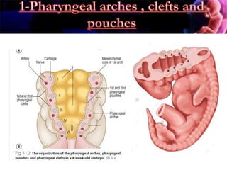
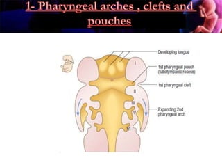
1) The head and neck develop from pharyngeal arches, clefts and pouches which form structures like the tongue, thyroid gland and face.
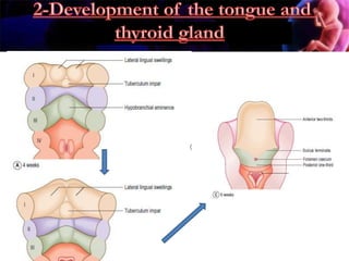
2) The tongue develops from swellings in the pharyngeal arches, while the thyroid gland develops from the foramen caecum.
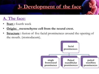
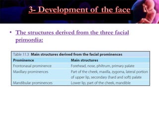
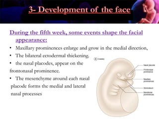
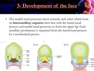
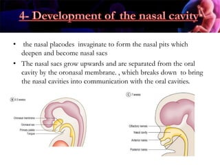
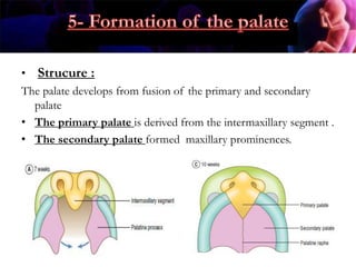
3) The face develops from the fusion of five facial prominences which give rise to structures of the nose, mouth and eyes.