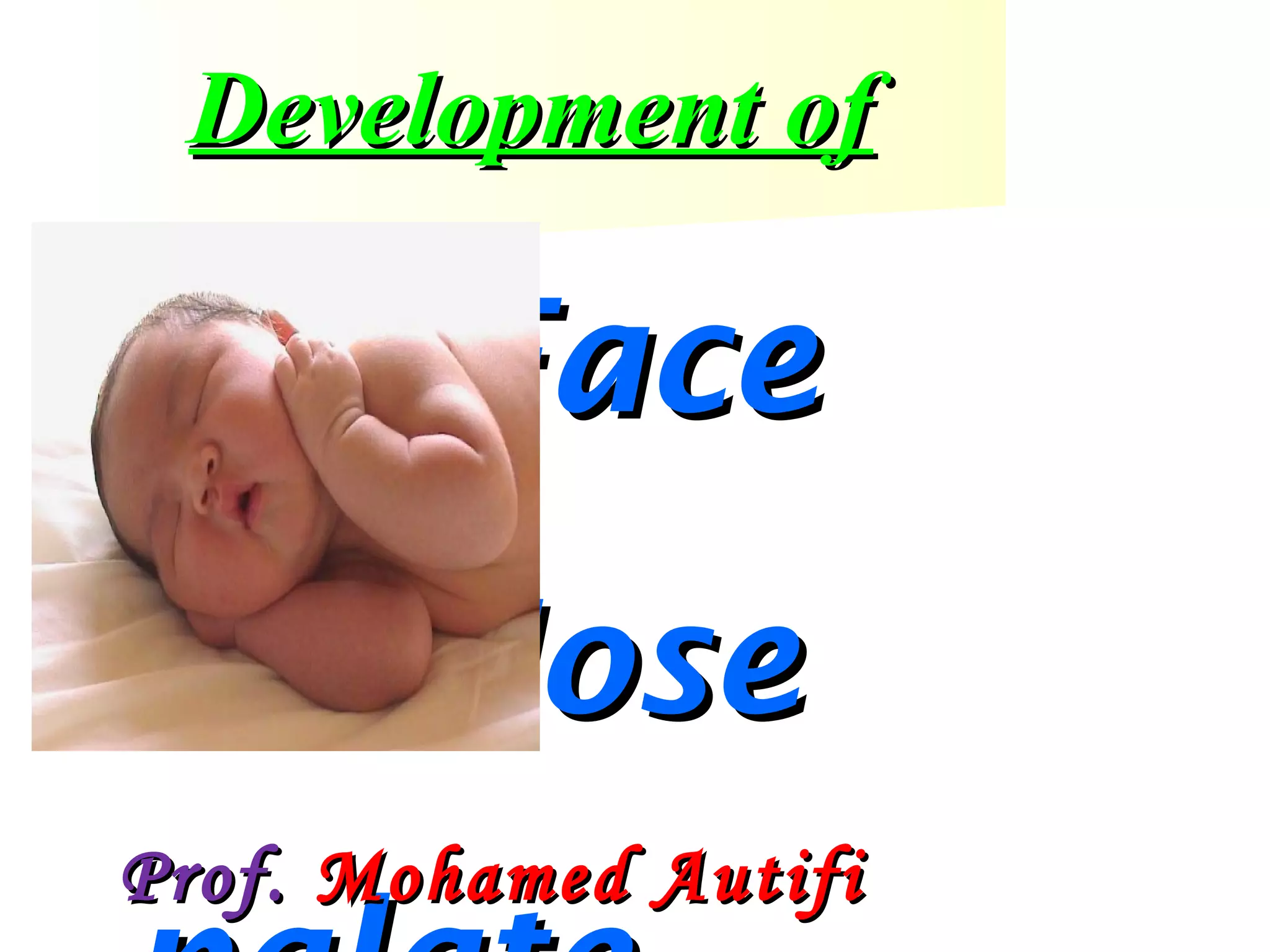
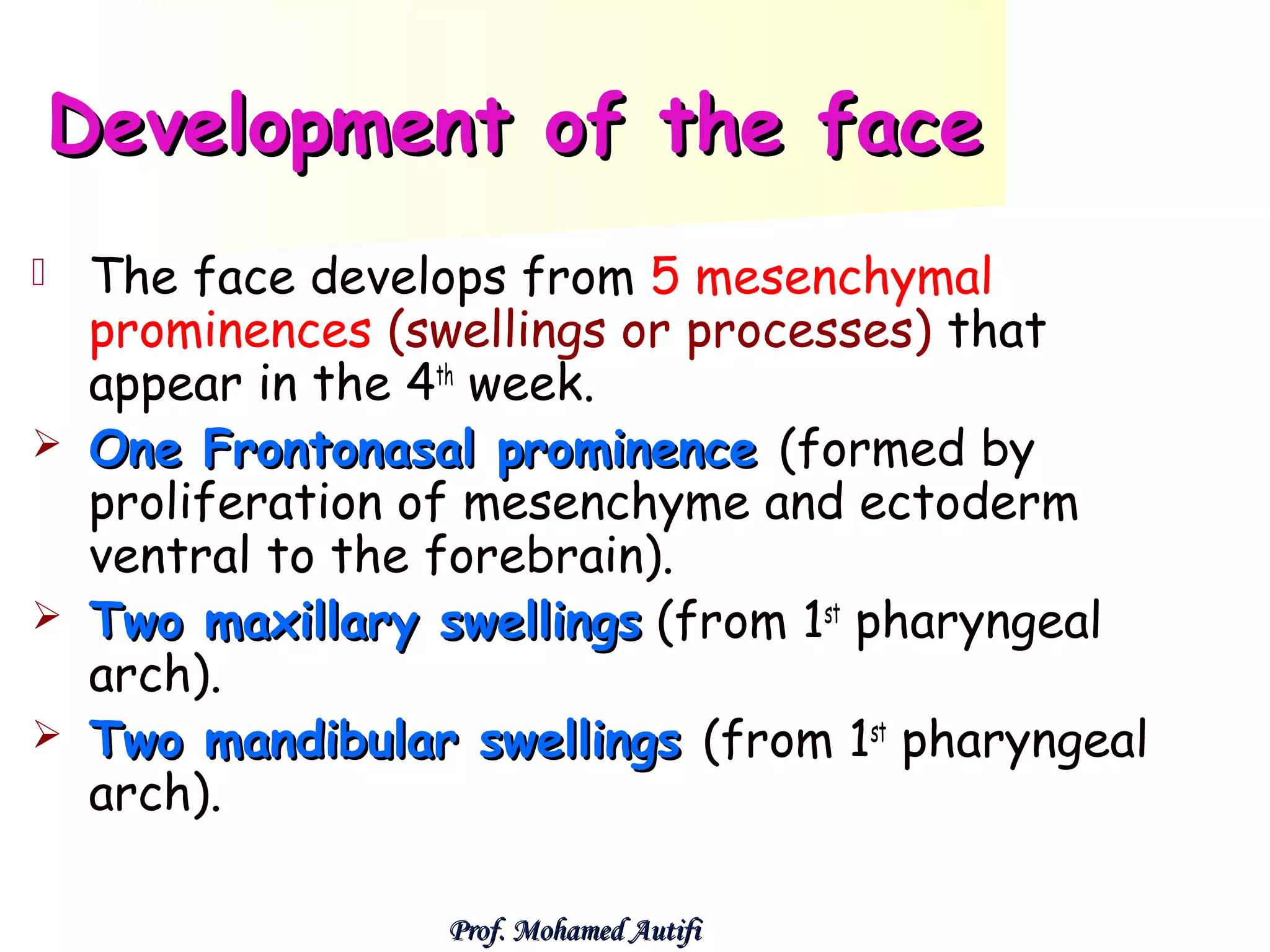
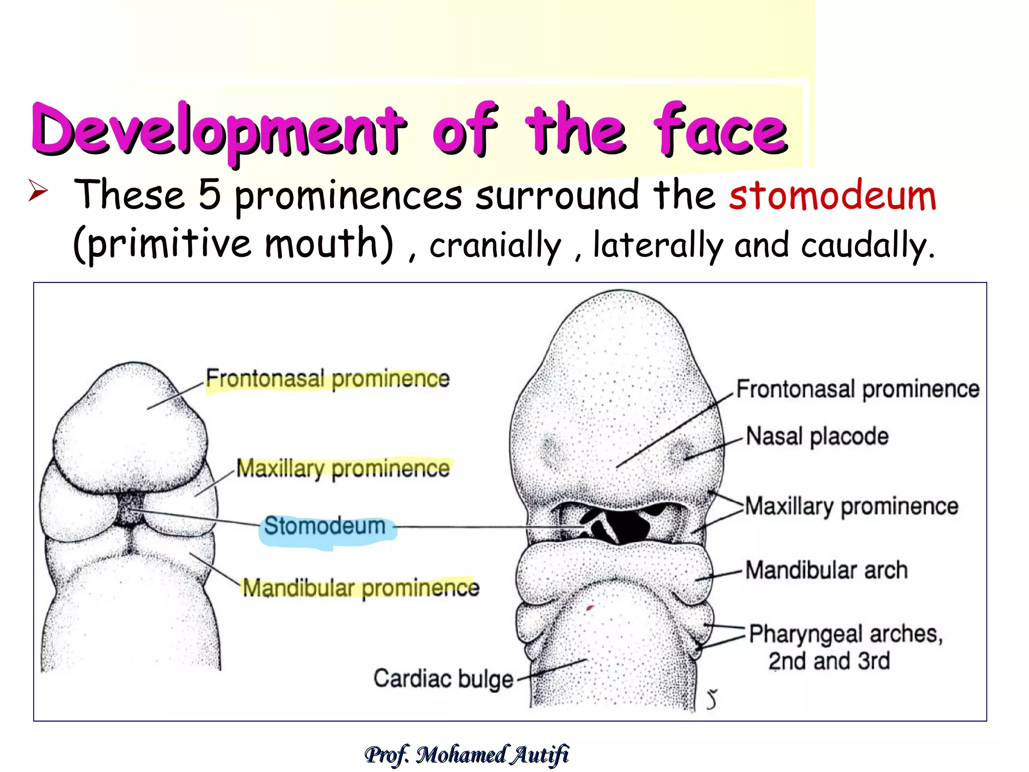
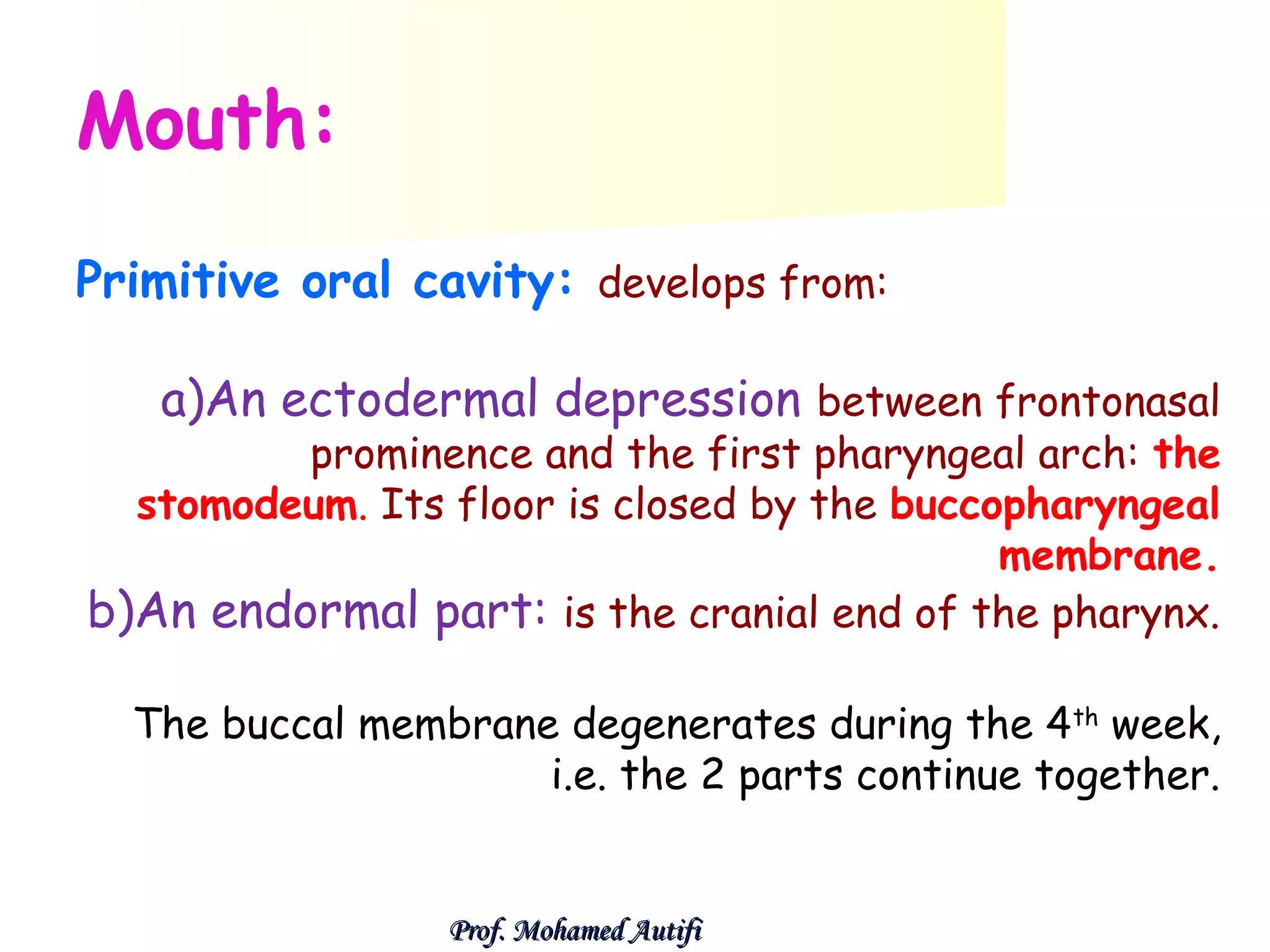
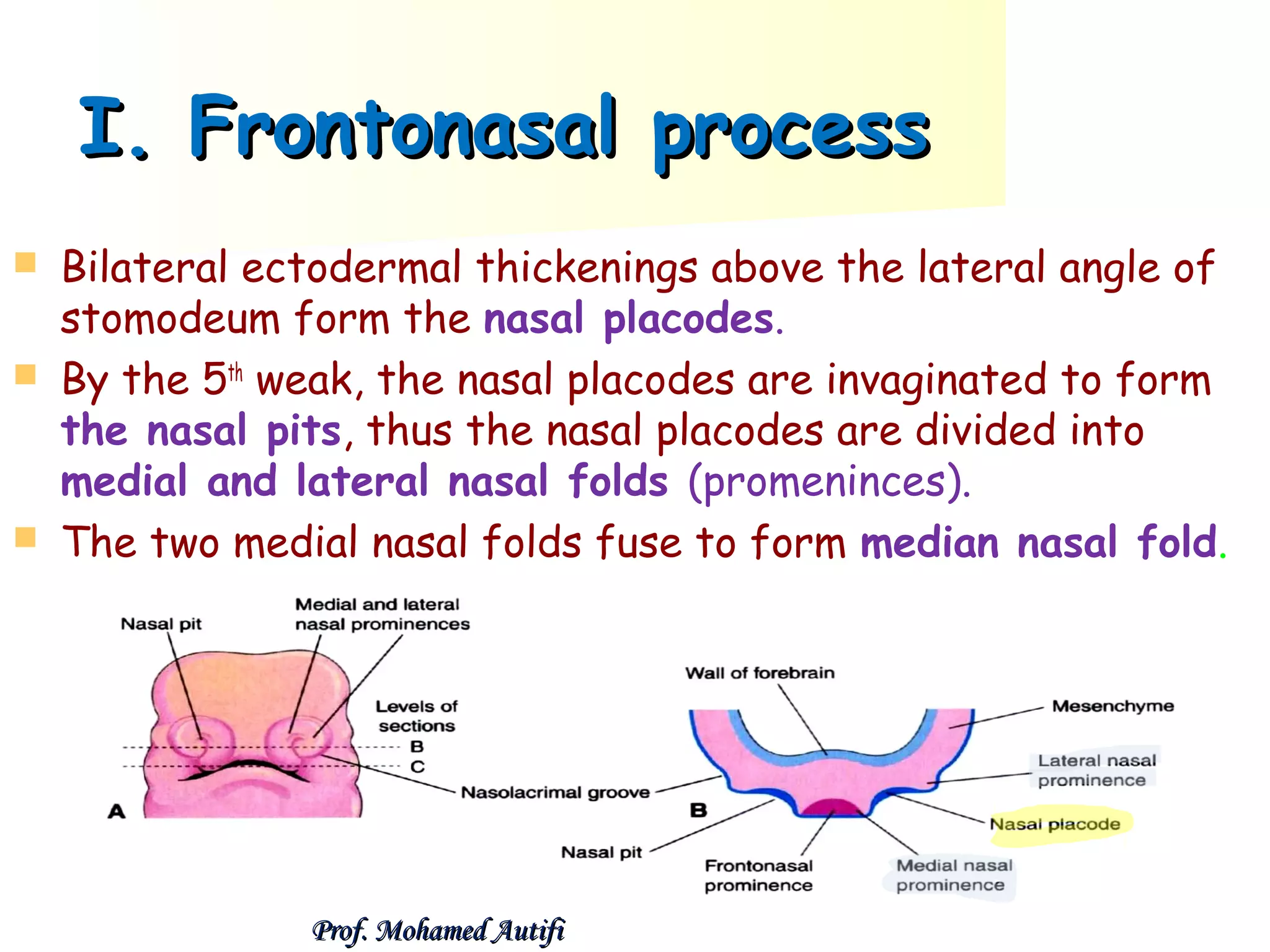
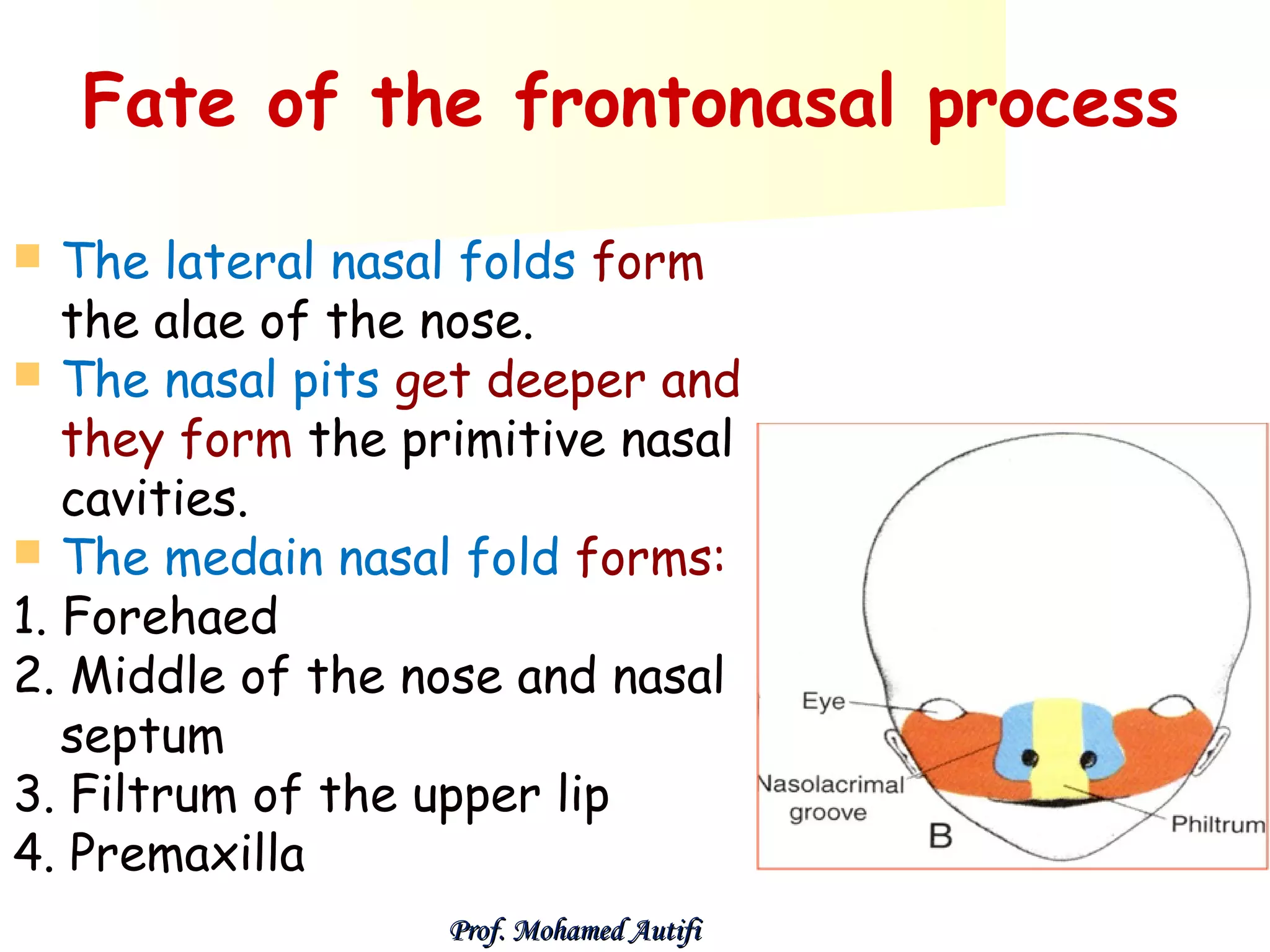
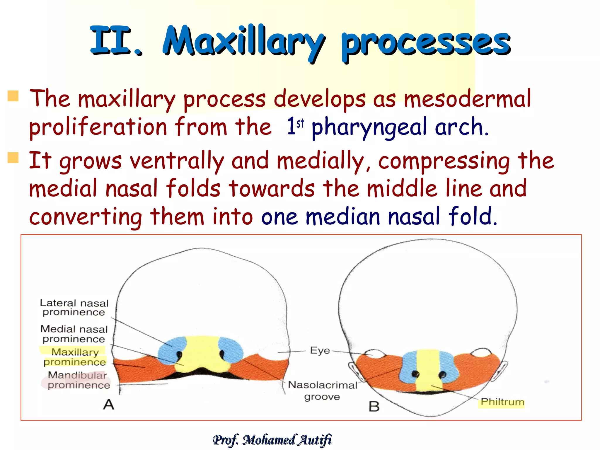
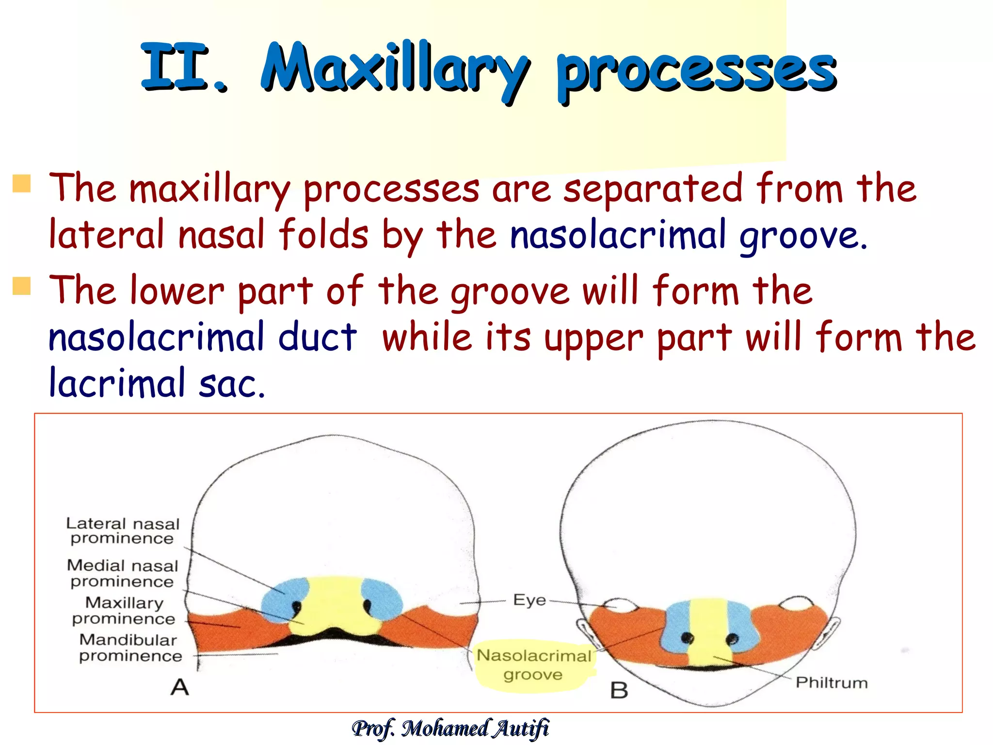
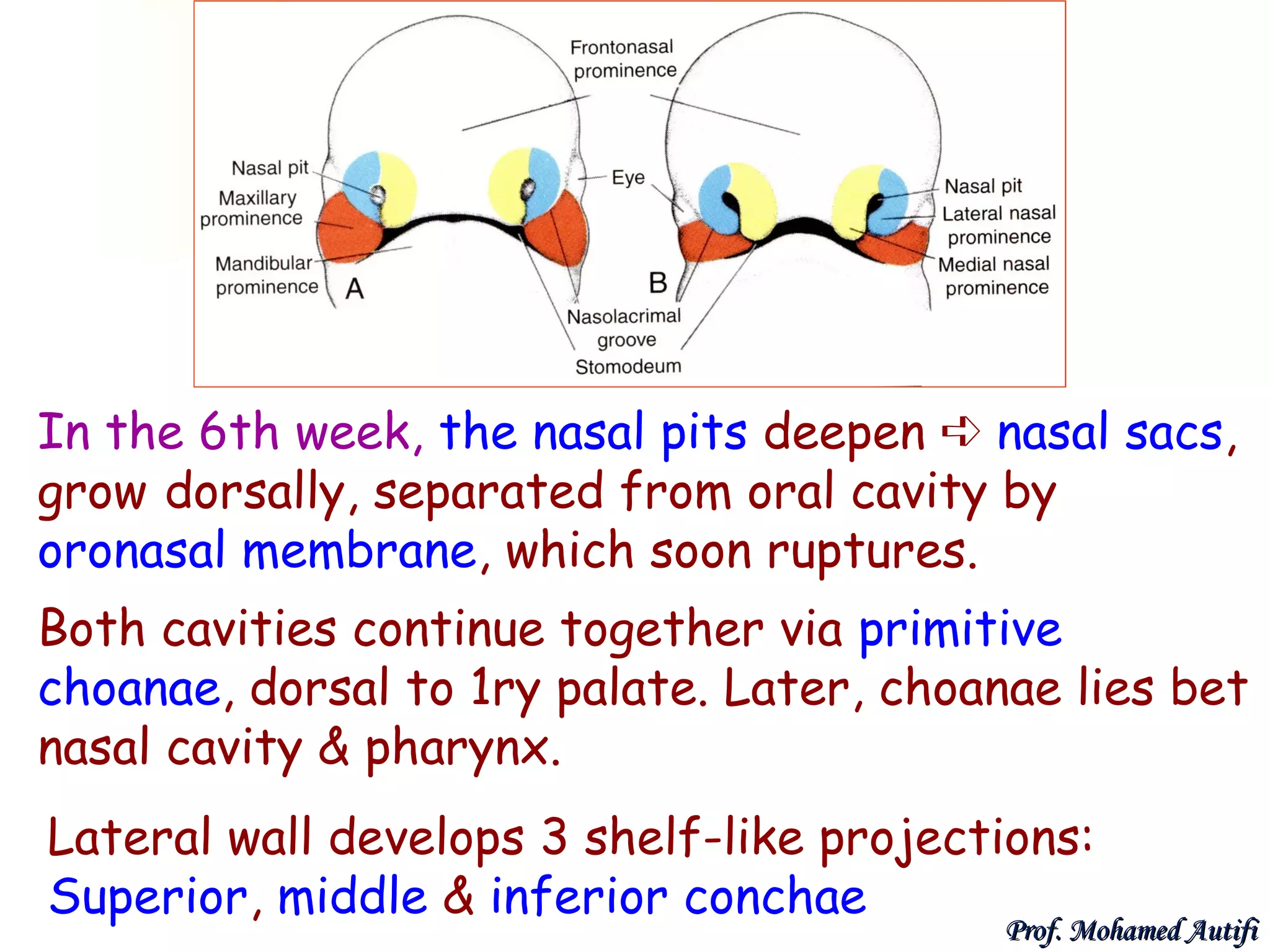
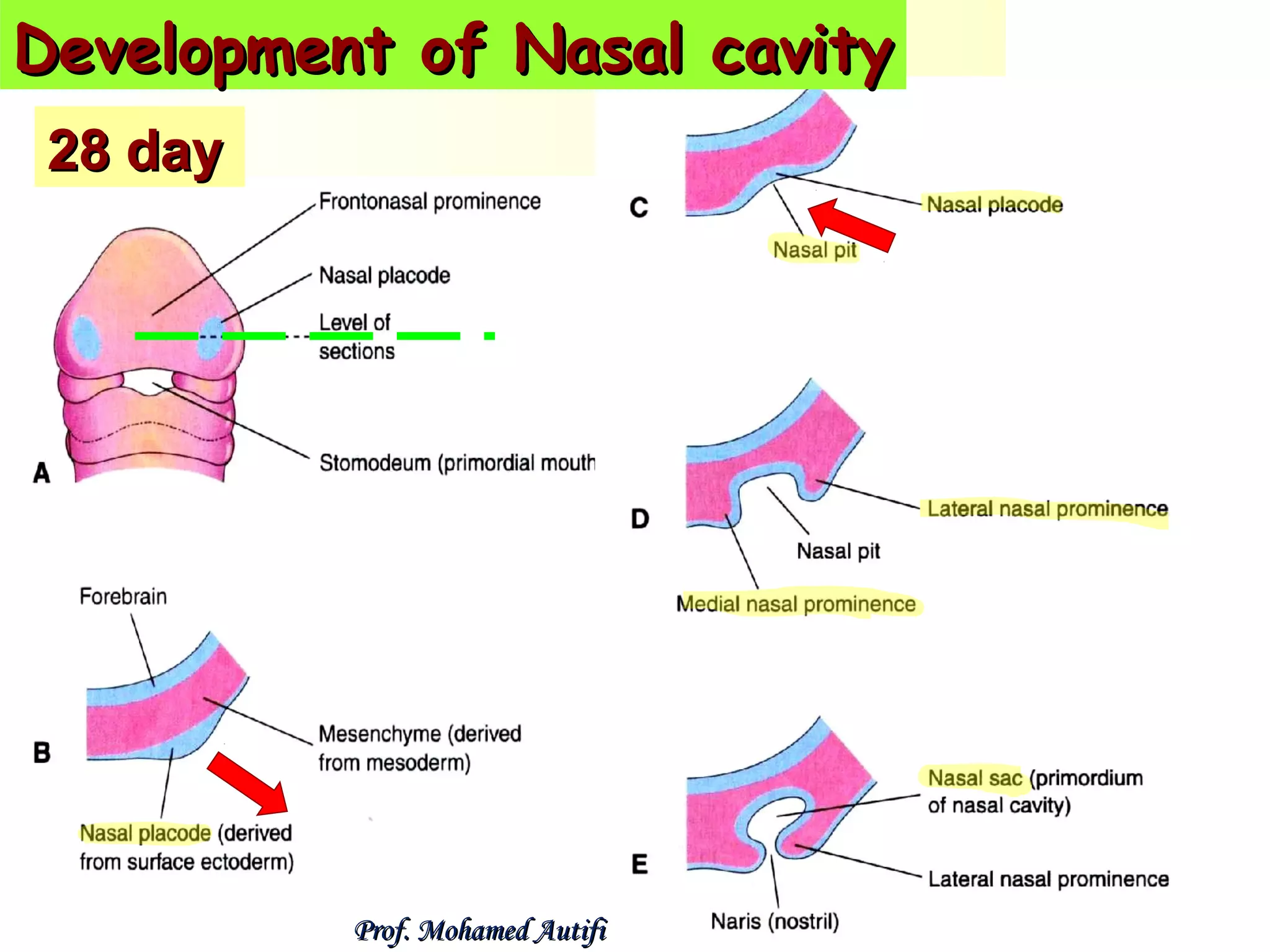
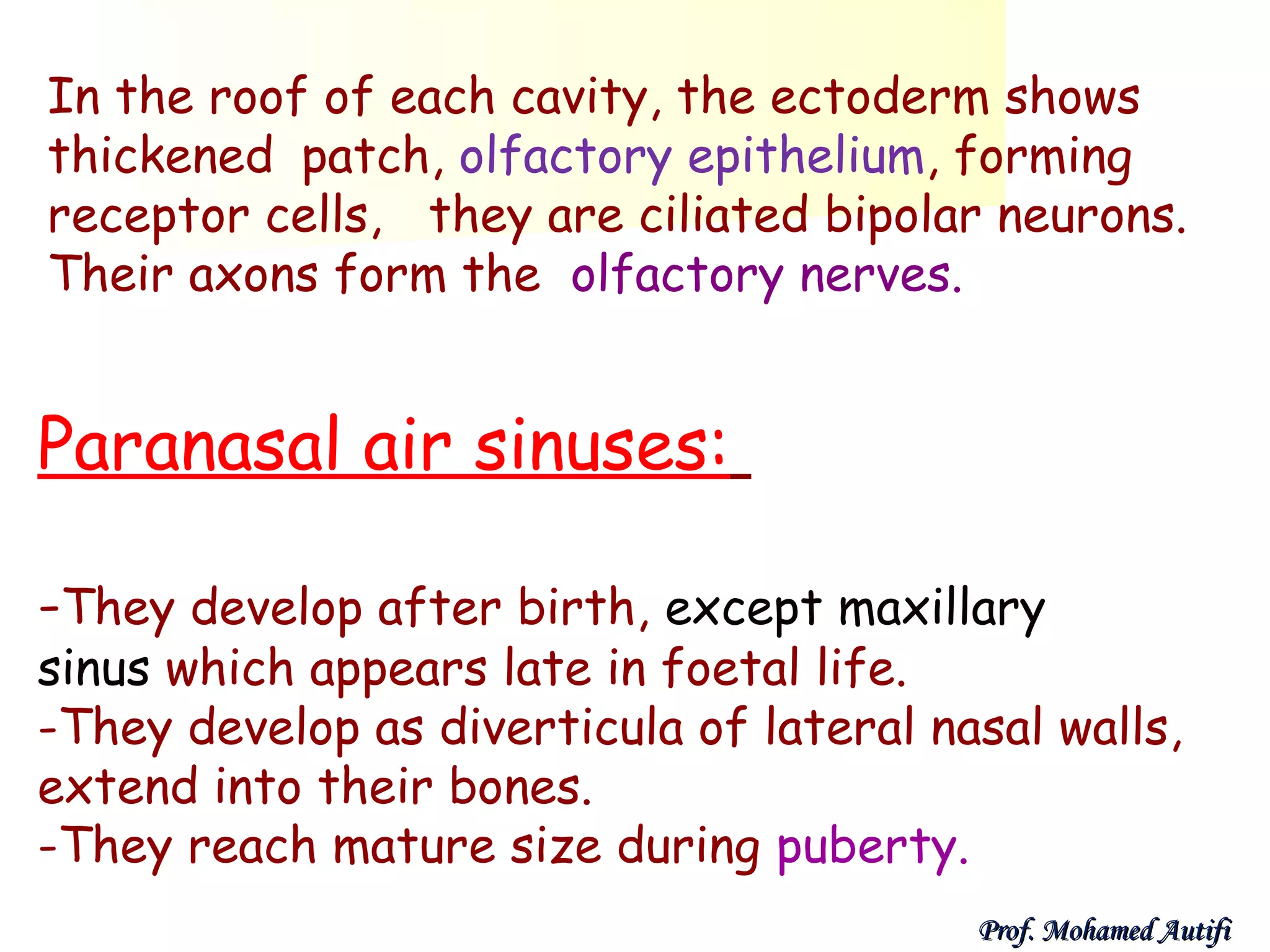
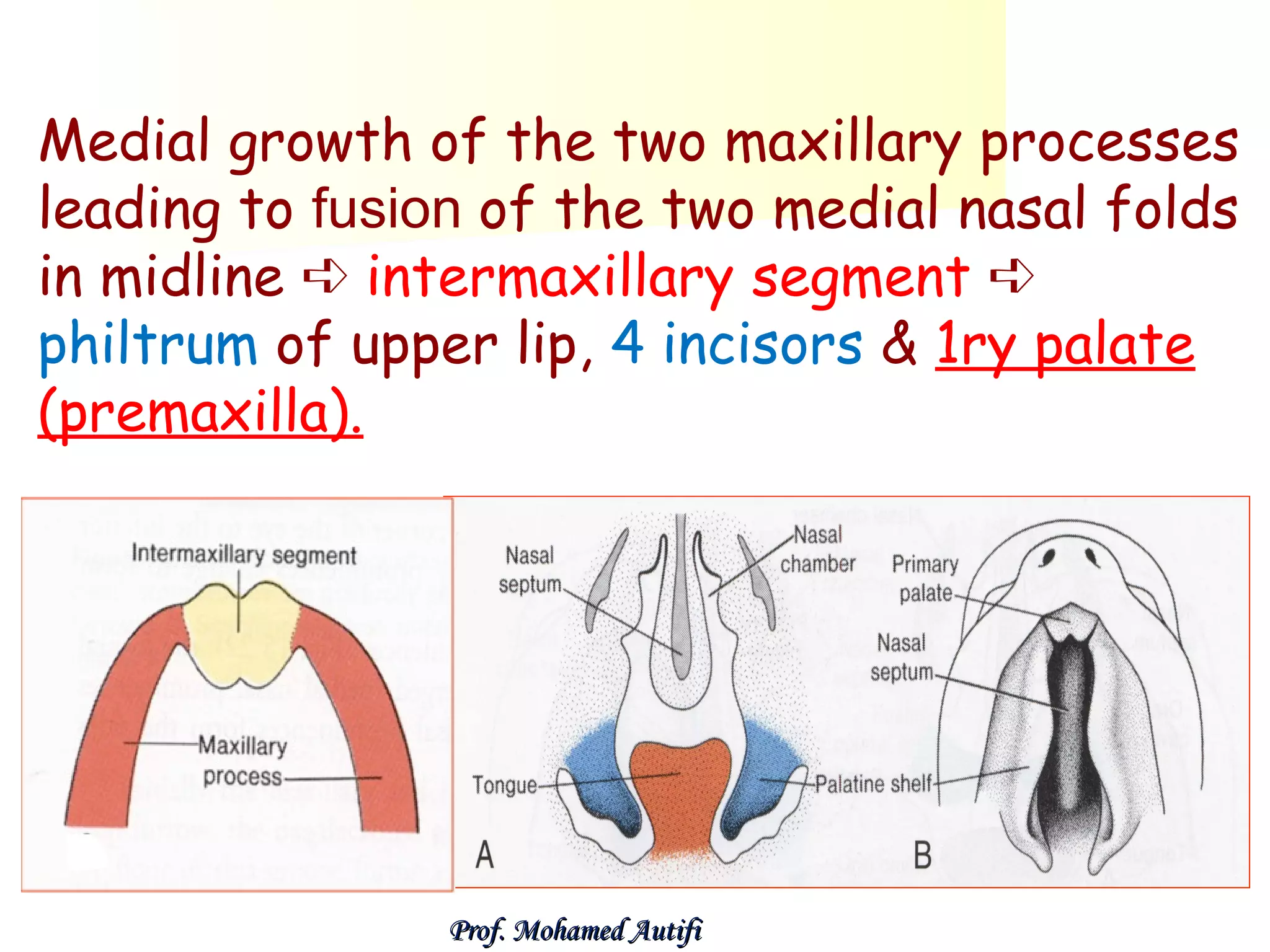
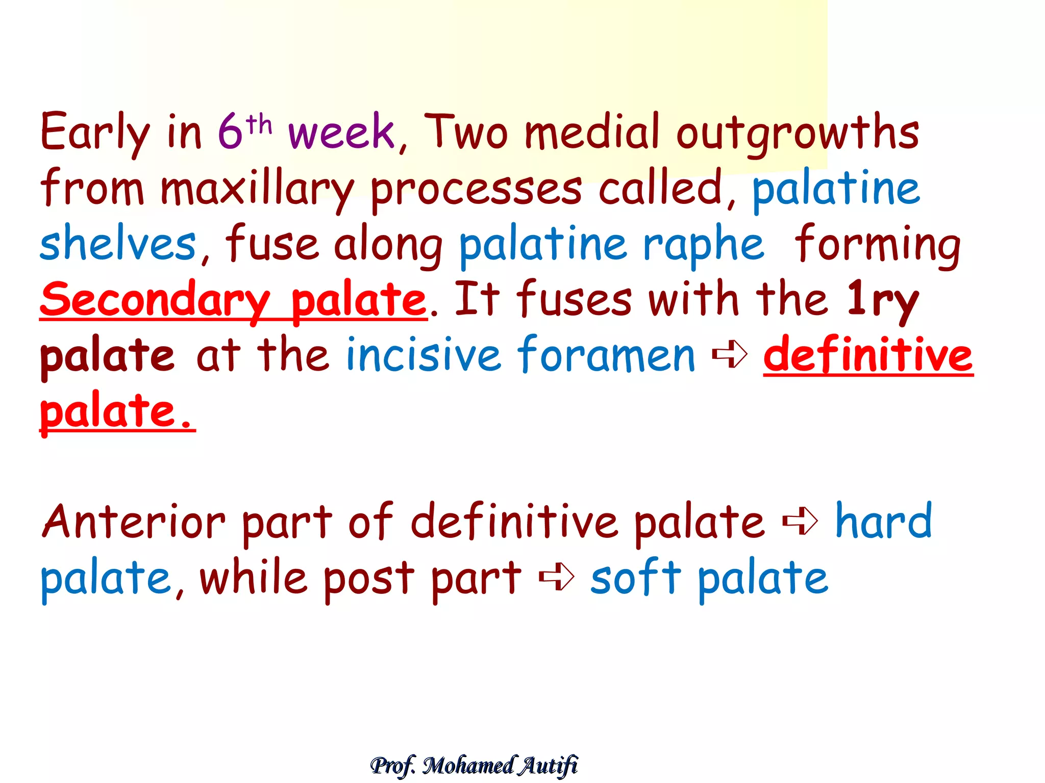
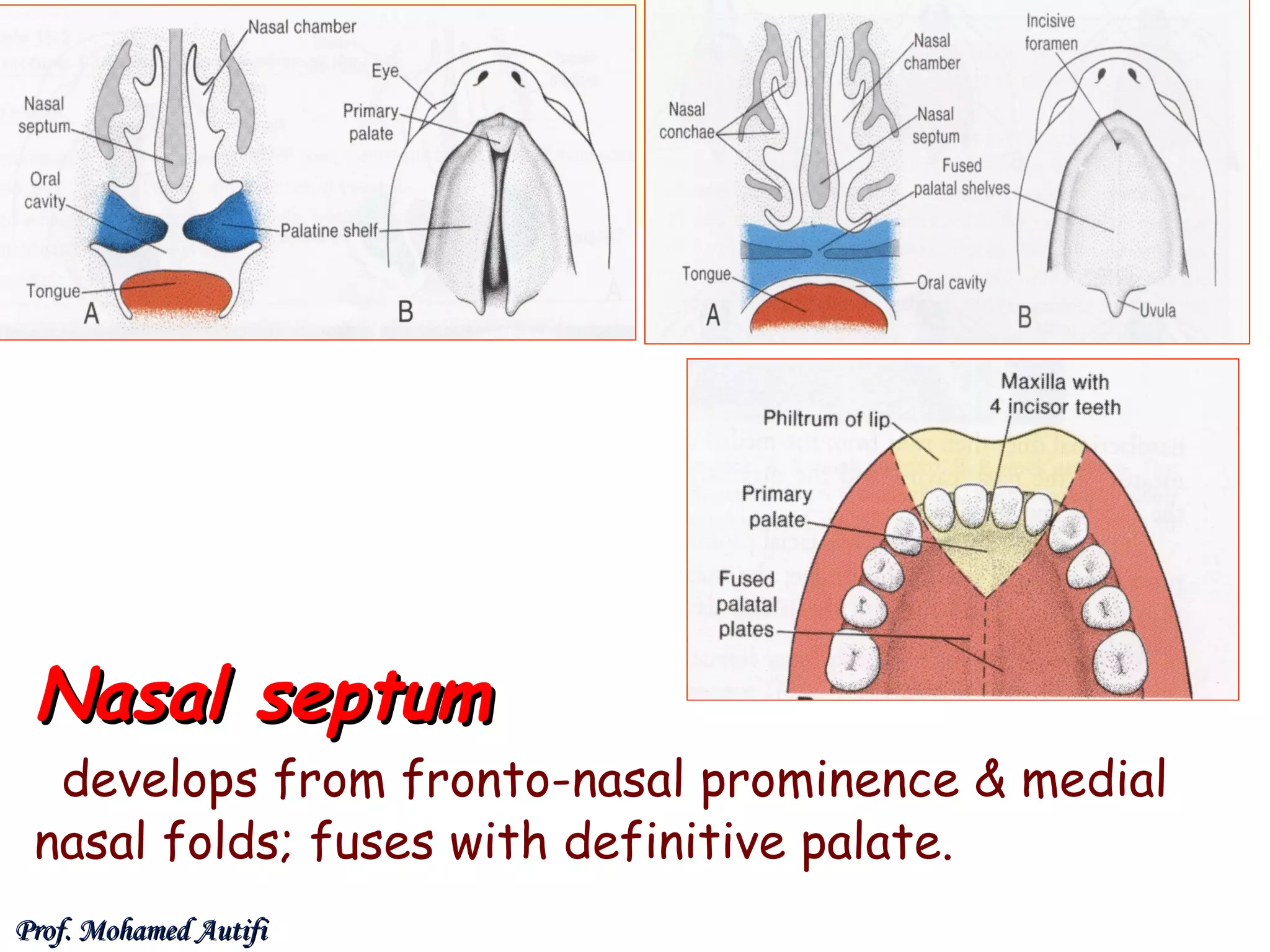
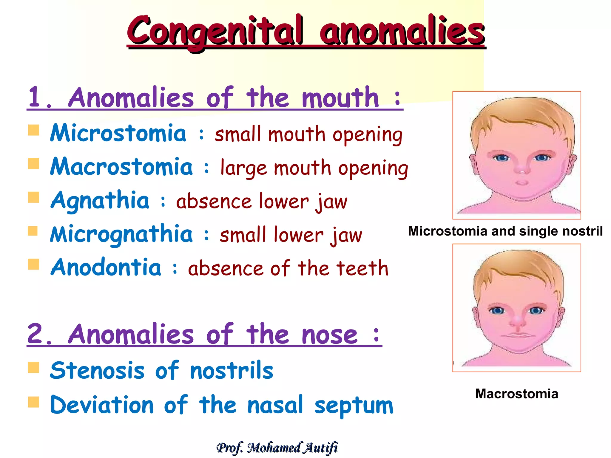
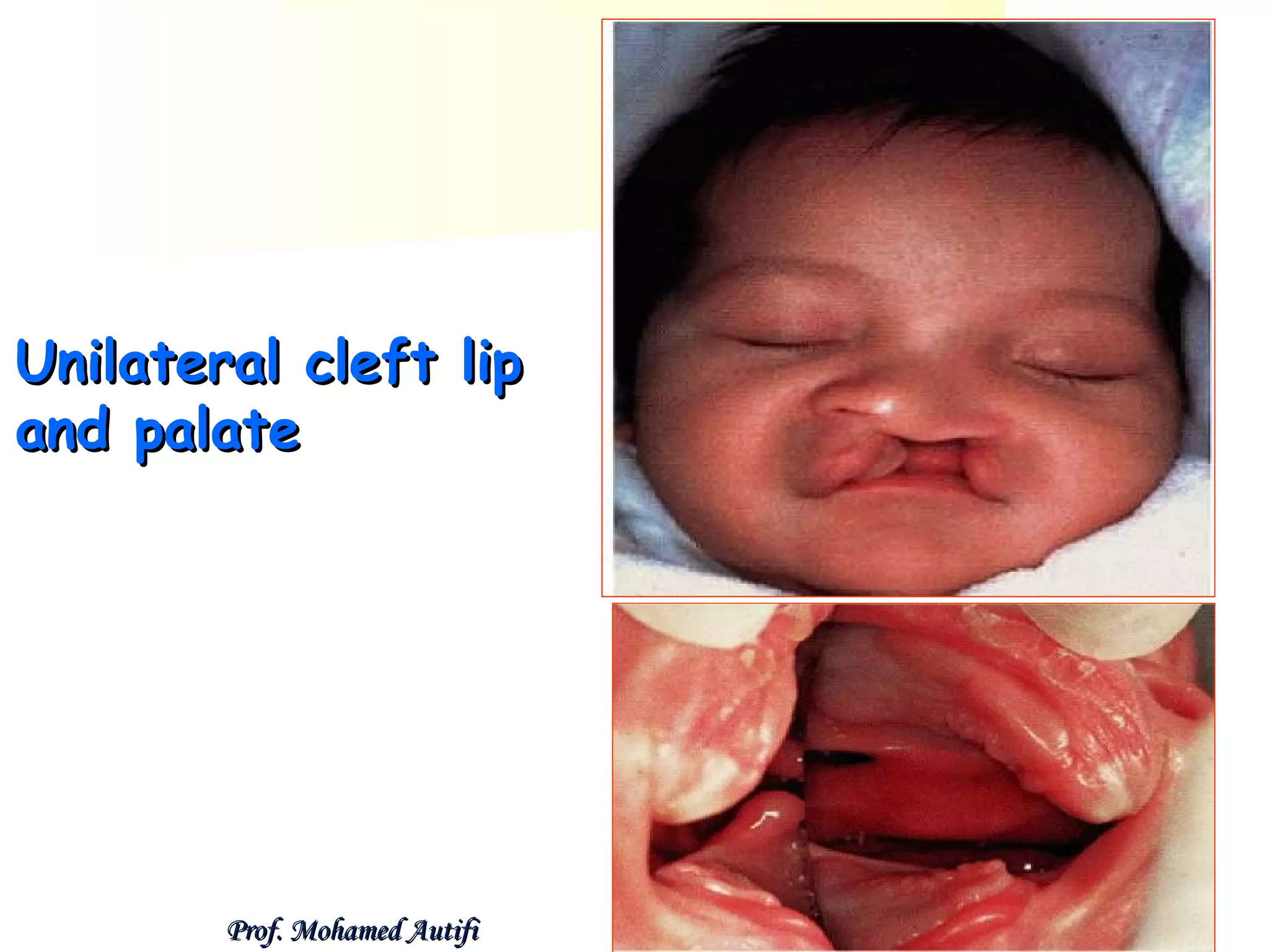
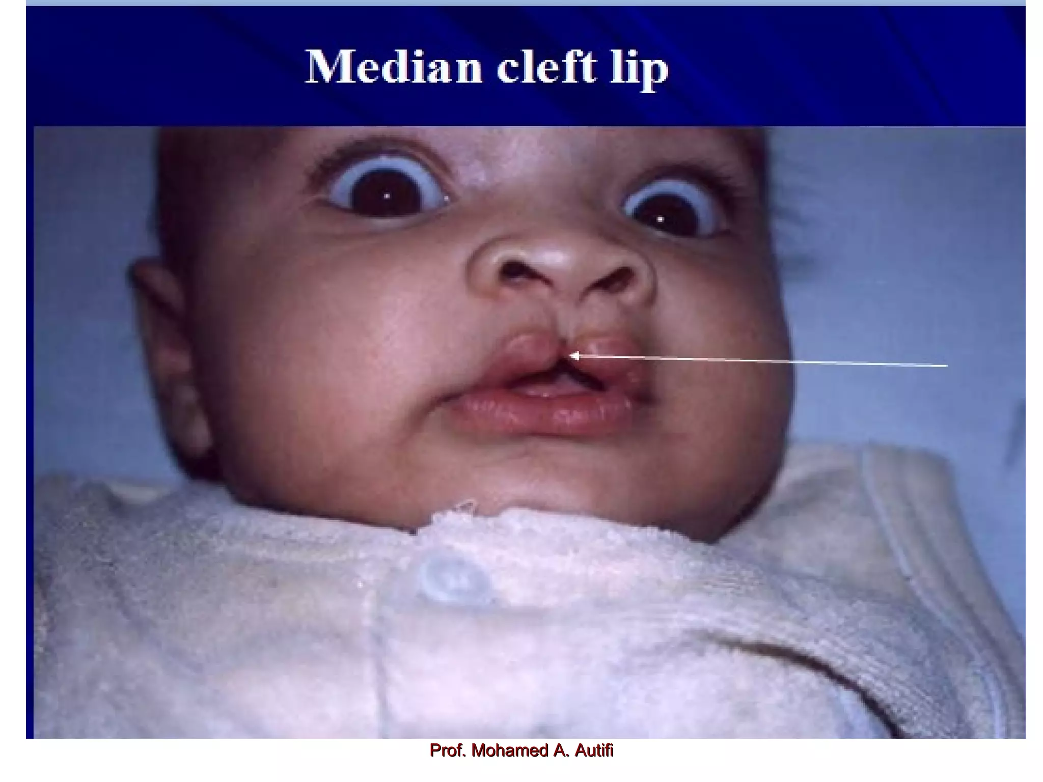
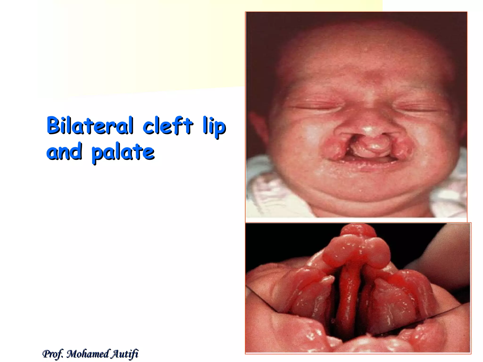
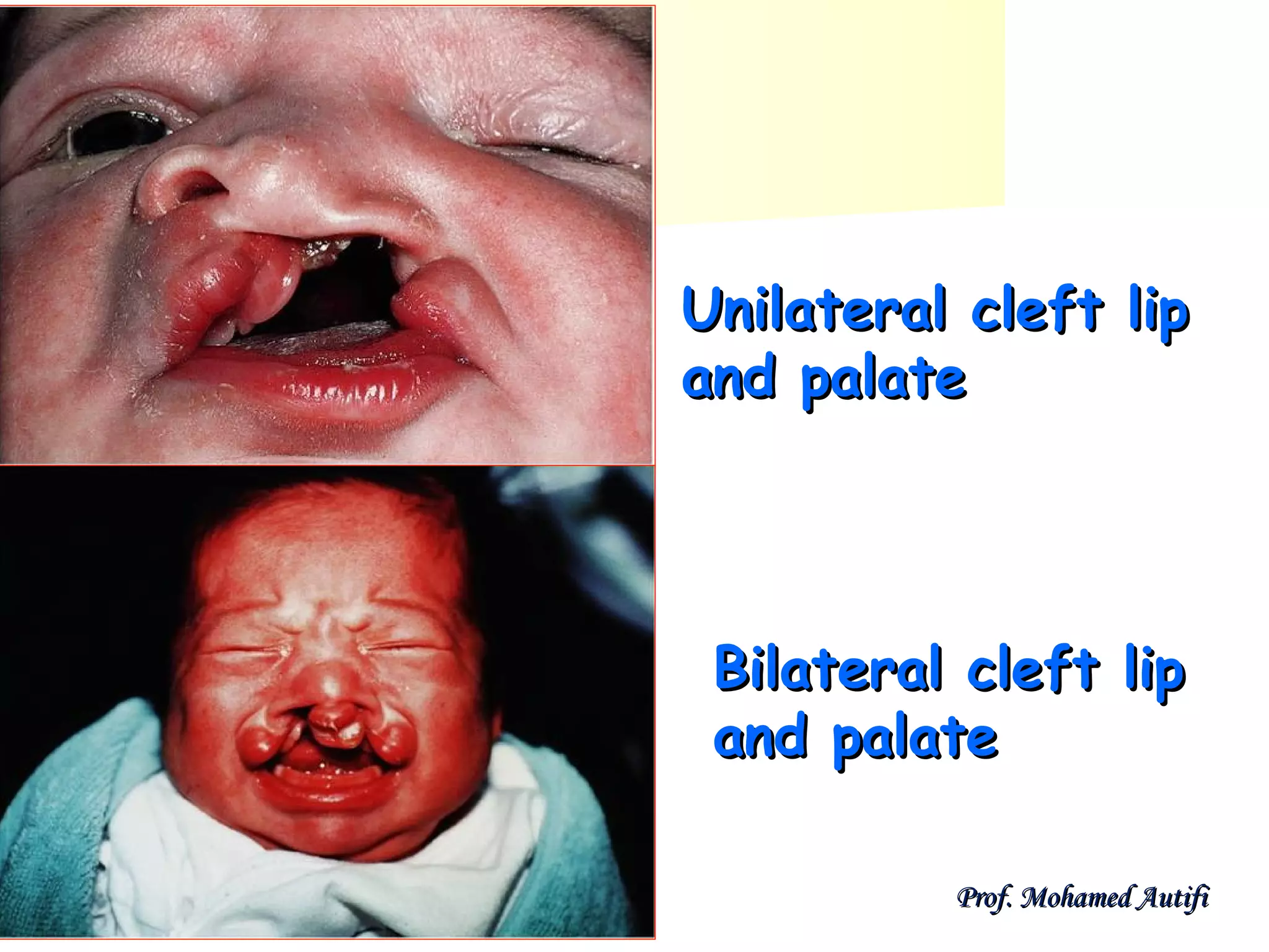
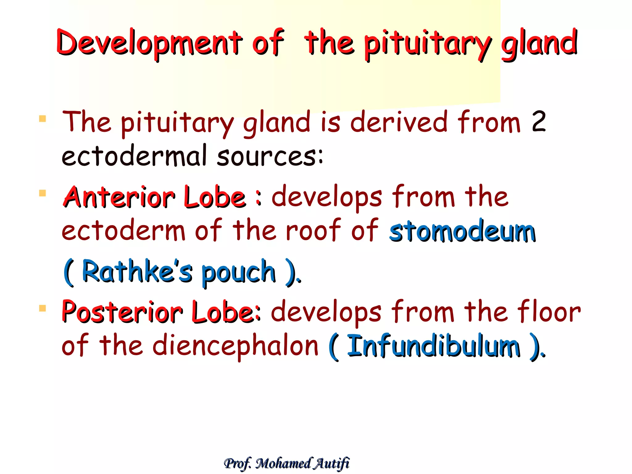
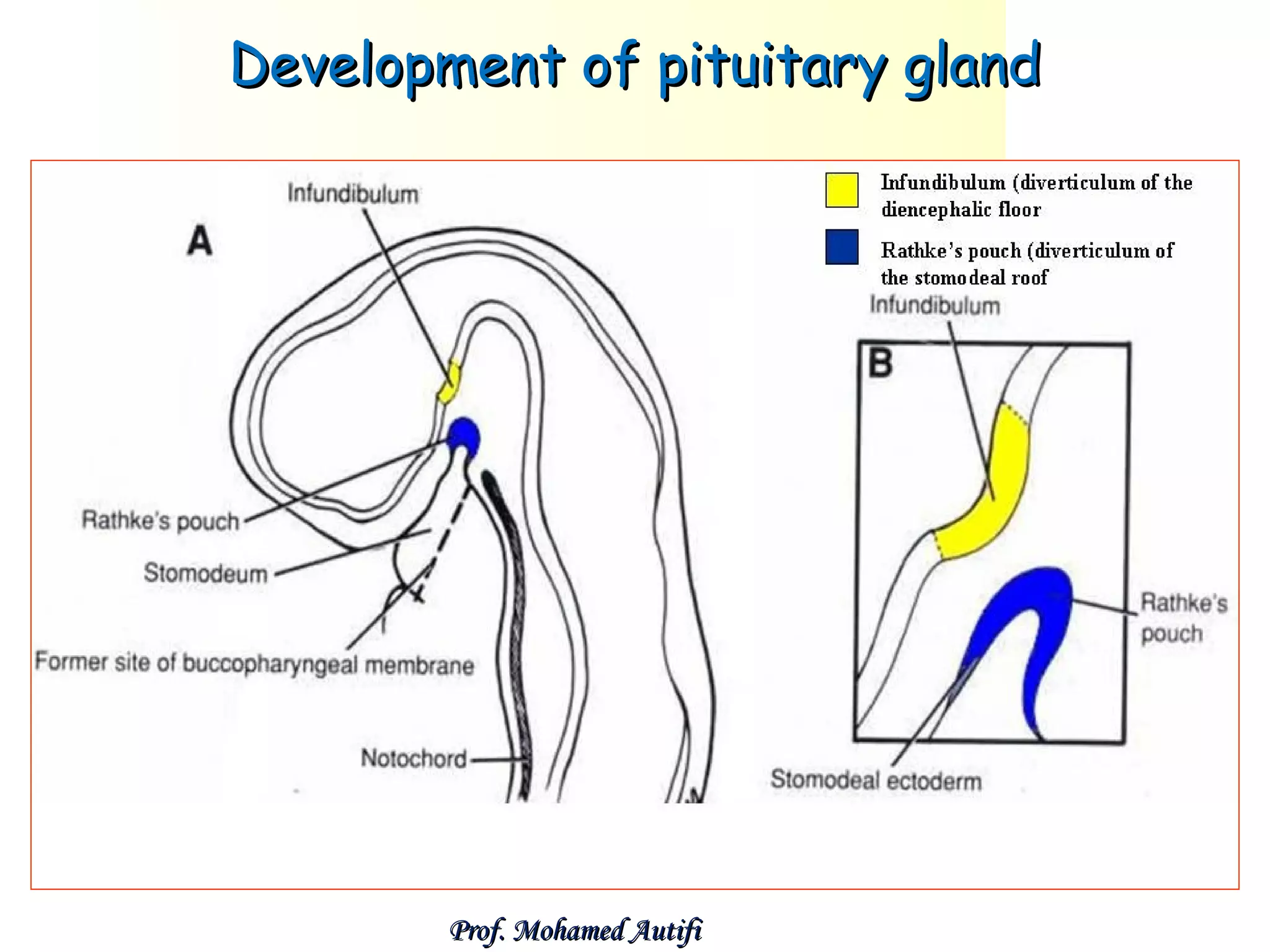
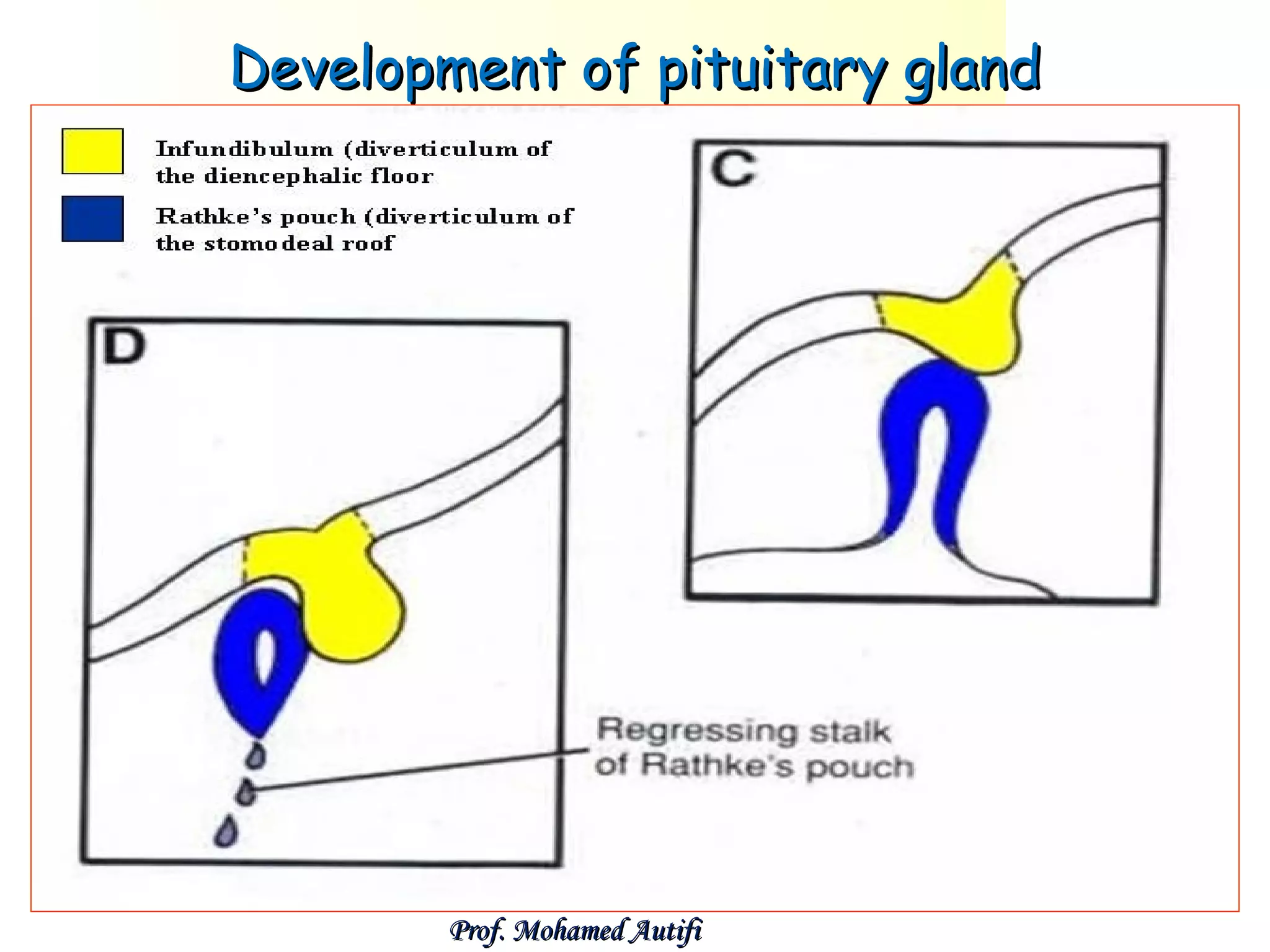
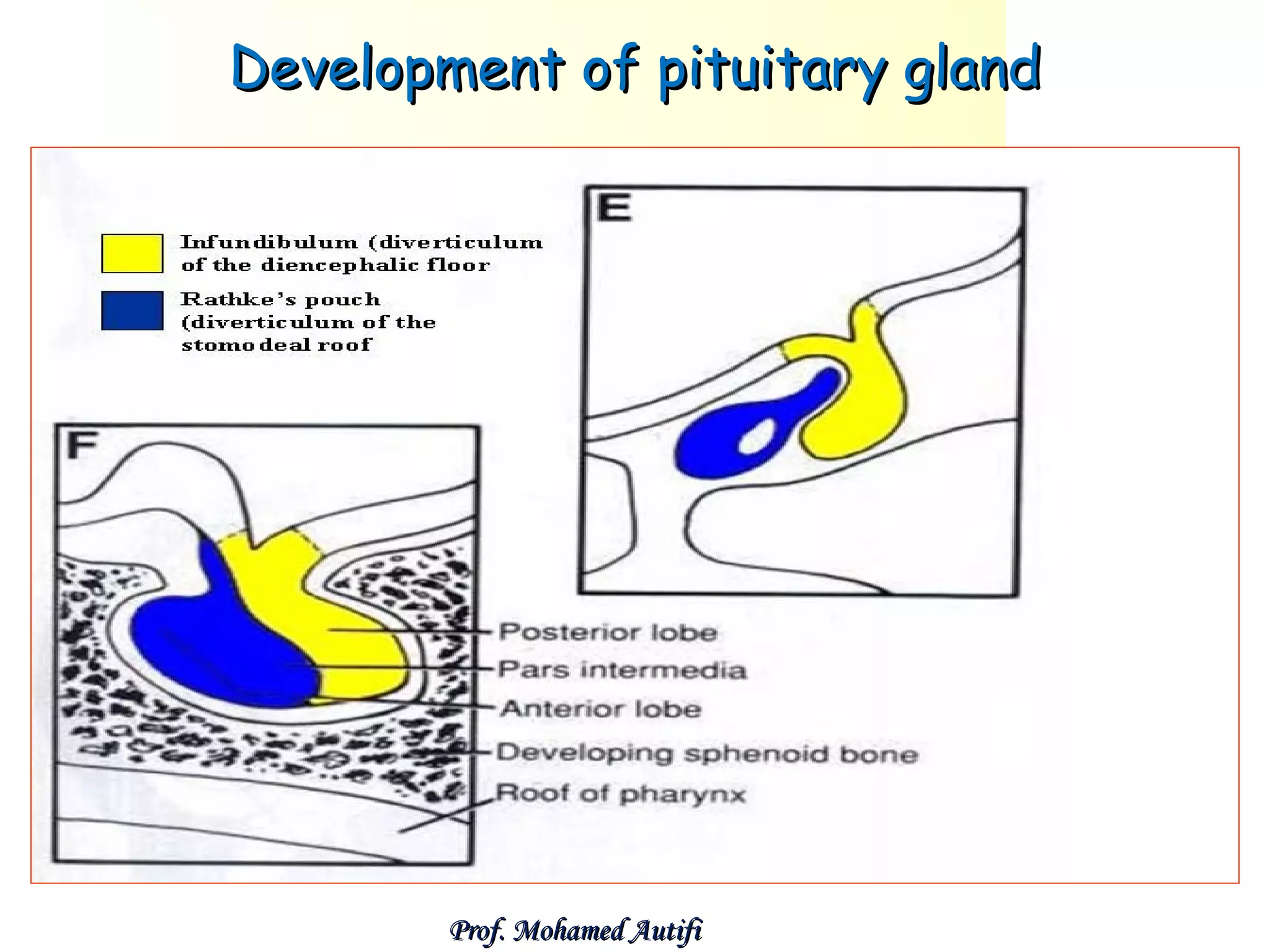
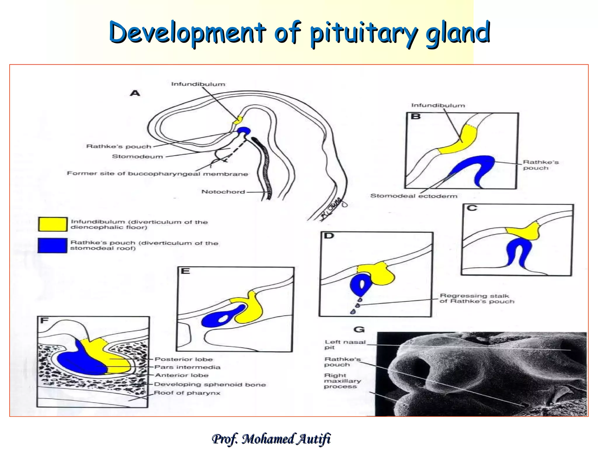
The document summarizes the development of the face, nose, palate, nasal cavities, and pituitary gland from the 4th week of development. It describes how the face develops from 5 mesenchymal prominences that surround the primitive mouth. It then discusses the development of specific structures such as the lips, salivary glands, nose, palate, and nasal cavities from these prominences. The pituitary gland is noted to develop from the ectoderm of the roof of the stomodeum and floor of the diencephalon. Some congenital anomalies are also briefly mentioned.
Central face begins to develop by 4th week, when olfactory placodes appear on both sides of the frontonasal process.
Gradually both placodes develop to form the median and lateral nasal process.
Upper lip is formed by 6th week by fusion of two median nasal processes in midline and the maxilllary process of the 1st branchial arch.
PRE-NATAL GROWTH AND DEVELOPMENT OF PALATEFormation of primary and secondary palate
Elevation of palatal shelves
Fusion of palatal shelves
Since the formation of various parts of the face involves fusion of diverse components.
Occasionally this fusion can be incomplete give rise to various anomalies
MANDIBULOFACIAL DYSOSTOSIS OR FIRST ARCH SYNDROME
- Entire first arch may remain underdeveloped on one or both sides, affecting
Lower eyelid
Maxilla
Mandible
External ear.
- Prominence of the cheek is absent
- Ear is displaced ventrally and caudally
Title: Sense of Taste
Presenter: Dr. Faiza, Assistant Professor of Physiology
Qualifications:
MBBS (Best Graduate, AIMC Lahore)
FCPS Physiology
ICMT, CHPE, DHPE (STMU)
MPH (GC University, Faisalabad)
MBA (Virtual University of Pakistan)
Learning Objectives:
Describe the structure and function of taste buds.
Describe the relationship between the taste threshold and taste index of common substances.
Explain the chemical basis and signal transduction of taste perception for each type of primary taste sensation.
Recognize different abnormalities of taste perception and their causes.
Key Topics:
Significance of Taste Sensation:
Differentiation between pleasant and harmful food
Influence on behavior
Selection of food based on metabolic needs
Receptors of Taste:
Taste buds on the tongue
Influence of sense of smell, texture of food, and pain stimulation (e.g., by pepper)
Primary and Secondary Taste Sensations:
Primary taste sensations: Sweet, Sour, Salty, Bitter, Umami
Chemical basis and signal transduction mechanisms for each taste
Taste Threshold and Index:
Taste threshold values for Sweet (sucrose), Salty (NaCl), Sour (HCl), and Bitter (Quinine)
Taste index relationship: Inversely proportional to taste threshold
Taste Blindness:
Inability to taste certain substances, particularly thiourea compounds
Example: Phenylthiocarbamide
Structure and Function of Taste Buds:
Composition: Epithelial cells, Sustentacular/Supporting cells, Taste cells, Basal cells
Features: Taste pores, Taste hairs/microvilli, and Taste nerve fibers
Location of Taste Buds:
Found in papillae of the tongue (Fungiform, Circumvallate, Foliate)
Also present on the palate, tonsillar pillars, epiglottis, and proximal esophagus
Mechanism of Taste Stimulation:
Interaction of taste substances with receptors on microvilli
Signal transduction pathways for Umami, Sweet, Bitter, Sour, and Salty tastes
Taste Sensitivity and Adaptation:
Decrease in sensitivity with age
Rapid adaptation of taste sensation
Role of Saliva in Taste:
Dissolution of tastants to reach receptors
Washing away the stimulus
Taste Preferences and Aversions:
Mechanisms behind taste preference and aversion
Influence of receptors and neural pathways
Impact of Sensory Nerve Damage:
Degeneration of taste buds if the sensory nerve fiber is cut
Abnormalities of Taste Detection:
Conditions: Ageusia, Hypogeusia, Dysgeusia (parageusia)
Causes: Nerve damage, neurological disorders, infections, poor oral hygiene, adverse drug effects, deficiencies, aging, tobacco use, altered neurotransmitter levels
Neurotransmitters and Taste Threshold:
Effects of serotonin (5-HT) and norepinephrine (NE) on taste sensitivity
Supertasters:
25% of the population with heightened sensitivity to taste, especially bitterness
Increased number of fungiform papillae
These lecture slides, by Dr Sidra Arshad, offer a quick overview of the physiological basis of a normal electrocardiogram.
Learning objectives:
1. Define an electrocardiogram (ECG) and electrocardiography
2. Describe how dipoles generated by the heart produce the waveforms of the ECG
3. Describe the components of a normal electrocardiogram of a typical bipolar lead (limb II)
4. Differentiate between intervals and segments
5. Enlist some common indications for obtaining an ECG
6. Describe the flow of current around the heart during the cardiac cycle
7. Discuss the placement and polarity of the leads of electrocardiograph
8. Describe the normal electrocardiograms recorded from the limb leads and explain the physiological basis of the different records that are obtained
9. Define mean electrical vector (axis) of the heart and give the normal range
10. Define the mean QRS vector
11. Describe the axes of leads (hexagonal reference system)
12. Comprehend the vectorial analysis of the normal ECG
13. Determine the mean electrical axis of the ventricular QRS and appreciate the mean axis deviation
14. Explain the concepts of current of injury, J point, and their significance
Study Resources:
1. Chapter 11, Guyton and Hall Textbook of Medical Physiology, 14th edition
2. Chapter 9, Human Physiology - From Cells to Systems, Lauralee Sherwood, 9th edition
3. Chapter 29, Ganong’s Review of Medical Physiology, 26th edition
4. Electrocardiogram, StatPearls - https://www.ncbi.nlm.nih.gov/books/NBK549803/
5. ECG in Medical Practice by ABM Abdullah, 4th edition
6. Chapter 3, Cardiology Explained, https://www.ncbi.nlm.nih.gov/books/NBK2214/
7. ECG Basics, http://www.nataliescasebook.com/tag/e-c-g-basics
micro teaching on communication m.sc nursing.pdfAnurag Sharma
Microteaching is a unique model of practice teaching. It is a viable instrument for the. desired change in the teaching behavior or the behavior potential which, in specified types of real. classroom situations, tends to facilitate the achievement of specified types of objectives.
Local Advanced Lung Cancer: Artificial Intelligence, Synergetics, Complex Sys...Oleg Kshivets
Overall life span (LS) was 1671.7±1721.6 days and cumulative 5YS reached 62.4%, 10 years – 50.4%, 20 years – 44.6%. 94 LCP lived more than 5 years without cancer (LS=2958.6±1723.6 days), 22 – more than 10 years (LS=5571±1841.8 days). 67 LCP died because of LC (LS=471.9±344 days). AT significantly improved 5YS (68% vs. 53.7%) (P=0.028 by log-rank test). Cox modeling displayed that 5YS of LCP significantly depended on: N0-N12, T3-4, blood cell circuit, cell ratio factors (ratio between cancer cells-CC and blood cells subpopulations), LC cell dynamics, recalcification time, heparin tolerance, prothrombin index, protein, AT, procedure type (P=0.000-0.031). Neural networks, genetic algorithm selection and bootstrap simulation revealed relationships between 5YS and N0-12 (rank=1), thrombocytes/CC (rank=2), segmented neutrophils/CC (3), eosinophils/CC (4), erythrocytes/CC (5), healthy cells/CC (6), lymphocytes/CC (7), stick neutrophils/CC (8), leucocytes/CC (9), monocytes/CC (10). Correct prediction of 5YS was 100% by neural networks computing (error=0.000; area under ROC curve=1.0).
Basavarajeeyam is an important text for ayurvedic physician belonging to andhra pradehs. It is a popular compendium in various parts of our country as well as in andhra pradesh. The content of the text was presented in sanskrit and telugu language (Bilingual). One of the most famous book in ayurvedic pharmaceutics and therapeutics. This book contains 25 chapters called as prakaranas. Many rasaoushadis were explained, pioneer of dhatu druti, nadi pareeksha, mutra pareeksha etc. Belongs to the period of 15-16 century. New diseases like upadamsha, phiranga rogas are explained.
Here is the updated list of Top Best Ayurvedic medicine for Gas and Indigestion and those are Gas-O-Go Syp for Dyspepsia | Lavizyme Syrup for Acidity | Yumzyme Hepatoprotective Capsules etc
Antimicrobial stewardship to prevent antimicrobial resistanceGovindRankawat1
India is among the nations with the highest burden of bacterial infections.
India is one of the largest consumers of antibiotics worldwide.
India carries one of the largest burdens of drug‑resistant pathogens worldwide.
Highest burden of multidrug‑resistant tuberculosis,
Alarmingly high resistance among Gram‑negative and Gram‑positive bacteria even to newer antimicrobials such as carbapenems.
NDM‑1 ( New Delhi Metallo Beta lactamase 1, an enzyme which inactivates majority of Beta lactam antibiotics including carbapenems) was reported in 2008
DISSERTATION on NEW DRUG DISCOVERY AND DEVELOPMENT STAGES OF DRUG DISCOVERYNEHA GUPTA
The process of drug discovery and development is a complex and multi-step endeavor aimed at bringing new pharmaceutical drugs to market. It begins with identifying and validating a biological target, such as a protein, gene, or RNA, that is associated with a disease. This step involves understanding the target's role in the disease and confirming that modulating it can have therapeutic effects. The next stage, hit identification, employs high-throughput screening (HTS) and other methods to find compounds that interact with the target. Computational techniques may also be used to identify potential hits from large compound libraries.
Following hit identification, the hits are optimized to improve their efficacy, selectivity, and pharmacokinetic properties, resulting in lead compounds. These leads undergo further refinement to enhance their potency, reduce toxicity, and improve drug-like characteristics, creating drug candidates suitable for preclinical testing. In the preclinical development phase, drug candidates are tested in vitro (in cell cultures) and in vivo (in animal models) to evaluate their safety, efficacy, pharmacokinetics, and pharmacodynamics. Toxicology studies are conducted to assess potential risks.
Before clinical trials can begin, an Investigational New Drug (IND) application must be submitted to regulatory authorities. This application includes data from preclinical studies and plans for clinical trials. Clinical development involves human trials in three phases: Phase I tests the drug's safety and dosage in a small group of healthy volunteers, Phase II assesses the drug's efficacy and side effects in a larger group of patients with the target disease, and Phase III confirms the drug's efficacy and monitors adverse reactions in a large population, often compared to existing treatments.
After successful clinical trials, a New Drug Application (NDA) is submitted to regulatory authorities for approval, including all data from preclinical and clinical studies, as well as proposed labeling and manufacturing information. Regulatory authorities then review the NDA to ensure the drug is safe, effective, and of high quality, potentially requiring additional studies. Finally, after a drug is approved and marketed, it undergoes post-marketing surveillance, which includes continuous monitoring for long-term safety and effectiveness, pharmacovigilance, and reporting of any adverse effects.
- Video recording of this lecture in English language: https://youtu.be/kqbnxVAZs-0
- Video recording of this lecture in Arabic language: https://youtu.be/SINlygW1Mpc
- Link to download the book free: https://nephrotube.blogspot.com/p/nephrotube-nephrology-books.html
- Link to NephroTube website: www.NephroTube.com
- Link to NephroTube social media accounts: https://nephrotube.blogspot.com/p/join-nephrotube-on-social-media.html
2. Development of the face
The face develops from 5 mesenchymal
prominences (swellings or processes) that
appear in the 4th week.
One Frontonasal prominence (formed by
proliferation of mesenchyme and ectoderm
ventral to the forebrain).
Two maxillary swellings (from 1st pharyngeal
arch).
Two mandibular swellings (from 1st pharyngeal
arch).
Prof. Mohamed Autifi
3. Development of the face
These 5 prominences surround the stomodeum
(primitive mouth) , cranially , laterally and caudally.
Prof. Mohamed Autifi
4. Mouth:
Primitive oral cavity: develops from:
a)An ectodermal depression between frontonasal
prominence and the first pharyngeal arch: the
stomodeum. Its floor is closed by the buccopharyngeal
membrane.
b)An endormal part: is the cranial end of the pharynx.
The buccal membrane degenerates during the 4 th week,
i.e. the 2 parts continue together.
Prof. Mohamed Autifi
5. Lips & gingivae:
They develop as a linear ectodermal thickenings
around the stomodeum labiogingival laminae.
They grow into mesenchyme, then degenerate
forming labiogingival grooves separating lips
from gingivae.
A small area of laminae persists in median plane
forming frenulum of the lip.
Prof. Mohamed Autifi
6. Salivary glands:
Appear as epithelial buds from oral cavity.
Parotid gland: The first to appear, early in 6th week, from oral
ectoderm, near angle of stomodeum. It forms a tube, extends into
cheek’s mesoderm.
Its Proximal part forming the parotid duct;
Its distal end breaks to form the glandular alveoli.
Capsule & connective septae develop from surrounding mesoderm.
The duct opening is carried to open inside the cheek.
Submandibular gland: Appear late in 6th week, from an endodermal
bud in floor of stomodeum (alveolo- lingual groove).
Develops in same way as parotid gland.
Sublingual gland: appear in 8th week, from multiple endodermal buds
in the alveolo-lingual groove.
Prof. Mohamed Autifi
7. I. Frontonasal process
Bilateral ectodermal thickenings above the lateral angle of
stomodeum form the nasal placodes.
By the 5th weak, the nasal placodes are invaginated to form
the nasal pits, thus the nasal placodes are divided into
medial and lateral nasal folds (promeninces).
The two medial nasal folds fuse to form median nasal fold.
Prof. Mohamed Autifi
9. Fate of the frontonasal process
The lateral nasal folds form
the alae of the nose.
The nasal pits get deeper and
they form the primitive nasal
cavities.
The medain nasal fold forms:
1. Forehaed
2. Middle of the nose and nasal
septum
3. Filtrum of the upper lip
4. Premaxilla
Prof. Mohamed Autifi
10. II. Maxillary processes
The maxillary process develops as mesodermal
proliferation from the 1st pharyngeal arch.
It grows ventrally and medially, compressing the
medial nasal folds towards the middle line and
converting them into one median nasal fold.
Prof. Mohamed Autifi
11. II. Maxillary processes
The maxillary processes are separated from the
lateral nasal folds by the nasolacrimal groove.
The lower part of the groove will form the
nasolacrimal duct while its upper part will form the
lacrimal sac.
Prof. Mohamed Autifi
12. II. Maxillary processes
A
palatine shelf arises from the medial
aspects of each maxillary process.
Both
shelves are approximated towards
each other and fuse together and with
the premaxilla forming the hard and
soft palate. Thus the nasal cavity
becomes separated from the oral cavity.
Prof. Mohamed Autifi
13. Fate of maxillary processes
1. Cheeks
2. Upper lip except the filtrum
3. Palate except the premaxilla
Prof. Mohamed Autifi
Frnotnasal
process
14. III. The mandibular processes
Develop
from the mesenchyme of the 1st
pharyngeal arch.
Fate
:
1. Lower jaw
2. Lower lip
3. Floor of the mouth
Prof. Mohamed Autifi
17. In the 6th week, the nasal pits deepen ➪ nasal sacs,
grow dorsally, separated from oral cavity by
oronasal membrane, which soon ruptures.
Both cavities continue together via primitive
choanae, dorsal to 1ry palate. Later, choanae lies bet
nasal cavity & pharynx.
Lateral wall develops 3 shelf-like projections:
Superior, middle & inferior conchae
Prof. Mohamed Autifi
21. In the roof of each cavity, the ectoderm shows
thickened patch, olfactory epithelium, forming
receptor cells, they are ciliated bipolar neurons.
Their axons form the olfactory nerves.
Paranasal air sinuses:
-They develop after birth, except maxillary
sinus which appears late in foetal life.
-They develop as diverticula of lateral nasal walls,
extend into their bones.
-They reach mature size during puberty.
Prof. Mohamed Autifi
23. Medial growth of the two maxillary processes
leading to fusion of the two medial nasal folds
in midline ➪ intermaxillary segment ➪
philtrum of upper lip, 4 incisors & 1ry palate
(premaxilla).
Prof. Mohamed Autifi
24. Early in 6th week, Two medial outgrowths
from maxillary processes called, palatine
shelves, fuse along palatine raphe forming
Secondary palate. It fuses with the 1ry
palate at the incisive foramen ➪ definitive
palate.
Anterior part of definitive palate ➪ hard
palate, while post part ➪ soft palate
Prof. Mohamed Autifi
25. Nasal septum
develops from fronto-nasal prominence & medial
nasal folds; fuses with definitive palate.
Prof. Mohamed Autifi
26. Congenital anomalies
1. Anomalies of the mouth :
Microstomia : small mouth opening
Macrostomia : large mouth opening
Agnathia : absence lower jaw
Microstomia and single nostril
Micrognathia : small lower jaw
Anodontia : absence of the teeth
2. Anomalies of the nose :
Stenosis of nostrils
Deviation of the nasal septum
Prof. Mohamed Autifi
Macrostomia
27. 3. Anomalies of the
face and palate :
1.Fusion dermoid: Cystic
swelling developed along
line of fusion of face
2. Oblique facial cleft:
Due to failure of fusion
between maxillary &
frontonasal processes
(at nasolacrimal groove).
May include cleft upper
lip.
Prof. Mohamed Autifi
28. 3. Cleft upper lip (harelip): Unilateral or
bilateral. The former is most common anomaly
of head & neck (1:1000). Due to failure of
fusion of maxillary process with medial nasal
fold.
4. Cleft lower lip: Usually central
5. Cleft uvula
Median cleft of the lower lip
Prof. Mohamed A. Autifi
29. 6. Cleft palate: (1:2500 births)
a) Anteriorly: Anterior to incisive Foramen, includes lateral
cleft lip, cleft upper jaw & cleft between 1ry & 2ry palates,
unilateral (C), bilateral (D)
b) Posteriorly: Behind incisive F, includes cleft of 2ry palate
& cleft uvula (E)
c) Antero-posteriorly: Combination, oblique/median (F)
Prof. Mohamed Autifi
37. Development of the pituitary gland
The pituitary gland is derived from 2
ectodermal sources:
Anterior Lobe : develops from the
ectoderm of the roof of stomodeum
( Rathke’s pouch ).
Posterior Lobe: develops from the floor
of the diencephalon ( Infundibulum ).
Prof. Mohamed Autifi
42. Congenital anomalies of pituitary
gland
Craniopharyngioma
Tumour formed from persistance of a
small portion of Rathk’s pouch in the
roof of the pharynx.
Prof. Mohamed Autifi