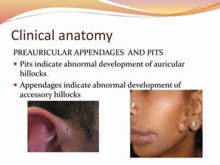
INTRODUCTION
 Anatomical unit serving both hearing and equilibrium
 In the embryo
1. the external ear
2. the middle ear
3. the inner ear
INTERNAL EAR
Internal ear
 Approximately 22 days – first indication
 Optic placodes invaginate and form otic or auditory
vesicles
 Each ventricle divides into
1. ventral component - saccule and cochlear duct
2. dorsal component – utricle, semicircular canals and
endolymphatic duct
Saccule, Cochlea and Organ of corti
 Tubular outpocketing at its lower pole
 It penetrates the surrounding mesenchyme in spiral
fashion. – 8 weeks, 2.5 turns.
 Differentiation into cartilage.
 Scala vestibuli and scala tympani are formed
 Vestibular memrane and basilar membrane
Saccule, Cochlea and Organ of corti
 Lateral wall – spiral ligament – surrounding cartilage
 Median angle – modiolus, the future axis of the bony
cochlear
 Inner ridge – the future spiral limbus
 Outer ridge – hair cells, the sensory cells of the
auditory system.
Organ of corti
 Sensory cells + tectorial membrane
 Impulses are transmitted to the spiral ganglion and
then to the nervous system by the auditory fibres
cranial nerve VIII
Utricle and semicircular canals
 During sixth week of development, semicircular canals
appear as flattened outpocketings of the reticular part
of the otic vesicle.
 Cels in ampulla – crista ampullaris containing sensory
cells for maintenance of equilibrium.
 Maculae acusticae develop in the walls of the utricle
and saccule.
Utricle and semicircular canals
 Impulses are carried to the brain by vestibular fibres of
cranial nerve VIII
 Statoacoustic ganglion – small group of cells breaks
away from its wall.
 Ganglion splits into cochlear and vestibular portions.
MIDDLE EAR
TYMPANIC CAVITY AND AUDITORY MEATUS
 1st pharyngeal pouch
 Contact with 1st pharyngeal cleft
 Distal part – primitive tympanic cavity
 Proximal part – auditory tube
Ossicles
 Malleus and incus - 1st pharyngeal pouch
 Stapes - 2nd pharyngeal pouch
 Remain embedded in the mesenchyme until 8th
month
 Tympanic cavity becomes twice its original size
 Endoderm epithelium connects ossicles to the wall of
the cavity in mesentary like fashion.
 Supporting ligaments of ossicles develop later.
Ossicles
 During late fetal life, by vacuolization of surrounding
tissue form tymapnic antrum.
 After birth – pneumatization
 Later, most of the mastoid air sinus come in contact
with the antrum and tympanic cavity – middle ear
infection.
EXTERNAL EAR
EXTERNAL AUDITORY MEATUS
 1st pharyngeal cleft
 At the beginning of 3rd month – medial plug is formed
 In the 7th month – plug dissolves and epithelial lining
of floor help in definitive eardrum formation
Eardrum or tympanic membrane
 Eardrum consists of
a) An ectodermal epithelial lining
b) An intermediate layer of mesenchyme
c) An endodermal lining which form the fibrous
strature.
Auricle
 Six mesenchymal proliferations
 These swellings, auricular hillocks later fuse and form
the definitive auricle.
 Developmental abnormalities of the auricle are
common
 Initially the external ears are in the lowerneck region
but later ascend to the side of the head at the level of
eye.
Clinical anatomy
CONGENITAL DEAFNESS
 Usually associated with deaf mutism
Clinical anatomy
EXTERNAL EAR DEFECTS
 They are common, they include minor and severe
abnormalities
Clinical anatomy
PREAURICULAR APPENDAGES AND PITS
 Pits indicate abnormal development of auricular
hillocks
 Appendages indicate abnormal development of
accessory hillocks
Development of ear

Development of ear

  • 2.
    INTRODUCTION  Anatomical unitserving both hearing and equilibrium  In the embryo 1. the external ear 2. the middle ear 3. the inner ear
  • 3.
  • 4.
    Internal ear  Approximately22 days – first indication  Optic placodes invaginate and form otic or auditory vesicles  Each ventricle divides into 1. ventral component - saccule and cochlear duct 2. dorsal component – utricle, semicircular canals and endolymphatic duct
  • 5.
    Saccule, Cochlea andOrgan of corti  Tubular outpocketing at its lower pole  It penetrates the surrounding mesenchyme in spiral fashion. – 8 weeks, 2.5 turns.  Differentiation into cartilage.  Scala vestibuli and scala tympani are formed  Vestibular memrane and basilar membrane
  • 7.
    Saccule, Cochlea andOrgan of corti  Lateral wall – spiral ligament – surrounding cartilage  Median angle – modiolus, the future axis of the bony cochlear  Inner ridge – the future spiral limbus  Outer ridge – hair cells, the sensory cells of the auditory system.
  • 8.
    Organ of corti Sensory cells + tectorial membrane  Impulses are transmitted to the spiral ganglion and then to the nervous system by the auditory fibres cranial nerve VIII
  • 9.
    Utricle and semicircularcanals  During sixth week of development, semicircular canals appear as flattened outpocketings of the reticular part of the otic vesicle.  Cels in ampulla – crista ampullaris containing sensory cells for maintenance of equilibrium.  Maculae acusticae develop in the walls of the utricle and saccule.
  • 11.
    Utricle and semicircularcanals  Impulses are carried to the brain by vestibular fibres of cranial nerve VIII  Statoacoustic ganglion – small group of cells breaks away from its wall.  Ganglion splits into cochlear and vestibular portions.
  • 12.
    MIDDLE EAR TYMPANIC CAVITYAND AUDITORY MEATUS  1st pharyngeal pouch  Contact with 1st pharyngeal cleft  Distal part – primitive tympanic cavity  Proximal part – auditory tube
  • 13.
    Ossicles  Malleus andincus - 1st pharyngeal pouch  Stapes - 2nd pharyngeal pouch  Remain embedded in the mesenchyme until 8th month  Tympanic cavity becomes twice its original size  Endoderm epithelium connects ossicles to the wall of the cavity in mesentary like fashion.  Supporting ligaments of ossicles develop later.
  • 14.
    Ossicles  During latefetal life, by vacuolization of surrounding tissue form tymapnic antrum.  After birth – pneumatization  Later, most of the mastoid air sinus come in contact with the antrum and tympanic cavity – middle ear infection.
  • 15.
    EXTERNAL EAR EXTERNAL AUDITORYMEATUS  1st pharyngeal cleft  At the beginning of 3rd month – medial plug is formed  In the 7th month – plug dissolves and epithelial lining of floor help in definitive eardrum formation
  • 16.
    Eardrum or tympanicmembrane  Eardrum consists of a) An ectodermal epithelial lining b) An intermediate layer of mesenchyme c) An endodermal lining which form the fibrous strature.
  • 17.
    Auricle  Six mesenchymalproliferations  These swellings, auricular hillocks later fuse and form the definitive auricle.  Developmental abnormalities of the auricle are common  Initially the external ears are in the lowerneck region but later ascend to the side of the head at the level of eye.
  • 18.
    Clinical anatomy CONGENITAL DEAFNESS Usually associated with deaf mutism
  • 19.
    Clinical anatomy EXTERNAL EARDEFECTS  They are common, they include minor and severe abnormalities
  • 20.
    Clinical anatomy PREAURICULAR APPENDAGESAND PITS  Pits indicate abnormal development of auricular hillocks  Appendages indicate abnormal development of accessory hillocks