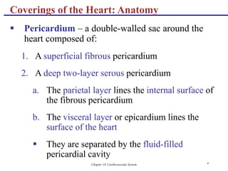
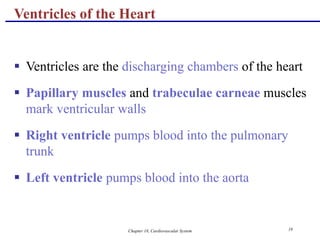
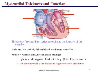
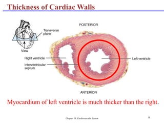
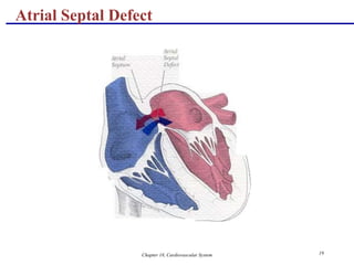
Download to read offline
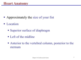
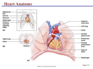
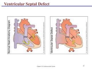
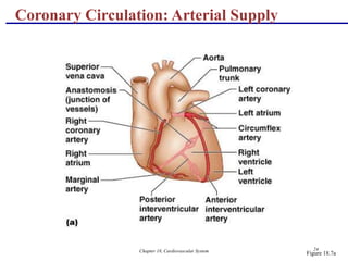
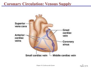
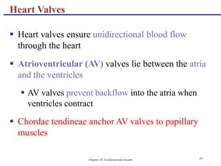
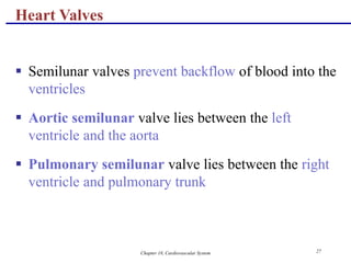
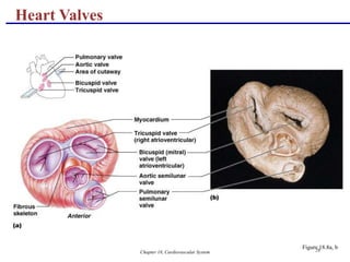
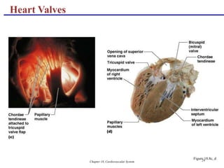
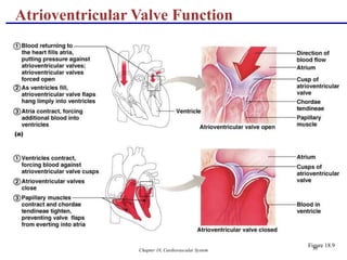
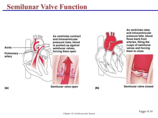
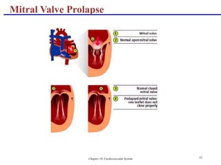
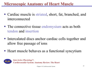
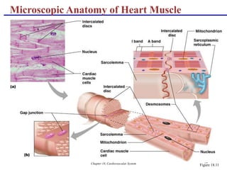

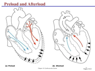


The document describes the anatomy and physiology of the cardiovascular system, with a focus on the heart. It discusses the internal and external structures of the heart, including its coverings, chambers, valves, vessels and blood supply. It also explains the cardiac cycle, electrical conduction system, heart sounds, and how the heart pumps blood through the lungs and body in a continuous loop. The document contains diagrams labeling the various parts of the heart and depicting the flow of blood and electrical impulses.