This document provides an outline for electrocardiogram (ECG or EKG) interpretation. It begins with an introduction to ECGs and their purpose. It then discusses ECG physiology, the different types of ECG tests, indications for ECGs, how to record an ECG, and an 8-step approach to ECG interpretation. Finally, it covers common ECG abnormalities and their significance. The overall document serves as a comprehensive guide for understanding ECGs, from the basics of what they measure to interpreting tracings and recognizing abnormal findings.
ECG Lecture: Sinus arrest, sinoatrial exit block, AV block and escape rhythmsMichael-Joseph Agbayani
Simple ECG lecture about sinus arrest, sinoatrial exit block, AV block and escape rhythms. Slideshow was made with an audience of medical professionals in mind.
Tachy Arrhythmias - Approach to ManagementArun Vasireddy
Tachyarrhythmias are disorders of heart rhythm which may present with a tachycardia i.e. a heart rate >100 bpm.
This article provides an overview of tachyarrhythmias in general and goes on to cover the most common tachyarrhythmias in more detail. The acute management of tachyarrhythmias, in an emergency setting, will be covered in the 'Acute' section of the fastbleep website.
Tachyarrhythmias are clinically important as they can precipitate cardiac arrest, cardiac failure, thromboembolic disease and syncopal events. As such, they crop up time and time again in exam papers and on the wards.
Tachyarrhythmias are classified based on whether they have broad or narrow QRS complexes on the ECG. Broad is defined as >0.12s (or more than 3 small squares on the standard ECG). Narrow is equal to or less than 0.12s. Broad QRS complexes are slower ventricular depolarisations that arise from the ventricles. Narrow complexes are ventricular depolarisations initiated from above the ventricles (known as supraventricular). One important exception is when there is a supraventricular depolarisation conducted through a diseased AV node. This will produce wide QRS complexes despite the rhythm being supraventricular in origin.
crème de la crème basics to understand electrocardiographic analysis in an easy & simple way with some specifications to its use in Emergency medicine/clinical toxicology practice.
crème de la crème basics to understand electrocardiographic analysis in an easy & simple way with some specifications to its use in Emergency medicine/clinical toxicology practice.
Instructions for Submissions thorugh G- Classroom.pptxJheel Barad
This presentation provides a briefing on how to upload submissions and documents in Google Classroom. It was prepared as part of an orientation for new Sainik School in-service teacher trainees. As a training officer, my goal is to ensure that you are comfortable and proficient with this essential tool for managing assignments and fostering student engagement.
Synthetic Fiber Construction in lab .pptxPavel ( NSTU)
Synthetic fiber production is a fascinating and complex field that blends chemistry, engineering, and environmental science. By understanding these aspects, students can gain a comprehensive view of synthetic fiber production, its impact on society and the environment, and the potential for future innovations. Synthetic fibers play a crucial role in modern society, impacting various aspects of daily life, industry, and the environment. ynthetic fibers are integral to modern life, offering a range of benefits from cost-effectiveness and versatility to innovative applications and performance characteristics. While they pose environmental challenges, ongoing research and development aim to create more sustainable and eco-friendly alternatives. Understanding the importance of synthetic fibers helps in appreciating their role in the economy, industry, and daily life, while also emphasizing the need for sustainable practices and innovation.
The Indian economy is classified into different sectors to simplify the analysis and understanding of economic activities. For Class 10, it's essential to grasp the sectors of the Indian economy, understand their characteristics, and recognize their importance. This guide will provide detailed notes on the Sectors of the Indian Economy Class 10, using specific long-tail keywords to enhance comprehension.
For more information, visit-www.vavaclasses.com
Unit 8 - Information and Communication Technology (Paper I).pdfThiyagu K
This slides describes the basic concepts of ICT, basics of Email, Emerging Technology and Digital Initiatives in Education. This presentations aligns with the UGC Paper I syllabus.
Operation “Blue Star” is the only event in the history of Independent India where the state went into war with its own people. Even after about 40 years it is not clear if it was culmination of states anger over people of the region, a political game of power or start of dictatorial chapter in the democratic setup.
The people of Punjab felt alienated from main stream due to denial of their just demands during a long democratic struggle since independence. As it happen all over the word, it led to militant struggle with great loss of lives of military, police and civilian personnel. Killing of Indira Gandhi and massacre of innocent Sikhs in Delhi and other India cities was also associated with this movement.
How to Split Bills in the Odoo 17 POS ModuleCeline George
Bills have a main role in point of sale procedure. It will help to track sales, handling payments and giving receipts to customers. Bill splitting also has an important role in POS. For example, If some friends come together for dinner and if they want to divide the bill then it is possible by POS bill splitting. This slide will show how to split bills in odoo 17 POS.
Model Attribute Check Company Auto PropertyCeline George
In Odoo, the multi-company feature allows you to manage multiple companies within a single Odoo database instance. Each company can have its own configurations while still sharing common resources such as products, customers, and suppliers.
We all have good and bad thoughts from time to time and situation to situation. We are bombarded daily with spiraling thoughts(both negative and positive) creating all-consuming feel , making us difficult to manage with associated suffering. Good thoughts are like our Mob Signal (Positive thought) amidst noise(negative thought) in the atmosphere. Negative thoughts like noise outweigh positive thoughts. These thoughts often create unwanted confusion, trouble, stress and frustration in our mind as well as chaos in our physical world. Negative thoughts are also known as “distorted thinking”.
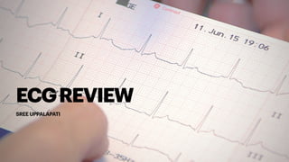
4. INTRODUCTION
• ‘ECG’ stands for electrocardiogram, or electrocardiograph. In some countries, the
abbreviation used is ‘EKG’.
• An ECG is a paper recording of the electrical activity. It records where electrical
impulses start and how they
f
low through the heart. It does not measure how well the
heart is pumping.
• It is the most important test for interpretation of the cardiac rhythm, conduction
system abnormalities, and the detection of myocardial ischemia.
5. TYPESOFECG
• 12-lead ECG provides 12 displays that are derived by using 10 electrodes. It is considered a
resting ECG, so patients are simply required to lay down, or sit up for the duration of the test.
• Exercise ECG also called a stress test, monitors the heart’s capabilities and activity under
physically demanding conditions, such as exercise. This test is performed with the patient
attached to an ECG and asked to walk on a treadmill or pedal on a stationary bike.
• Pharmacological stress test is indicated for patients who cannot exercise adequately
• Holter Monitor (portable ECG) is an option for those who need to be monitored for an
extended period of time. Using adhesive-backed electrodes connected to a monitor (which
can be strapped to a waistband)
• This device will record any irregularities that may not be picked up during shorter ECG
tests.
6. PHYSIOLOGY
• Think of the heart as having internal
wiring. The internal pacemaker is
the sinoatrial node situated in the
right atrium.
• In a normal heart, the sinoatrial
node
f
ires regularly and the
electrical impulse spreads through
an anatomical path to the ventricles
resulting in ventricular contraction.
The ventricular contraction is felt as
the pulse or the heartbeat.
• Each heartbeat is represented by
one ECG complex.
7. • An ECG complex is composed of
f
ive parts
• P wave represents electrical activation, called depolarization, of the
atrial muscle.
• PR interval is the time taken for the electrical impulse to spread from
the atria to the ventricles through the atrioventricular node and the
high-speed conducting pathway called the bundle of His.
• QRS complex records the impulse spreading throughout the ventricles
resulting in ventricular contraction. In the normal heart, this does not
take more than 3 small squares on an ECG.
• ST segment is the period when the ventricles are completely activated.
• T wave is the return (repolarization) of the ventricular muscle to its
resting electrical state.
• A normal beat is represented by one P wave followed by one QRS complex
and then one T wave.
PHYSIOLOGY
8. INDICATIONS
• Chest pain
• Palpitations
• Breathlessness
• Dizziness
• Episode of syncope (blackout)
• Unexplained fall
• Stroke or a transient ischemic attack (TIA)
• Remember that the patient's symptoms and physical signs will guide interpretation of the
ECG.
9. • Electrodes are placed on the chest and
limbs of the patient to record di
ff
erent views
of the heart's electrical activity.
• One electrode is attached to each limb.
These four electrodes provide six ‘limb
leads’ or six di
ff
erent views of the heart in a
vertical plane.
• These are called leads I, II, III, aVL, aVF
and aVR.
• Six electrodes are attached to the chest,
recording leads V 1 to V 6
RECORDINGANECG
11. • If a patient has a regular heart rhythm their heart rate
can be calculated using the following method:
• Count the number of large squares present within
one R-R interval.
• Divide 300 by this number to calculate heart rate.
• If a patient’s heart rhythm is irregular
• Count the number of complexes on the rhythm strip
(each rhythm strip is typically 10 seconds long).
• Multiply the number of complexes by 6 (giving you
the average number of complexes in 1 minute).
• Dissociated rhythms require independent (atrial/
ventricular) rates determined
STEP1:RATE
Sinus Rhythm: 60-100 bpm
Sinus Tachycardia: > 100 bpm
Sinus Bradycardia: < 60 bpm
12. STEP2:RHYTHM
• A patient’s heart rhythm can be regular or irregular.
• A regular rhythm means there is the same number of squares between each QRS
complex
• Irregular rhythms can be either:
• Regularly irregular (i.e. a recurrent pattern of irregularity)
• Irregularly irregular (i.e. completely disorganised)
• Variable number of squares between each QRS complex
• Mark out several consecutive R-R intervals on a piece of paper, then move them along the
rhythm strip to check if the subsequent intervals are similar.
13.
14. STEP3:AXIS
• Cardiac axis describes the overall direction of electrical spread within the heart.
• Normal cardiac axis: Lead II has the most positive de
f
lection compared to leads I and III.
• Left axis deviation: Lead I has the most positive de
f
lection, Leads II and III are negative.
• Right axis deviation: Lead III has the most positive de
f
lection and lead I should be
negative.
• Right axis deviation is associated with right ventricular hypertrophy.
15.
16. • look at the P waves and answer the following questions:
1. Are P waves present?
• If absent and there is an irregular rhythm it may suggest
a diagnosis of A. Fib
2. P wave followed by a QRS complex?
• If there is more than one P wave before each QRS
complex, then conduction to the ventricles is abnormal.
This is called heart block (AV block).
3. Do the P waves look normal? – check duration, direction
and shape
4. If P waves are absent, is there any atrial activity?
• Sawtooth baseline →
f
lutter waves
• Chaotic baseline →
f
ibrillation waves
• Flat line → no atrial activity at all
STEP4:PWAVE
17. Atrial
f
ibrillation-
Chaotic and erratic baseline with no discrete P waves in
between irregularly spaced QRS complexes.
Atrial
f
lutter- rapid succession of identical, back-to-
back atrial depolarization waves (Sawtooth).
Ventricular
f
ibrillation-A completely erratic rhythm
with no identi
f
iable waves.
18. STEP5:INTERVALS- PR INTERVAL
• PR interval should be between 120-200 ms (3-5 small squares).
• Short PR intervals may suggest of Wol
ff
-Parkinson-White syndrome
• Long PR interval suggests the presence of AV block (heart block)
• First-degree AV block involves a
f
ixed prolonged PR interval (>200 ms).
19. STEP5:INTERVALS- PR INTERVAL
• Second-degree AV block (Mobitz type 1 or Wenckebach phenomenon) is progressive
lengthening of PR interval until a beat is “dropped” (a P wave not followed by a QRS
complex).
• Second-degree (type 2) AV block has dropped beats that are not preceded by a change
in the length of the PR interval (as in type I).
• Third-degree (complete) AV block is when the atria and ventricles beat independently
of each other. P waves and QRS complexes not rhythmically associated (Atrial rate >
ventricular rate).
21. STEP5:INTERVALS- QT INTERVAL
• The QT interval is dependent upon the heart rate; it is shorter at faster heart rates and
longer when the rate is slower. Thus, a QT interval that is corrected for heart rate (QTc)
has been classically calculated based on Bazett's widely used formula:
• QTc = QT interval / square root of the RR interval (in seconds)
• Normal value for the QTc in men is usually given as about ≤440 ms and in women as
about ≤450 to 460 ms.
• Prolonged QTc causes premature action potentials during the late phases of
depolarization. This increases the risk of developing ventricular arrhythmias, including
fatal ventricular
f
ibrillation.
• It is also associated with Torsades de Pointes
22. DRUGINDUCEDLONGQT
• Prolongation of the QT interval may be due to an adverse drug reaction (ABCDEF):
• AntiArrhythmics (class IA, III)
• AntiBiotics (eg, macrolides,
f
luoroquinolones)
• Anti“C”ychotics (eg, haloperidol, ziprasidone)
• AntiDepressants (eg, TCAs)
• AntiEmetics (eg, ondansetron)
• AntiFungals (eg, azoles)
23. STEP6:QRSCOMPLEX
• When assessing a QRS complex, you need to pay attention to the following
characteristics:
• Width
• A narrow (< 0.12 seconds) QRS complex occurs when the impulse is conducted down
the bundle of His and the Purkinje
f
ibre to the ventricles.
• A broad (> 0.12 seconds) QRS complex occurs if there is an abnormal depolarization
sequence – for example, a bundle branch block because the impulse gets to one
ventricle rapidly down the intrinsic conduction system then has to spread slowly
across the myocardium to the other ventricle.
24. STEP6:QRSCOMPLEX
• Height
• Small complexes are de
f
ined as < 5mm in the limb leads or < 10 mm in the chest
leads.
• Tall complexes imply ventricular hypertrophy (although can be due to body habitus
e.g. tall slim people).
• The Sokolow-Lyon index: S wave in V1 + R wave in V5 or V6 (whichever is larger) ≥
35 mm (≥ 7 large squares); R wave in aVL ≥ 11 mm
25. • Morphology
• To assess morphology, you need to
assess the individual waves of the QRS
complex.
• Q-waves
• Isolated Q waves can be normal.
• A pathological Q wave is > 25% the size of
the R wave that follows it or > 2mm in
height and > 40ms in width.
STEP6:QRSCOMPLEX
26. • R and S waves
• Assess the R wave progression across the
chest leads (from small in V1 to large in
V6).
• The transition from S > R wave to R > S
wave should occur in V3 or V4.
• Poor progression (i.e. S > R through to
leads V5 and V6) can be a sign of
previous MI but can also occur in very
large people due to poor lead position.
STEP6:QRSCOMPLEX
27. STEP6:QRSCOMPLEX
• J point segment is where the S wave joins the ST segment.
• This point can be elevated resulting in the ST segment that follows it also being raised
(this is known as “high take-o
ff
”).
• Delta wave is a sign that the ventricles are being activated earlier than normal from a
point distant to the AV node. The early activation then spreads slowly across the
myocardium causing the slurred upstroke of the QRS complex.
28. STEP7:STSEGMENT-TWAVE
• ST segment is the part of the ECG between the end of the S wave and the start of the T
wave.
• In a healthy individual, it should be an isoelectric line (neither elevated nor
depressed).
• ST-elevation is signi
f
icant when it is greater than 1 mm (1 small square) in 2 or more
contiguous limb leads or >2mm in 2 or more chest leads.
• It is most commonly caused by acute full-thickness myocardial infarction.
• ST depression ≥ 0.5 mm in ≥ 2 contiguous leads indicates myocardial ischaemia.
29. • Tall T wave
• T waves are considered tall if they are > 5mm in the limb
leads and > 10mm in the chest leads (the same criteria as
‘small’ QRS complexes)
• Tall T waves can be associated with: Hyperkalaemia (“tall
tented T waves”) & Hyperacute STEMI
• Inverted T waves are normal in V1 and lead III
• Inverted T waves in other leads are a nonspeci
f
ic sign of a
wide variety of conditions such as Ischemia, Bundle
branch blocks (V4-6 in LBBB and V1-V3 in RBBB)
• Pulmonary embolism, Left ventricular hypertrophy (in the
lateral leads), Hypertrophic cardiomyopathy (widespread).
STEP7:STSEGMENT-T
WAVE
30. BiphasicTwaves have two peaks and can be
indicative of ischaemia and hypokalaemia.
FlattenedTwaves are a non-speci
f
ic sign, that may
represent ischaemia or electrolyte imbalance.
31. • U waves are not a common
f
inding.
• The U wave is a > 0.5mm de
f
lection after the
T wave best seen in V2 or V3.
• These become larger the slower the
bradycardia – classically U waves are seen in
various electrolyte imbalances,
hypothermia and secondary to
antiarrhythmic therapy (such as digoxin,
procainamide or amiodarone).
UWAVE
33. STEP8:OVERALLINTERPRETATION
• Only after the prior steps have been completed should an overall description be given,
followed by an interpretation and possible diagnoses.
• For instance, the description may state that the rate is rapid and irregular with no P
waves and ST elevation in leads II, III, and aVF with ST depression in leads I, aVL, and
V4-6.
• The interpretation would be that there is rapid atrial
f
ibrillation and an inferior ST-
elevation myocardial infarction. This ensures assimilation of all information in the ECG
and that no detail will be overlooked.
35. ECGREDFLAGS
• The following ECG abnormalities could be clinically important, but always consider the
patients' clinical state
f
irst. Any of these changes could present as chest pain,
breathlessness, palpitations or collapse.
• Ventricular rate above 120bpm or below 45bpm
• Atrial
f
ibrillation
• Complete heart block
• ST segment elevation or depression
• Abnormal T wave inversion
• Wide QRS width
36. • S1Q3T3 – wide S in I, large Q and inverted T
in III
• Acute Right BBB (transient, often
incomplete)
• R.A.D. and rightward rotation (horizontal
plane)
• Inverted T waves V1 ➞ V4 and ST depression
in II
PULMONARY
EMBOLISM
37. • Modern arti
f
icial pacemakers have sensing
capabilities and also provide a regular pacing
stimulus. This electrical stimulus records on
EKG as a tiny vertical spike that appears just
before the “captured” cardiac response.
• triggered (activated) when the patient’s own
rhythm ceases or slows markedly.
• inhibited (cease pacing) if the patient’s own
rhythm resumes at a reasonable rate.
• reset pacing (at same rate) to synchronize
with a premature beat.
ARTIFICIAL
PACEMAKERS
38. HEREDITARYCHANNELOPATHIES
• These are inherited mutations of cardiac ion channels leading to abnormal myocardial
action potential which increases risk of ventricular tachyarrhythmias and sudden cardiac
death (SCD).
• Brugada syndrome (loss of function mutation of Na+
channels.)- Autosomal dominant.
• ECG pattern of pseudo-right bundle branch block and ST-segment elevations in leads
V1-V2.
• Congenital long QT syndrome (loss of function mutation of K+
channels)
• Romano-Ward syndrome- autosomal dominant, pure cardiac phenotype (no deafness).
• Jervell and Lange-Nielsen syndrome- autosomal recessive, sensorineural deafness.
39. • Most common type of ventricular pre-
excitation syndrome.
• Abnormal fast accessory conduction
pathway from atria to ventricle (bundle of
Kent) bypasses the rate-slowing AV node.
• Ventricles begin to partially depolarize
earlier characteristic delta wave with
widened QRS complex and shortened PR
interval on ECG.
• May result in reentry circuit supraventricular
tachycardia.
WOLFF-PARKINSON-
WHITESYNDROME
40. • Polymorphic ventricular tachycardia,
characterized by shifting sinusoidal
waveforms on ECG; can progress to
ventricular
f
ibrillation.
• Long QT interval predisposes to torsades de
pointes.
• Caused by drugs, hypokalemia+
, decreased
Mg, Hypocalcemia, congenital abnormalities.
• Treatment includes magnesium sulfate.
TORSADESDEPOINTES
41. • Hyperkalemia
• Wide QRS and peaked T waves on ECG
• Hypokalemia
• U waves and
f
lattened T waves on ECG,
arrhythmias
• Hypercalcemia- Short QT
• Hypocalcemia- QT prolongation
ELECTROLYTE
ABNORMALITIES
43. REFERENCES
•DR MATTHEW JACKSON·DATA INTERPRETATION·LAST UPDATED:JULY 23, 2022. (2022, JULY
23). HOW TO READ AN ECG: ECG INTERPRETATION: EKG. GEEKY MEDICS. RETRIEVED
OCTOBER 6, 2022, FROM HTTPS://GEEKYMEDICS.COM/HOW-TO-READ-AN-ECG/
•DUBIN, D. (N.D.). RAPID INTERPRETATION OF EKG’S.
•ECG TUTORIAL: BASIC PRINCIPLES OF ECG ANALYSIS. UPTODATE. (N.D.). RETRIEVED
OCTOBER 6, 2022, FROM HTTPS://WWW.UPTODATE.COM/CONTENTS/ECG-TUTORIAL-
BASIC-PRINCIPLES-OF-ECG-ANALYSIS?SOURCE=HISTORY_WIDGET
•HAMPTON, J. R., ADLAM, D., & HAMPTON, J. R. (2019). THE ECG MADE PRACTICAL. ELSEVIER.