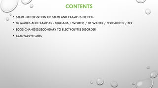
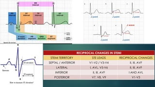
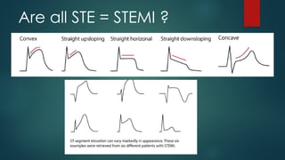
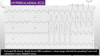
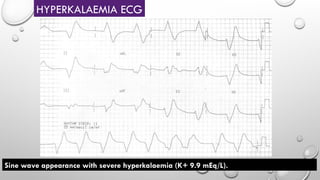
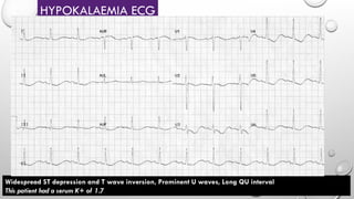
ECG RECOGNITION : ECGS THAT
YOU MUST NOT MISSED
PRESENTED BY
DR MOHD HABROL AFZAM BIN ABD WAHAB
SUPERVISED BY
NORASLAWATI BINTI RAZAK
CONTENTS
• STEMI : RECOGNITION OF STEMI AND EXAMPLES OF ECG
• MI MIMICS AND EXAMPLES : BRUGADA / WELLENS / DE WINTER / PERICARDITIS / BER
• ECGS CHANGES SECONDARY TO ELECTROLYTES DISORDER
• BRADYARRYTHMIAS
ST ELEVATION MYOCARDIAC
INFARCTION
RECIPROCAL CHANGES IN STEMI
STEMI TERRITORY STE LEADS RECIPROCAL CHANGES
SEPTAL / ANTERIOR V1-V2 / V3-V4 II, III, AVF
LATERAL I, AVL, V5-V6 II, III, AVF
INFERIOR II, III, AVF I AND AVL
POSTERIOR V7, V8, V9 V1-V3
Are all STE = STEMI ?
40 YEARS OLD, DM, CHEST PAIN SINCE 2HS
• ST elevation and hyperacute T waves in V2-4
• ST elevation in I and aVL with reciprocal ST depression in lead III
• Q waves are present in the septal leads V1-2
• These features indicate a hyperacute anteroseptal STEMI
40 YEARS OLD, DM, CHEST PAIN SINCE 2HS
• ST elevation is present in the high lateral leads (I and aVL).
• There is also subtle ST elevation with hyperacute T waves in V5-6.
• There is reciprocal ST depression in the inferior leads (III and aVF) with associated ST depression in V1-3
(which could represent anterior ischaemia or reciprocal change). This is high lateral STEMI
45 YEARS OLD, HPT, SOB AND DIAPHORESIS SINCE 4H
In this ECG, posterior MI is suggested by the presence of:
• ST depression in V2-3 , Tall, broad R waves (> 30ms) in V2-3
• Dominant R wave (R/S ratio > 1) in V2, Upright terminal portions of the T waves in V2-3
• Posterior ECG done on next slide
• Posterior infarction is diagnosed based on the presence of ST segment elevation >0.5mm in leads V7-9.
45 YEARS OLD, HPT, SOB AND DIAPHORESIS SINCE 4H
Reference : LITFL ST Segment
STEMI MIMICs
Reference : LITFL ST Segment
Brugada Syndrome
• Brugada Syndrome is an
ECG abnormality with a high
incidence of sudden death
in patients with structurally
normal heart
• The mean age of sudden
death is 41, with the age at
diagnosis ranging from 2
days to 84 years.
• Incidence high in Southeast
Asia where it had been
previously described as
Sudden Unexplained
Nocturnal Death Syndrome
(SUNDS).
• High risk VT / VF
• Definitive treatment = ICD.
• Brugada sign in isolation is
of questionable
significance.
Type 1
• Coved ST segment
elevation >2mm in
>1 of V1-V3 followed
by a negative T
wave.
Type 2
• Has >2mm of
saddleback shaped ST
elevation.
Reference : LITFL ST Segment
Brugada Syndrome
Reference : LITFL ST Segment
Type 2
• Has >2mm of saddleback shaped ST elevation.
Reference : LITFL ST Segment
Wellens Syndrome
• Clinical syndrome characterised
by biphasic or deeply inverted T waves
in V2-3, plus a history of recent chest pain
now resolved.
• It is highly specific for critical stenosis of
the left anterior descending artery (LAD)
Reference : LITFL ST Segment
Wellens Syndrome (Type A Pattern)
• Biphasic precordial T waves with terminal negativity,
most prominent in V2-3
• Minor precordial ST elevation
• Preserved R wave progression (R wave in V3 > 3mm)
Reference : LITFL ST Segment
Wellens Syndrome (Type B Pattern)
• There are deep, symmetrical T wave inversions
throughout the anterolateral leads (V1-6, I, aVL)
BENIGN EARLY REPOLARIZATION VS PERICARDITIS
Reference : LITFL ST Segment
Typical morphology of BER
The ST segment-T wave complex in BER has a characteristic appearance:
• There is elevation of the J point
• The T wave is peaked and slightly asymmetrical
• The ST segment and the ascending limb of the T wave form an upward
concavity
• The descending limb of the T wave is straighter and slightly steeper than
the ascending limb
One characteristic feature of BER is the presence of a notched or irregular
J point: the so-called “fish hook” pattern. This is often best seen in lead V4.
Reference : LITFL ST Segment
• ST segment height = 1 mm
• T wave height = 6 mm
• ST / T wave ratio = 0.16
• The ST / T wave ratio < 0.25 is
consistent with BER
• ST segment height = 2 mm
• T wave height = 4 mm
• ST / T wave ratio = 0.5
• The ST / T wave ratio > 0.25 is
consistent with pericarditis
Reference : LITFL ST Segment
Acute Pericarditis:
•Widespread concave ST elevation and PR depression
is present throughout the precordial (V2-6) and limb
leads (I, II, aVL, aVF).
•There is reciprocal ST depression and PR elevation in
aVR.
PR depression and ST elevation in V5
Reference : LITFL ST Segment
Acute Pericarditis:
• Sinus tachycardia
• Widespread concave STE and PR depression (I, II,
III, aVF,
V4-6)
• Reciprocal ST depression and PR elevation in V1
and aVR
• Spodick’s sign best visualised in lead II
Downsloping TP segment seen as an early ECG manifestation in ~30% of patients
with pericarditis, best visualised in leads II and the lateral precordial leads
Reference : LITFL ST Segment
ECG of Benign Early Repolarisation (BER), demonstrating:
1) Generalised concave ST elevation in precordial (V2-6) and limb leads (I,
II, III, aVF)
2) J-point notching / Slurred is evident in the inferior leads (II, III and aVF)
3) ST elevation : T wave ratio < 0.25 in V6
Reference : LITFL ST Segment
ST elevation and J-point notching are more prominent at a slower heart rate.
Reference : LITFL ST Segment
Traumatic Brain Injury
• This ECG was taken from a previously healthy 18-year old girl with severe traumatic brain injury and
massively raised intracranial pressure (30-40 mmHg)
• There is widespread ST elevation with a pericarditis-like morphology and no reciprocal change (except in
aVR and V1)
• She had no cardiac injury / abnormality to explain the ST elevation
• The ST segments normalised as the intracranial pressure came under control (following treatment with
Reference : LITFL ST Segment
De Winter T Wave
-All three previous ECG are De winter T wave ECG = Anterior STEMI equivalent
ECG Diagnostic Criteria
• Tall, prominent, symmetrical T waves in the precordial leads
• Upsloping ST segment depression > 1mm at the J point in the precordial leads
• Absence of ST elevation in the precordial leads
• Reciprocal ST segment elevation (0.5mm – 1mm) in aVR
• Typical STEMI morphology may precede or follow the De Winter pattern
•The de Winter pattern is seen in ~2% of acute LAD occlusions and is often under-
recognised by clinicians
• ST elevation in aVR with co-existent multi-lead ST depression indicates subendocardial ischaemia due to
O2 supply/demand mismatch. Clinical causes include:
• Left main coronary artery (LMCA) stenosis
• Proximal left anterior descending artery (LAD) stenosis
• Severe triple vessel disease
• Hypoxia or hypotension, for example following resuscitation from cardiac arrest
STE in AVR
• PR interval < 120ms
• Delta wave: slurring slow rise of initial portion of the QRS
• QRS prolongation > 110ms
• Same features but without delta wave = Lown-Ganong-Levine (LGL) Syndrome
Wolff-Parkinson-White aka WPW Syndrome
Electrical Alternans in Massive Pericardiac Effusion
ELECTROLYTE DISORDER ECG
CHANGES
Bazett formula: QTC = QT / √ RR
Fridericia formula: QTC = QT / RR 1/3
Framingham formula: QTC = QT + 0.154 (1 – RR)
Hodges formula: QTC = QT + 1.75 (heart rate – 60)
The RR interval is given in seconds (RR interval = 60 / heart rate).
Normal QTc values
• QTc is prolonged if > 440ms in men or > 460ms in
women
• QTc > 500 is associated with an increased risk of
torsades de pointes
• QTc is abnormally short if < 350ms
• A useful rule of thumb is that a normal QT is less than
half the preceding RR interval
HYPOCALCAEMIA ECG
QTc 500ms in a patient with hypoparathyroidism (post thyroidectomy) and serum corrected calcium of 1.40
mmol/L
• Prolonged PR interval. Broad, bizarre QRS complexes — these merge with both the preceding P wave and
subsequent T wave. Peaked T waves.
HYPERKALAEMIA ECG
Sine wave appearance with severe hyperkalaemia (K+ 9.9 mEq/L).
HYPERKALAEMIA ECG
HYPOKALAEMIA ECG
Widespread ST depression and T wave inversion, Prominent U waves, Long QU interval
This patient had a serum K+ of 1.7
BRADYARRYTHMIAS
• HR < 60BPM
• REGULAR R TO R INTERVAL
• NO PROLONGED PR INTERVAL
• NO DROP BEAT
SINUS BRADYCARDIA
SINUS BRADYCARDIA
• REGULAR R TO R INTERVAL
• CONSTANT PROLONGED PR INTERVAL (>0.2s OR 5 SMALL BOXES OR 1 BIG BOX)
• NO DROP BEAT
1ST
DEGREE HEART BLOCK
2ND DEGREE HEART BLOCK
3RD DEGREE HEART BLOCK
2ND
DEGREE HEART BLOCK, MOBITZ TYPE II
2ND
DEGREE HEART BLOCK, MOBITZ TYPE II
2ND
DEGREE HEART BLOCK, MOBITZ TYPE II
Acute Inferolateral, possible posterior involvement STEMI with complete heart block
HIGH GRADE AV BLOCK
• High-grade AV block (4:1 conduction ratio).
• Atrial rate is approximately 140 bpm.
• Ventricular rate is approximately 35 bpm.
THANK YOU

Ecg that you cannot missed as emergency doctors

  • 1.
    ECG RECOGNITION :ECGS THAT YOU MUST NOT MISSED PRESENTED BY DR MOHD HABROL AFZAM BIN ABD WAHAB SUPERVISED BY NORASLAWATI BINTI RAZAK
  • 2.
    CONTENTS • STEMI :RECOGNITION OF STEMI AND EXAMPLES OF ECG • MI MIMICS AND EXAMPLES : BRUGADA / WELLENS / DE WINTER / PERICARDITIS / BER • ECGS CHANGES SECONDARY TO ELECTROLYTES DISORDER • BRADYARRYTHMIAS
  • 3.
  • 4.
    RECIPROCAL CHANGES INSTEMI STEMI TERRITORY STE LEADS RECIPROCAL CHANGES SEPTAL / ANTERIOR V1-V2 / V3-V4 II, III, AVF LATERAL I, AVL, V5-V6 II, III, AVF INFERIOR II, III, AVF I AND AVL POSTERIOR V7, V8, V9 V1-V3
  • 5.
    Are all STE= STEMI ?
  • 7.
    40 YEARS OLD,DM, CHEST PAIN SINCE 2HS • ST elevation and hyperacute T waves in V2-4 • ST elevation in I and aVL with reciprocal ST depression in lead III • Q waves are present in the septal leads V1-2 • These features indicate a hyperacute anteroseptal STEMI
  • 8.
    40 YEARS OLD,DM, CHEST PAIN SINCE 2HS • ST elevation is present in the high lateral leads (I and aVL). • There is also subtle ST elevation with hyperacute T waves in V5-6. • There is reciprocal ST depression in the inferior leads (III and aVF) with associated ST depression in V1-3 (which could represent anterior ischaemia or reciprocal change). This is high lateral STEMI
  • 9.
    45 YEARS OLD,HPT, SOB AND DIAPHORESIS SINCE 4H In this ECG, posterior MI is suggested by the presence of: • ST depression in V2-3 , Tall, broad R waves (> 30ms) in V2-3 • Dominant R wave (R/S ratio > 1) in V2, Upright terminal portions of the T waves in V2-3 • Posterior ECG done on next slide
  • 10.
    • Posterior infarctionis diagnosed based on the presence of ST segment elevation >0.5mm in leads V7-9. 45 YEARS OLD, HPT, SOB AND DIAPHORESIS SINCE 4H
  • 11.
  • 12.
  • 13.
    Reference : LITFLST Segment Brugada Syndrome • Brugada Syndrome is an ECG abnormality with a high incidence of sudden death in patients with structurally normal heart • The mean age of sudden death is 41, with the age at diagnosis ranging from 2 days to 84 years. • Incidence high in Southeast Asia where it had been previously described as Sudden Unexplained Nocturnal Death Syndrome (SUNDS). • High risk VT / VF • Definitive treatment = ICD. • Brugada sign in isolation is of questionable significance. Type 1 • Coved ST segment elevation >2mm in >1 of V1-V3 followed by a negative T wave. Type 2 • Has >2mm of saddleback shaped ST elevation.
  • 14.
    Reference : LITFLST Segment Brugada Syndrome
  • 15.
    Reference : LITFLST Segment Type 2 • Has >2mm of saddleback shaped ST elevation.
  • 16.
    Reference : LITFLST Segment Wellens Syndrome • Clinical syndrome characterised by biphasic or deeply inverted T waves in V2-3, plus a history of recent chest pain now resolved. • It is highly specific for critical stenosis of the left anterior descending artery (LAD)
  • 17.
    Reference : LITFLST Segment Wellens Syndrome (Type A Pattern) • Biphasic precordial T waves with terminal negativity, most prominent in V2-3 • Minor precordial ST elevation • Preserved R wave progression (R wave in V3 > 3mm)
  • 18.
    Reference : LITFLST Segment Wellens Syndrome (Type B Pattern) • There are deep, symmetrical T wave inversions throughout the anterolateral leads (V1-6, I, aVL)
  • 19.
  • 20.
    Reference : LITFLST Segment Typical morphology of BER The ST segment-T wave complex in BER has a characteristic appearance: • There is elevation of the J point • The T wave is peaked and slightly asymmetrical • The ST segment and the ascending limb of the T wave form an upward concavity • The descending limb of the T wave is straighter and slightly steeper than the ascending limb One characteristic feature of BER is the presence of a notched or irregular J point: the so-called “fish hook” pattern. This is often best seen in lead V4.
  • 21.
    Reference : LITFLST Segment • ST segment height = 1 mm • T wave height = 6 mm • ST / T wave ratio = 0.16 • The ST / T wave ratio < 0.25 is consistent with BER • ST segment height = 2 mm • T wave height = 4 mm • ST / T wave ratio = 0.5 • The ST / T wave ratio > 0.25 is consistent with pericarditis
  • 22.
    Reference : LITFLST Segment Acute Pericarditis: •Widespread concave ST elevation and PR depression is present throughout the precordial (V2-6) and limb leads (I, II, aVL, aVF). •There is reciprocal ST depression and PR elevation in aVR. PR depression and ST elevation in V5
  • 23.
    Reference : LITFLST Segment Acute Pericarditis: • Sinus tachycardia • Widespread concave STE and PR depression (I, II, III, aVF, V4-6) • Reciprocal ST depression and PR elevation in V1 and aVR • Spodick’s sign best visualised in lead II Downsloping TP segment seen as an early ECG manifestation in ~30% of patients with pericarditis, best visualised in leads II and the lateral precordial leads
  • 24.
    Reference : LITFLST Segment ECG of Benign Early Repolarisation (BER), demonstrating: 1) Generalised concave ST elevation in precordial (V2-6) and limb leads (I, II, III, aVF) 2) J-point notching / Slurred is evident in the inferior leads (II, III and aVF) 3) ST elevation : T wave ratio < 0.25 in V6
  • 25.
    Reference : LITFLST Segment ST elevation and J-point notching are more prominent at a slower heart rate.
  • 26.
    Reference : LITFLST Segment Traumatic Brain Injury • This ECG was taken from a previously healthy 18-year old girl with severe traumatic brain injury and massively raised intracranial pressure (30-40 mmHg) • There is widespread ST elevation with a pericarditis-like morphology and no reciprocal change (except in aVR and V1) • She had no cardiac injury / abnormality to explain the ST elevation • The ST segments normalised as the intracranial pressure came under control (following treatment with
  • 27.
  • 28.
  • 29.
  • 30.
  • 31.
    Reference : LITFLST Segment De Winter T Wave -All three previous ECG are De winter T wave ECG = Anterior STEMI equivalent ECG Diagnostic Criteria • Tall, prominent, symmetrical T waves in the precordial leads • Upsloping ST segment depression > 1mm at the J point in the precordial leads • Absence of ST elevation in the precordial leads • Reciprocal ST segment elevation (0.5mm – 1mm) in aVR • Typical STEMI morphology may precede or follow the De Winter pattern •The de Winter pattern is seen in ~2% of acute LAD occlusions and is often under- recognised by clinicians
  • 32.
    • ST elevationin aVR with co-existent multi-lead ST depression indicates subendocardial ischaemia due to O2 supply/demand mismatch. Clinical causes include: • Left main coronary artery (LMCA) stenosis • Proximal left anterior descending artery (LAD) stenosis • Severe triple vessel disease • Hypoxia or hypotension, for example following resuscitation from cardiac arrest STE in AVR
  • 33.
    • PR interval< 120ms • Delta wave: slurring slow rise of initial portion of the QRS • QRS prolongation > 110ms • Same features but without delta wave = Lown-Ganong-Levine (LGL) Syndrome Wolff-Parkinson-White aka WPW Syndrome
  • 34.
    Electrical Alternans inMassive Pericardiac Effusion
  • 35.
  • 39.
    Bazett formula: QTC= QT / √ RR Fridericia formula: QTC = QT / RR 1/3 Framingham formula: QTC = QT + 0.154 (1 – RR) Hodges formula: QTC = QT + 1.75 (heart rate – 60) The RR interval is given in seconds (RR interval = 60 / heart rate). Normal QTc values • QTc is prolonged if > 440ms in men or > 460ms in women • QTc > 500 is associated with an increased risk of torsades de pointes • QTc is abnormally short if < 350ms • A useful rule of thumb is that a normal QT is less than half the preceding RR interval
  • 40.
    HYPOCALCAEMIA ECG QTc 500msin a patient with hypoparathyroidism (post thyroidectomy) and serum corrected calcium of 1.40 mmol/L
  • 41.
    • Prolonged PRinterval. Broad, bizarre QRS complexes — these merge with both the preceding P wave and subsequent T wave. Peaked T waves. HYPERKALAEMIA ECG
  • 42.
    Sine wave appearancewith severe hyperkalaemia (K+ 9.9 mEq/L). HYPERKALAEMIA ECG
  • 43.
    HYPOKALAEMIA ECG Widespread STdepression and T wave inversion, Prominent U waves, Long QU interval This patient had a serum K+ of 1.7
  • 44.
  • 47.
    • HR <60BPM • REGULAR R TO R INTERVAL • NO PROLONGED PR INTERVAL • NO DROP BEAT SINUS BRADYCARDIA
  • 48.
  • 49.
    • REGULAR RTO R INTERVAL • CONSTANT PROLONGED PR INTERVAL (>0.2s OR 5 SMALL BOXES OR 1 BIG BOX) • NO DROP BEAT 1ST DEGREE HEART BLOCK
  • 50.
  • 51.
  • 53.
  • 54.
  • 55.
    2ND DEGREE HEART BLOCK,MOBITZ TYPE II Acute Inferolateral, possible posterior involvement STEMI with complete heart block
  • 56.
    HIGH GRADE AVBLOCK • High-grade AV block (4:1 conduction ratio). • Atrial rate is approximately 140 bpm. • Ventricular rate is approximately 35 bpm.
  • 57.