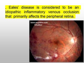
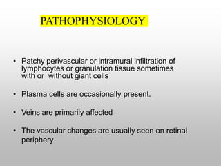
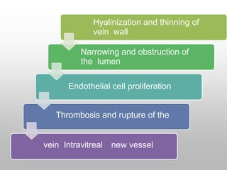
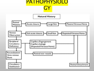
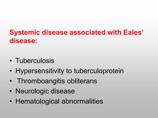
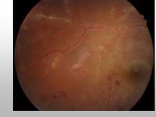
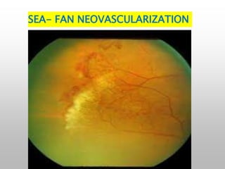
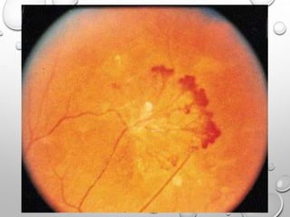
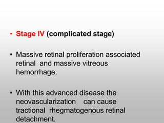
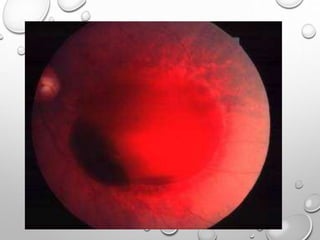
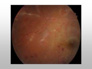
EALE’S DISEASE
DR.ASMATULLAH
PGrOphthalmology
KTH-MTI,PESHAWAR.
, Eales’ disease is considered to be an
idiopathic inflammatory venous occlusion
that primarily affects the peripheral retina.
Perivasculiti
s, mainly
periphlebitis
RETINAL CHANGES
INCLUDE
Pheripher
al non
perfusion
• This inflammation induced vascular occlusion
can lead to a proliferative vascular
retinopathy, with sequelae such as
1) recurrent vitreous
hemorrhage
2) traction retinal detachment.
PATHOPHYSIOLOGY
• Patchy perivascular or intramural infiltration of
lymphocytes or granulation tissue sometimes
with or without giant cells
• Plasma cells are occasionally present.
• Veins are primarily affected
• The vascular changes are usually seen on retinal
periphery
Hyalinization and thinning of
vein wall
Narrowing and obstruction of
the lumen
Endothelial cell proliferation
Thrombosis and rupture of the
vein Intravitreal new vessel
PATHOPHYSIOLO
GY
Systemic disease associated with Eales’
disease:
• Tuberculosis
• Hypersensitivity to tuberculoprotein
• Thromboangitis obliterans
• Neurologic disease
• Hematological abnormalities
• The assumption of tubercular
aetiology is based on active or
healed tuberculosis in some patient
with Eales’ disease.
• Ophthalmoscopic evaluation in patient
with active or healed TB showed 1.3%
had Eales’ disease .
Hypersensitivity to
tuberculoprotein: Allergic reaction to
tuberculosis has been reported by
many authors till date.
Positive Mantoux reaction which is as
high as 90% in some series.
STAGES OF EALES’
DISEASE
Stage I: (Inflammatory stage)
• Localized areas of peripheral retinal edema
with sheathing of the smaller caliber vascular
branches.
• Minute retinal hemorrhages as well as
minute vascular connection b/w two
adjoining vessels.
STAGE II (ISCHEMIC
STAGE)
• Involvement of larger vessels and extend
more posteriorly .
• Veins as well as arterioles may be
sheathed
• Widespread retinal hemorrhages and
vitreous looks hazy .
• Stage III (stage of neovascularisation)
• Peripheral new blood vessels with
numerous vitreous and retinal
hemorrhages.
• The hemorrhages frequently recurs.
SEA- FAN NEOVASCULARIZATION
• Stage IV (complicated stage)
• Massive retinal proliferation associated
retinal and massive vitreous
hemorrhage.
• With this advanced disease the
neovascularization can cause
tractional rhegmatogenous retinal
detachment.
DIFFERENTIAL
DIAGNOSIS:
• Vasculitis mimicking Eales’
diseaseSystemic Ocular
Behcet’s disease Birdshot retinochoroidopathy
Coat’s disease
Leukemia Pars planitis
Multiple sclerosis Viral retinitis
Systemic lupus erythematosus
Toxocariasis
Toxoplasmosis
Tuberculosis
Wegener’s granulomatosis
• Proliferative vascular retinopathy
mimicking Eales’ disease:
Systemic Ocular
Diabetes mellitus BRVO
Sarcoidosis CRVO
Sickle cell disease ROP
Pars planitis
Coats’ disease
CLINICAL FEATURES:
• Usually occurs in young , healthy people, with a
peak incidence between the ages of 30 and 40
years.
•
• It occurs more frequently in males 80-90%.
•
• 75% cases it presents before 49 years.
• Can be unilateral or bilateral.90% bilateral (Duke
Elder) retinal vasculitis
Vitreous floaters or blurring of vision,
symptomsattributable to recurrent
vitreous hemorrhages.
80% between the age of 20-40 years and
95% were male (O.K Malla and co
workers)
54.34% between 20-30 years and 94.73%
male
• More commonly reported from Indian
subcontinent. The reported incidence in India is
1 in 200-250 patient
• Anterior uveitis/Vitritis.
• Active perivasculitis with exudates around the
veins in one or more quadrants. Arterioles may
be affected.
HEALED
PERIVASCULITIS AS
SHEATHING OF THE
VEINS
Macular changes
uncommon
Peripheral retinal
neovascularisation
reported in 36-84% of
RECURRENT VITREOUS
HEMORRHAGES, THE
HALL MARK OF THE
DISEASE
Some vitreous hemorrhages
resolve, some do not ( organize
with multiple VR adhesions &
RRD/TRD
Some patient specially with
multiple sclerosis are
F F A :
• To delineate areas of capillary nonperfusion,
peripheral retinal nonperfusion is present in all
patients with Eales’ disease.
• Retinal or disc neovascularisation
•
• Macular edema
• Helps in monitoring the regression and
disappearance of new vessels during treatment
and follow up.
TREATMENT??
• Symptomatic treatment.
• Treatment aim :
reducing retinal perivasculitis and associated
vitritis ;
reducing risk of vitreous hemorrhage from new
vessels by retinal ablation and
surgical removal of non resolving vitreous
hemorrhage and/or vitreous membranes.
• Observation.
• Medical
Corticosteroids
Antituberculosis drugs
Immunosuppressive
drugs.
• Retinal ablation
Photocoagulatio
n cryotherapy
• Surgical
Observation:
• Patient with inactive retinal vasculitis
• Follow up 6 months to 1 year interval.
• Patient with fresh vitreous hemorrhage if
retina is found to be attached.
• Such vitreous hemorrhage usually clears by 6
to 8 weeks.
MEDICAL
THERAPY
• Corticosteroids are mainstay of therapy in
active perivasculitis stage of Eales’ disease.
• Majority of cases 1mg/kg body weight,
tapered to 10mg/week over 6 to 8 weeks.
• Maintenance 15 to 20mg/day for 1 to 2
months.
• Periocular depot steroid injection may be
added for associated macular edema.
• Systemic and Periocular steroid useful in
patient having 3 quadrants involvement
with macular edema.
• Systemic steroid only if less than 3
quadrant involvement.
• No difference in response between
Mantoux positive and negative cases.
• Immunosuppressive therapy in patient
unresponsive or have unacceptable side
effects. (Azathioprine and cyclosporine)
• Some investigators have recommended
ATT (Rifampicin and Isoniazid) for 9
months.
PHOTOCOAGULATION
• Mainstay of therapy in proliferative stage of
Eales’ disease.
• The aim
Regulate the circulation
To obliterate surface neovascularisation and
Close leaking intraretinal
microvascular abnormalities.
• Sectoral laser for capillary
non perfusion and PRP for
neovascularisation of disc.
• Occasional massive hemorrhage
can occur.
• After laser, regressing
neovascularisation can cause
macular distortion and retinal
tear.
• Laser not advised in
active inflammatory
VITREORETINAL SURGERY
• Vitrectomy alone or combined with
other vitreoretinal surgical
procedures is often required.
• Nonresolving vitreous hemorrhage
with obscuration of central vision of 3
months duration may be subjected to
vitrectomy.
• Vitrectomy done between 3 to 6 months
has better results than done after 6 months
(Kumar et al).
• Early vitrectomy in patient with TRD,
extensive vitreous membranes or
epimacular membranes.
• Endolaser can be given along with
vitrectomy.
TRACTIONAL RETINAL FOLD AFTER
VITRECTOMY
SUMMARY AND
CONCLUSIONS:
• Characteristic clinical findings and
angiographic pattern.
• Mimic several ocular or systemic disease
presenting as retinal vasculitis or proliferative
retinal vasculopathy.
• Hypersensitivity to tubercular protein has
been considered a prime cause of Eales’
• Probable multifactorial etiology.
• HLA, retinal autoimmunity,
mycobacterium genome, free radical
mediated damage.
• Corticosteroids in active disease and laser
photocoagulation in ischemic and
proliferative stage.
• Results of vitrectomy in non resolving
vitreous hemorrhage with or without retinal
detachment are satisfactory.
• Thank u for
listening

Eales disease by dr.asmat

  • 1.
  • 2.
    , Eales’ diseaseis considered to be an idiopathic inflammatory venous occlusion that primarily affects the peripheral retina.
  • 3.
  • 4.
    • This inflammationinduced vascular occlusion can lead to a proliferative vascular retinopathy, with sequelae such as 1) recurrent vitreous hemorrhage 2) traction retinal detachment.
  • 5.
    PATHOPHYSIOLOGY • Patchy perivascularor intramural infiltration of lymphocytes or granulation tissue sometimes with or without giant cells • Plasma cells are occasionally present. • Veins are primarily affected • The vascular changes are usually seen on retinal periphery
  • 6.
    Hyalinization and thinningof vein wall Narrowing and obstruction of the lumen Endothelial cell proliferation Thrombosis and rupture of the vein Intravitreal new vessel
  • 7.
  • 8.
    Systemic disease associatedwith Eales’ disease: • Tuberculosis • Hypersensitivity to tuberculoprotein • Thromboangitis obliterans • Neurologic disease • Hematological abnormalities
  • 9.
    • The assumptionof tubercular aetiology is based on active or healed tuberculosis in some patient with Eales’ disease. • Ophthalmoscopic evaluation in patient with active or healed TB showed 1.3% had Eales’ disease .
  • 10.
    Hypersensitivity to tuberculoprotein: Allergicreaction to tuberculosis has been reported by many authors till date. Positive Mantoux reaction which is as high as 90% in some series.
  • 13.
    STAGES OF EALES’ DISEASE StageI: (Inflammatory stage) • Localized areas of peripheral retinal edema with sheathing of the smaller caliber vascular branches. • Minute retinal hemorrhages as well as minute vascular connection b/w two adjoining vessels.
  • 15.
    STAGE II (ISCHEMIC STAGE) •Involvement of larger vessels and extend more posteriorly . • Veins as well as arterioles may be sheathed • Widespread retinal hemorrhages and vitreous looks hazy .
  • 17.
    • Stage III(stage of neovascularisation) • Peripheral new blood vessels with numerous vitreous and retinal hemorrhages. • The hemorrhages frequently recurs.
  • 19.
  • 24.
    • Stage IV(complicated stage) • Massive retinal proliferation associated retinal and massive vitreous hemorrhage. • With this advanced disease the neovascularization can cause tractional rhegmatogenous retinal detachment.
  • 29.
    DIFFERENTIAL DIAGNOSIS: • Vasculitis mimickingEales’ diseaseSystemic Ocular Behcet’s disease Birdshot retinochoroidopathy Coat’s disease Leukemia Pars planitis Multiple sclerosis Viral retinitis Systemic lupus erythematosus Toxocariasis Toxoplasmosis Tuberculosis Wegener’s granulomatosis
  • 30.
    • Proliferative vascularretinopathy mimicking Eales’ disease: Systemic Ocular Diabetes mellitus BRVO Sarcoidosis CRVO Sickle cell disease ROP Pars planitis Coats’ disease
  • 31.
    CLINICAL FEATURES: • Usuallyoccurs in young , healthy people, with a peak incidence between the ages of 30 and 40 years. • • It occurs more frequently in males 80-90%. • • 75% cases it presents before 49 years. • Can be unilateral or bilateral.90% bilateral (Duke Elder) retinal vasculitis
  • 32.
    Vitreous floaters orblurring of vision, symptomsattributable to recurrent vitreous hemorrhages. 80% between the age of 20-40 years and 95% were male (O.K Malla and co workers) 54.34% between 20-30 years and 94.73% male
  • 33.
    • More commonlyreported from Indian subcontinent. The reported incidence in India is 1 in 200-250 patient • Anterior uveitis/Vitritis. • Active perivasculitis with exudates around the veins in one or more quadrants. Arterioles may be affected.
  • 35.
    HEALED PERIVASCULITIS AS SHEATHING OFTHE VEINS Macular changes uncommon Peripheral retinal neovascularisation reported in 36-84% of
  • 36.
    RECURRENT VITREOUS HEMORRHAGES, THE HALLMARK OF THE DISEASE Some vitreous hemorrhages resolve, some do not ( organize with multiple VR adhesions & RRD/TRD Some patient specially with multiple sclerosis are
  • 37.
    F F A: • To delineate areas of capillary nonperfusion, peripheral retinal nonperfusion is present in all patients with Eales’ disease. • Retinal or disc neovascularisation • • Macular edema • Helps in monitoring the regression and disappearance of new vessels during treatment and follow up.
  • 40.
  • 41.
    • Symptomatic treatment. •Treatment aim : reducing retinal perivasculitis and associated vitritis ; reducing risk of vitreous hemorrhage from new vessels by retinal ablation and surgical removal of non resolving vitreous hemorrhage and/or vitreous membranes.
  • 42.
    • Observation. • Medical Corticosteroids Antituberculosisdrugs Immunosuppressive drugs. • Retinal ablation Photocoagulatio n cryotherapy • Surgical
  • 43.
    Observation: • Patient withinactive retinal vasculitis • Follow up 6 months to 1 year interval. • Patient with fresh vitreous hemorrhage if retina is found to be attached. • Such vitreous hemorrhage usually clears by 6 to 8 weeks.
  • 44.
    MEDICAL THERAPY • Corticosteroids aremainstay of therapy in active perivasculitis stage of Eales’ disease. • Majority of cases 1mg/kg body weight, tapered to 10mg/week over 6 to 8 weeks. • Maintenance 15 to 20mg/day for 1 to 2 months. • Periocular depot steroid injection may be added for associated macular edema.
  • 45.
    • Systemic andPeriocular steroid useful in patient having 3 quadrants involvement with macular edema. • Systemic steroid only if less than 3 quadrant involvement. • No difference in response between Mantoux positive and negative cases.
  • 46.
    • Immunosuppressive therapyin patient unresponsive or have unacceptable side effects. (Azathioprine and cyclosporine) • Some investigators have recommended ATT (Rifampicin and Isoniazid) for 9 months.
  • 47.
    PHOTOCOAGULATION • Mainstay oftherapy in proliferative stage of Eales’ disease. • The aim Regulate the circulation To obliterate surface neovascularisation and Close leaking intraretinal microvascular abnormalities.
  • 48.
    • Sectoral laserfor capillary non perfusion and PRP for neovascularisation of disc. • Occasional massive hemorrhage can occur. • After laser, regressing neovascularisation can cause macular distortion and retinal tear. • Laser not advised in active inflammatory
  • 50.
    VITREORETINAL SURGERY • Vitrectomyalone or combined with other vitreoretinal surgical procedures is often required. • Nonresolving vitreous hemorrhage with obscuration of central vision of 3 months duration may be subjected to vitrectomy.
  • 51.
    • Vitrectomy donebetween 3 to 6 months has better results than done after 6 months (Kumar et al). • Early vitrectomy in patient with TRD, extensive vitreous membranes or epimacular membranes. • Endolaser can be given along with vitrectomy.
  • 52.
    TRACTIONAL RETINAL FOLDAFTER VITRECTOMY
  • 53.
    SUMMARY AND CONCLUSIONS: • Characteristicclinical findings and angiographic pattern. • Mimic several ocular or systemic disease presenting as retinal vasculitis or proliferative retinal vasculopathy. • Hypersensitivity to tubercular protein has been considered a prime cause of Eales’
  • 54.
    • Probable multifactorialetiology. • HLA, retinal autoimmunity, mycobacterium genome, free radical mediated damage. • Corticosteroids in active disease and laser photocoagulation in ischemic and proliferative stage. • Results of vitrectomy in non resolving vitreous hemorrhage with or without retinal detachment are satisfactory.
  • 55.
    • Thank ufor listening