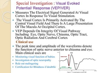
OPTIC NERVE DISEASE
 9th July 2020 ,
DR M SAQUIB
Vice Principal
MBBS,MS , FSCEH DELHI,FHVDESAI PUNE,
EX REGISTRARA JNMCH,AMU
CONSULTANT OPHTHALMOLOGIST
HOD D/O OPHTHALMOLOGY
G.S .MEDICAL COLLEGE
Founder sec: MEDICS India ,
Mail-dms2k5@gmail.com , 9634123800
Optic Neuropathy
Clinical feature
 Visual Acuity –
Reduced
 Impaired Color
Vision
 Decrease contrast
sensitivity
 RAPD
 Optic Disc Edema
,Hyperemia ,
Paleness and
Atrophy
 Visual field Defect
 VEP –Defective
Examination & Investigation
 Distance And Near Vision ( UCVA/BCVA )
 Colour Vision
 Field Of Vision – Central /Peripheral
 Pupillary Reaction
 Intraocular Pressure (I.O.P)
 Ophthalmoscopy
 Slit Lamp Biomicroscopy
 Flourescein Angiography
 X Ray Skull /PNS, OPTIC FORAMEN / CANAL
AND ORBIT
 ULTRASONOGRAPHY
 CT SCAN
 MRI
 OCT
 VISUAL EVOKED RESPONSE (VER )
Special Investigation : Visual Evoked
Potential/ Response (VEP/VER)
 Measures The Electrical Signal Generated At Visual
Cortex In Response To Visual Stimulation.
 The Visual Cortex Is Primarily Activated By The
Central Visual Field And There Is A Large Presentation
Of The Macula At Occipital Cortex.
 VEP Depends On Integrity Of Visual Pathway
Including Eye, Optic Nerve, Chiasma, Optic Tract,
Optic Radiation And Cerebral Cortex
Clinical use
 The peak time and amplitude of the waveforms denote
the function of optic nerve anterior to chiasma and eye.
Main clinical uses are
 Monitoring visual function of babies
 Investigation of optic neuropathy
 Rule out malingering
 Certification for Blindness if doubtful
Perimetry
 Visual field testing can be performed by various
methods, including confrontation technique, amsler grid,
tangent screen, kinetic perimetry, or static perimetry.
 The normal eye can detect stimuli over a 120º range
vertically and a nearly 160 degree range horizontally.
From the point of fixation, stimuli can typically be
detected 60º superiorly, 70º inferiorly, 60º nasally, and
100 degrees temporally
Perimetry
Perimetry
 Detect Dysfunction in
 Central and peripheral vision .
 Glaucoma
 Stroke
 Pituitry disease
 Brain Tumor
 Optic Neuropathy ( AION)
Disease of Optic Nerve
 Congenital anomalies
 Inflammatory lesion
 Optic Neuritis
 Nutritional Optic Neuropathy /Toxic
 Vascular Disturbance ;
 Anterior Ischaemic neuropathy
 Papilloedema
 Degeneration :
 Optic Atrophy
 Traumatic lesion
 Tumours
Congenital anomalies
 Anomalies of Optic Disc
 Crescent sites inverse
 Congenital Pigmentation
 Coloboma
 Drusen
 Hypoplasia of optic Disc
Anomalies of the Nerve Fibres
Medullated (Opaque Nerve fibres )
 Papillitis: Characterised By Disc Oedema And
Hypermaemia That May Be Associated With Flame –
Shaped Peripapillary Haemorhage .Unilateral
>>>Bilaterla
 Retrobulbar Neuritis (Acute) : Optic Nerve Head Is
Not Primarily Involved So Appear Normal.
 Neuro Retinitis :Is An Inflammation Of The Neural
Retina ,Macula Optic Nerve.
OPTIC NEURITIS
 Inflammatory Demyelinating
 Etiology :
 Idiopathic –
 Hereditary Optic Neuritis-Leber’s disease
 Demyelinating disorders- Multiple Sclerosis , Devic’s
disease
 Parainfectious Optic Neuritis- Viral ,Post Immunization
 Infectious optic Neuritis- Acute Ethmoiditis, TB , cat
scrath fever , Syphilis ,AIDS
 Autoimmune disorder- Sarcoidosis , PAN,SLE GB
SYNDROME
 Toxic Optic Neuritis
Optic Neuritis- Clinical feature
Phosphenes - Glowing sensation without stimuli
Uhthoff’s symptoms- Temporary worsening of Multiple
sclerosis symptoms , Optic Neuritis with increase
temperature , fatigue ,Pain ,weakness.
Pulfrich’s phenomenon – Depth perception impaired
specially for moving object .
Ophthalmoscopic Features :
 Hyperaemia of disc
 Blurring of Disc Margin
 Oedematous Disc
 Physiological cup obliterated
 Retinal vein congested ,Tortuous
 Splinter haemorhage
 Fine Exudates on the Disc
 Vitreous – Inflammatory cells
 Temporal Pallor of Disc
Optic Neuritis – Demyelinating
 Demyelination
 Nerve Fibres Loss Their Insulating Myelin
Fibres Lose Their Insulating Myelin Layer ,
Disrupting Nervous Conduction Within The
White Matter Tracts Of The Brain , Brainstem
And Spinal Cord
 ISOLATED OPTIC NEURITIS – Generalised
Disease Develop S, Overall 10 Year Risk Of
Developing Multiple Sclerosis Following Optic
Neuritis In 40% Cases
 Multiple sclerosis Most common
 DEVICE DISEASE (NEUROMYELITIS OPTICA) :
Bilateral optic neuritis and subsequent transverse myelitis
(spinal cord demyelination )
DIAGNOSIS :
 Presentation
 3rd – 6th Decades
 Monoocular blurring
 Tiny light flashes( Phosphenes)
 Ocular pain , exacerbated with ocular movement
 Tender Globe
 VA- 6/18 – 6/60 ,Occasionally worse
 Fundus Normal – Retrobulbar Optic Neuritis
 PERIMETRY –DIFFUSE VISUAL FIELD
DEFECT
 CENTRAL ALTITUDINAL OR ARCUATE
 COURSE- Vision worsens over several days to 2
weeks then begins to improve
 Initial recovery is rapid ----slow improvement
 PROGNOSIS – 75% RECOVER TO 6/9
RESIDUAL
 CLINICAL FEATURES – ABNORMAL COLOUR
VISION ,LIGHT BRIGHTNESS,OPTIC
ATROPHY , MILD RAPD
TREATMENT
 Treatment speed recovery but does not
influence eventual visual outcome
 STEROID - INTRAVENOUS
METHYLPREDNISOLONE – 3 DAYS
FOLLOWED BY REDUCING COURSE
OF ORAL PREDNISOLONE
 IMMUNOMODULATION THERAPY – (
INTERFERON –BETA) AT THE FIRST
EPISODE OF OPTIC NEURITIS MAY
REDUCE THE RISK OF CLINICAL MS
IN HIGHER RISK PATIENT .
LEBER’S HEREDITARY OPTIC NEUROPATHY
(LHON)
 Sequential subacute optic Neuropathy
 Male ( 11-30 Years)
 Genetics- Point Mutation in mitochondrial DNA .
 Mitochondrial DNA is exclusively from Mother .
 Clinical Features :
 Early phase –Asymptomatic
 Blurring affecting the central visual field loss
 Painless bilateral Visual loss ( Young to Adult age
group)
 Visual Field – Centrocaecal scotoma
 Pupillary Reaction – Normal
 Ophthalmoscopy :
 Disc swelling
 Peripapillary Retinal Telangiectasia
 Advanced case – Pale ,Atrophic Optic Disc
NUTRITIONAL OPTIC NEUROPATHY
/Toxic
 Chronic Retrobulbar Neuritis
 Damage to Optic Nerve Fibres due to Exogenous or
Endogenous toxic products .
 Bilateral , Chronic
 Drugs-Quinine,Chloroquine ,Ethambutol, Isoniazid ,
Streptomycin ,Amiodaron
 Tabacco Amblyopia
 Methyl Alcohol
 Ethambutol
Tabacco Amblyopia
 Pipe smokers ,
 Heavy Drinkers with Diet deficient in Protein Vitamin B
Complex – Tobacco –Alcohol Amblyopia
 Excessive Tobacco + Decrease Cyanide Detoxification ( Alcoholic Dietry
Deficiency Of Sulphur Rich Protein)….Excessive Cyanide In Blood
…Degeneration Of Macular Ganglion Cells …Degeneration Of Papillomacular
Bundle Nerve Fiber..Toxic Amblyopia
 Clinical Feature :
 Male
 40-60 Years
 Bilateral Gradual Progressive
 Central Vision Impairment
 Difficulty in Doing Near work /Fogging
 Bilateral Centrocaecal Scotoma
 Fundus - Normal to Temporal pallor of
Optic Disc
 Treatment – Cessation of Tobacco and
Alcohol
 Hydroxocobalamin 1000 ug I.M weekly for
10 weeks .
 Care of general Health and Nutrition
 Prognosis- Good , Slow visual recovery
 Anterior Ischemic Optic Neuropathy (AION) Involves
The 1mm Segment Of The Optic Nerve Head, Also
Known As The Optic Disc, And Results In Visible Disc
Swelling.
 AION Has Two Varieties. The First Is Non-arteritic
(NAION) And The Second Is Arteritic (AAION) And Is
Almost Always Associated With Giant Cell Arteritis.
 Posterior Ischemic Optic Neuropathy (PION)
Encompasses Those Conditions That Result In Ischemia
To Any Portion Of The Optic Nerve Posterior To The
Optic Disc. By Definition, PION Will Not Cause Disc
Edema.
ANTERIOR ISCHEMIC OPTIC NEUROPATHY ( AION )
 A. Arteritic Anterior Ischemic
optic Neuropathy
 Inflammatory , Thrombotic
Occlusion of short posterior
ciliary artery .
 Giant Cell Arteritis
 70 Years
 < 10% cases of AION.
 C/F: Giant cell arteritis features –
Headach tenderness of temporal
arteries and scalp . Pain on
speaking or chewing ( Jaw
Claudication )
 Fever ,Malaise ,Anorexia weight
loss
 OCULAR :
 VA < 6/60
 RAPD : +
 Pale Optic Disc ,Oedema
,Cotton wool spot ,Splinter
Haemorhage .
 Peripapillary Pallor and
oedema , Choroidal Ischemia
 Amaurosis fugax
 CRAO
 Ocular Ischemic syndrome –
ophthalmic artery
involvement
 Diplopia
Investigation :
 FFA : Delayed Choroidal
Filling
 Perimetry : Visual field –
Altitudinal Hemianopia
 ESR , Creactive Protein ,
Marked increase
 Temporal Artery Biopsy –
Diagnostic
 Treatment : Corticosteroid
 Intravenous – 1gm/day 3-5
days
 Oral Prednisolone – 80 mg
/day ..Taper to 10 mg till 12
months
B. Non - Arteritic Anterior Ischemic optic Neuropathy
 > 90% case of Ischemic
Optic Neuropathy .
 Occlusion of posterior
cilliary artery
 Etiology –Unknown
 Risk factor – Ocular :
Crowded Disc and cUp ,
Cataract Surgery , Acute
Primary Angle Closure Not
Treated
 Systemic: Hypertension ,
Diabetes, Smoking ,
Collagen Vascular Diasease,
Sudden Hypotension
 Drugs- Sildenafil
 Age - < 60 years
 Sex- Male = Female (
AAION more in
female)
 Visual loss- Less
marked
 Optic Disc –
Hyperaemia
 ESR – 20 -40 MM 1st
hour ( AAION 70mm)
 C reactive protein –
Normal
 FFA- Only Optic Disc
Delay
Treatment ;
 No proven Treatment
 Address Systemic causes
 Aspirin reduce TIA
PAPILLOEDEMA
 Passive hydrostatic non inflammatory
swelling of optic nerve head secondary to
raised intracranial pressure.
 ▪ Usually bilateral ; may be unilateral.
 ▪ Optic disc swelling in the absence of
raised intracranial pressure is referred to
as optic disc edema
CAUSES of Disc Oedema
 Inflammation : Papillitis ,Neuroretinitis ,
Papillophlebitis ,Uveitis
 Vascular Causes: CRVO, Diabetes, Papillopathy ,
Uremia , AION
 Orbital cause- Tumor , Graves orbitopathy , Orbital
cellulitis
 Infiltrative- Lymphomas,Leukaemia
PAPILLOEDEMA
Etiopathogenesis Of Papilloedema
 Non Inflammatory oedema of optic Disc
 1.Congenital : Aqueductal stenosis , Craniosyntosis
 2.Space Occupying Lesions :ICSOL of cerebellum
,midbrain,parieto occipital region . Abscess , Tuberculoma
,Aneurysm .Poterior fossa tumor ..Aqueduct of sylvius
 3.Intra cranial infection : Meningitis , Encephalitis
 4.Intra cranial Haemorhage : Cerebral , Subarachnoid
Haemorhage
 5.Obstruction of CSF absorption : By Damaged Arachnoid
Villi
 6.Tumor of Spinal Cord
 7. Idiopathic Intracranial Hypertension : Pseudotumour
Cerebri ,Obese Young women ,Chronic Headach
 8.Systemic Condition : Malignant Hypertension ,
Pregnancy Induced Hypertension ,etc
 9.Diffuse Cerebral Oedema : Blunt Trauma
 Cerebral Venous Sinus Thrombosis ;
 Unilateral /Or Asymmetric Papilloedema
 FOSTER KENNEDY SYNDROME – Olfactory /Sphenoid
meningoma and frontal lobe tumours. Pressure optic atrophy on side
of tumour and pailloedema other side
Pathogenesis
 Hayreh’s Theory
 Statsis of Axoplasm in the prelaminar region of
optic Disc due Altered Pressure gradient across
lamina cribrosa,
 Increased Intracranial Pressure – Malignant
Hypertension ,Orbital lesions
 Axonal swelling – Venous Congestion –
Extracellular edema
 Develop 1-7 Days of raised IOP. SAH – 2-8
hours ,Subsides 6-8 weeks after normal ICT
Clinical Feature-
 Recurrent Transient Visual Loss
 Frontal Headach
 Vomiting
 Vision ,Pupillary Reaction - Normal in Acute Stage
Signs(Mechanical)
 Elevation of the optic disc.
 Blurring of the optic disc margin.
 Filling in of the physiological cup. Edema of
the peripapillary nerve fiber layer.
 Retinal or choroidal folds(Paton’s lines)
 Macular fan.
Signs(Vascular)
 Hyperemia of the optic disc.
 Vascular congestion.
 Peripapillary haemorrhage.
 Exudates in the disc or peripapillary area.
 Nerve fiber layer infarcts
Early Papilledema
 Disc elevation.
 Venous distention and tortuosity.
 Obscuration of the normal disc margin
and overlying retinal vessels.
 Absence of spontaneous venous pulsation
6-9 month of chronic papilloedema
Gliosis Atrophy of neurons
Retinal arteriol narrowing , white sheathing around
vessels
Diagnosis , Treatment
,Prognosis
 Differential Diagnosis –
 Papillitis
 Pseudopapailloedema- Hypermetropia ,
Drusen
 Neurological Emergency ,
 Imaging - CT Scan , MRI with Contrast
 Cerebral Decompression required
..Chronic ..Prognosis bad
OPTIC NERVE DISEASE

OPTIC NERVE DISEASE

  • 1.
    OPTIC NERVE DISEASE 9th July 2020 , DR M SAQUIB Vice Principal MBBS,MS , FSCEH DELHI,FHVDESAI PUNE, EX REGISTRARA JNMCH,AMU CONSULTANT OPHTHALMOLOGIST HOD D/O OPHTHALMOLOGY G.S .MEDICAL COLLEGE Founder sec: MEDICS India , Mail-dms2k5@gmail.com , 9634123800
  • 2.
    Optic Neuropathy Clinical feature Visual Acuity – Reduced  Impaired Color Vision  Decrease contrast sensitivity  RAPD  Optic Disc Edema ,Hyperemia , Paleness and Atrophy  Visual field Defect  VEP –Defective
  • 3.
    Examination & Investigation Distance And Near Vision ( UCVA/BCVA )  Colour Vision  Field Of Vision – Central /Peripheral  Pupillary Reaction  Intraocular Pressure (I.O.P)  Ophthalmoscopy  Slit Lamp Biomicroscopy  Flourescein Angiography  X Ray Skull /PNS, OPTIC FORAMEN / CANAL AND ORBIT  ULTRASONOGRAPHY  CT SCAN  MRI  OCT  VISUAL EVOKED RESPONSE (VER )
  • 4.
    Special Investigation :Visual Evoked Potential/ Response (VEP/VER)  Measures The Electrical Signal Generated At Visual Cortex In Response To Visual Stimulation.  The Visual Cortex Is Primarily Activated By The Central Visual Field And There Is A Large Presentation Of The Macula At Occipital Cortex.  VEP Depends On Integrity Of Visual Pathway Including Eye, Optic Nerve, Chiasma, Optic Tract, Optic Radiation And Cerebral Cortex Clinical use  The peak time and amplitude of the waveforms denote the function of optic nerve anterior to chiasma and eye. Main clinical uses are  Monitoring visual function of babies  Investigation of optic neuropathy  Rule out malingering  Certification for Blindness if doubtful
  • 5.
    Perimetry  Visual fieldtesting can be performed by various methods, including confrontation technique, amsler grid, tangent screen, kinetic perimetry, or static perimetry.  The normal eye can detect stimuli over a 120º range vertically and a nearly 160 degree range horizontally. From the point of fixation, stimuli can typically be detected 60º superiorly, 70º inferiorly, 60º nasally, and 100 degrees temporally
  • 7.
  • 8.
    Perimetry  Detect Dysfunctionin  Central and peripheral vision .  Glaucoma  Stroke  Pituitry disease  Brain Tumor  Optic Neuropathy ( AION)
  • 9.
    Disease of OpticNerve  Congenital anomalies  Inflammatory lesion  Optic Neuritis  Nutritional Optic Neuropathy /Toxic  Vascular Disturbance ;  Anterior Ischaemic neuropathy  Papilloedema  Degeneration :  Optic Atrophy  Traumatic lesion  Tumours
  • 10.
    Congenital anomalies  Anomaliesof Optic Disc  Crescent sites inverse  Congenital Pigmentation  Coloboma  Drusen  Hypoplasia of optic Disc Anomalies of the Nerve Fibres Medullated (Opaque Nerve fibres )
  • 11.
     Papillitis: CharacterisedBy Disc Oedema And Hypermaemia That May Be Associated With Flame – Shaped Peripapillary Haemorhage .Unilateral >>>Bilaterla  Retrobulbar Neuritis (Acute) : Optic Nerve Head Is Not Primarily Involved So Appear Normal.  Neuro Retinitis :Is An Inflammation Of The Neural Retina ,Macula Optic Nerve.
  • 12.
    OPTIC NEURITIS  InflammatoryDemyelinating  Etiology :  Idiopathic –  Hereditary Optic Neuritis-Leber’s disease  Demyelinating disorders- Multiple Sclerosis , Devic’s disease  Parainfectious Optic Neuritis- Viral ,Post Immunization  Infectious optic Neuritis- Acute Ethmoiditis, TB , cat scrath fever , Syphilis ,AIDS  Autoimmune disorder- Sarcoidosis , PAN,SLE GB SYNDROME  Toxic Optic Neuritis
  • 14.
    Optic Neuritis- Clinicalfeature Phosphenes - Glowing sensation without stimuli Uhthoff’s symptoms- Temporary worsening of Multiple sclerosis symptoms , Optic Neuritis with increase temperature , fatigue ,Pain ,weakness. Pulfrich’s phenomenon – Depth perception impaired specially for moving object .
  • 15.
    Ophthalmoscopic Features : Hyperaemia of disc  Blurring of Disc Margin  Oedematous Disc  Physiological cup obliterated  Retinal vein congested ,Tortuous  Splinter haemorhage  Fine Exudates on the Disc  Vitreous – Inflammatory cells  Temporal Pallor of Disc
  • 16.
    Optic Neuritis –Demyelinating  Demyelination  Nerve Fibres Loss Their Insulating Myelin Fibres Lose Their Insulating Myelin Layer , Disrupting Nervous Conduction Within The White Matter Tracts Of The Brain , Brainstem And Spinal Cord  ISOLATED OPTIC NEURITIS – Generalised Disease Develop S, Overall 10 Year Risk Of Developing Multiple Sclerosis Following Optic Neuritis In 40% Cases
  • 18.
     Multiple sclerosisMost common  DEVICE DISEASE (NEUROMYELITIS OPTICA) : Bilateral optic neuritis and subsequent transverse myelitis (spinal cord demyelination ) DIAGNOSIS :  Presentation  3rd – 6th Decades  Monoocular blurring  Tiny light flashes( Phosphenes)  Ocular pain , exacerbated with ocular movement  Tender Globe
  • 19.
     VA- 6/18– 6/60 ,Occasionally worse  Fundus Normal – Retrobulbar Optic Neuritis  PERIMETRY –DIFFUSE VISUAL FIELD DEFECT  CENTRAL ALTITUDINAL OR ARCUATE  COURSE- Vision worsens over several days to 2 weeks then begins to improve  Initial recovery is rapid ----slow improvement  PROGNOSIS – 75% RECOVER TO 6/9 RESIDUAL  CLINICAL FEATURES – ABNORMAL COLOUR VISION ,LIGHT BRIGHTNESS,OPTIC ATROPHY , MILD RAPD
  • 20.
    TREATMENT  Treatment speedrecovery but does not influence eventual visual outcome  STEROID - INTRAVENOUS METHYLPREDNISOLONE – 3 DAYS FOLLOWED BY REDUCING COURSE OF ORAL PREDNISOLONE  IMMUNOMODULATION THERAPY – ( INTERFERON –BETA) AT THE FIRST EPISODE OF OPTIC NEURITIS MAY REDUCE THE RISK OF CLINICAL MS IN HIGHER RISK PATIENT .
  • 22.
    LEBER’S HEREDITARY OPTICNEUROPATHY (LHON)  Sequential subacute optic Neuropathy  Male ( 11-30 Years)  Genetics- Point Mutation in mitochondrial DNA .  Mitochondrial DNA is exclusively from Mother .  Clinical Features :  Early phase –Asymptomatic  Blurring affecting the central visual field loss  Painless bilateral Visual loss ( Young to Adult age group)
  • 23.
     Visual Field– Centrocaecal scotoma  Pupillary Reaction – Normal  Ophthalmoscopy :  Disc swelling  Peripapillary Retinal Telangiectasia  Advanced case – Pale ,Atrophic Optic Disc
  • 24.
    NUTRITIONAL OPTIC NEUROPATHY /Toxic Chronic Retrobulbar Neuritis  Damage to Optic Nerve Fibres due to Exogenous or Endogenous toxic products .  Bilateral , Chronic  Drugs-Quinine,Chloroquine ,Ethambutol, Isoniazid , Streptomycin ,Amiodaron  Tabacco Amblyopia  Methyl Alcohol  Ethambutol
  • 25.
    Tabacco Amblyopia  Pipesmokers ,  Heavy Drinkers with Diet deficient in Protein Vitamin B Complex – Tobacco –Alcohol Amblyopia  Excessive Tobacco + Decrease Cyanide Detoxification ( Alcoholic Dietry Deficiency Of Sulphur Rich Protein)….Excessive Cyanide In Blood …Degeneration Of Macular Ganglion Cells …Degeneration Of Papillomacular Bundle Nerve Fiber..Toxic Amblyopia  Clinical Feature :  Male  40-60 Years  Bilateral Gradual Progressive  Central Vision Impairment
  • 26.
     Difficulty inDoing Near work /Fogging  Bilateral Centrocaecal Scotoma  Fundus - Normal to Temporal pallor of Optic Disc  Treatment – Cessation of Tobacco and Alcohol  Hydroxocobalamin 1000 ug I.M weekly for 10 weeks .  Care of general Health and Nutrition  Prognosis- Good , Slow visual recovery
  • 27.
     Anterior IschemicOptic Neuropathy (AION) Involves The 1mm Segment Of The Optic Nerve Head, Also Known As The Optic Disc, And Results In Visible Disc Swelling.  AION Has Two Varieties. The First Is Non-arteritic (NAION) And The Second Is Arteritic (AAION) And Is Almost Always Associated With Giant Cell Arteritis.  Posterior Ischemic Optic Neuropathy (PION) Encompasses Those Conditions That Result In Ischemia To Any Portion Of The Optic Nerve Posterior To The Optic Disc. By Definition, PION Will Not Cause Disc Edema.
  • 28.
    ANTERIOR ISCHEMIC OPTICNEUROPATHY ( AION )  A. Arteritic Anterior Ischemic optic Neuropathy  Inflammatory , Thrombotic Occlusion of short posterior ciliary artery .  Giant Cell Arteritis  70 Years  < 10% cases of AION.  C/F: Giant cell arteritis features – Headach tenderness of temporal arteries and scalp . Pain on speaking or chewing ( Jaw Claudication )  Fever ,Malaise ,Anorexia weight loss  OCULAR :  VA < 6/60  RAPD : +  Pale Optic Disc ,Oedema ,Cotton wool spot ,Splinter Haemorhage .  Peripapillary Pallor and oedema , Choroidal Ischemia  Amaurosis fugax  CRAO  Ocular Ischemic syndrome – ophthalmic artery involvement  Diplopia
  • 30.
    Investigation :  FFA: Delayed Choroidal Filling  Perimetry : Visual field – Altitudinal Hemianopia  ESR , Creactive Protein , Marked increase  Temporal Artery Biopsy – Diagnostic  Treatment : Corticosteroid  Intravenous – 1gm/day 3-5 days  Oral Prednisolone – 80 mg /day ..Taper to 10 mg till 12 months
  • 31.
    B. Non -Arteritic Anterior Ischemic optic Neuropathy  > 90% case of Ischemic Optic Neuropathy .  Occlusion of posterior cilliary artery  Etiology –Unknown  Risk factor – Ocular : Crowded Disc and cUp , Cataract Surgery , Acute Primary Angle Closure Not Treated  Systemic: Hypertension , Diabetes, Smoking , Collagen Vascular Diasease, Sudden Hypotension  Drugs- Sildenafil  Age - < 60 years  Sex- Male = Female ( AAION more in female)  Visual loss- Less marked  Optic Disc – Hyperaemia  ESR – 20 -40 MM 1st hour ( AAION 70mm)  C reactive protein – Normal  FFA- Only Optic Disc Delay
  • 32.
    Treatment ;  Noproven Treatment  Address Systemic causes  Aspirin reduce TIA
  • 34.
    PAPILLOEDEMA  Passive hydrostaticnon inflammatory swelling of optic nerve head secondary to raised intracranial pressure.  ▪ Usually bilateral ; may be unilateral.  ▪ Optic disc swelling in the absence of raised intracranial pressure is referred to as optic disc edema
  • 35.
    CAUSES of DiscOedema  Inflammation : Papillitis ,Neuroretinitis , Papillophlebitis ,Uveitis  Vascular Causes: CRVO, Diabetes, Papillopathy , Uremia , AION  Orbital cause- Tumor , Graves orbitopathy , Orbital cellulitis  Infiltrative- Lymphomas,Leukaemia
  • 36.
  • 37.
    Etiopathogenesis Of Papilloedema Non Inflammatory oedema of optic Disc  1.Congenital : Aqueductal stenosis , Craniosyntosis  2.Space Occupying Lesions :ICSOL of cerebellum ,midbrain,parieto occipital region . Abscess , Tuberculoma ,Aneurysm .Poterior fossa tumor ..Aqueduct of sylvius  3.Intra cranial infection : Meningitis , Encephalitis  4.Intra cranial Haemorhage : Cerebral , Subarachnoid Haemorhage  5.Obstruction of CSF absorption : By Damaged Arachnoid Villi  6.Tumor of Spinal Cord
  • 38.
     7. IdiopathicIntracranial Hypertension : Pseudotumour Cerebri ,Obese Young women ,Chronic Headach  8.Systemic Condition : Malignant Hypertension , Pregnancy Induced Hypertension ,etc  9.Diffuse Cerebral Oedema : Blunt Trauma  Cerebral Venous Sinus Thrombosis ;  Unilateral /Or Asymmetric Papilloedema  FOSTER KENNEDY SYNDROME – Olfactory /Sphenoid meningoma and frontal lobe tumours. Pressure optic atrophy on side of tumour and pailloedema other side
  • 39.
    Pathogenesis  Hayreh’s Theory Statsis of Axoplasm in the prelaminar region of optic Disc due Altered Pressure gradient across lamina cribrosa,  Increased Intracranial Pressure – Malignant Hypertension ,Orbital lesions  Axonal swelling – Venous Congestion – Extracellular edema  Develop 1-7 Days of raised IOP. SAH – 2-8 hours ,Subsides 6-8 weeks after normal ICT
  • 40.
    Clinical Feature-  RecurrentTransient Visual Loss  Frontal Headach  Vomiting  Vision ,Pupillary Reaction - Normal in Acute Stage
  • 41.
    Signs(Mechanical)  Elevation ofthe optic disc.  Blurring of the optic disc margin.  Filling in of the physiological cup. Edema of the peripapillary nerve fiber layer.  Retinal or choroidal folds(Paton’s lines)  Macular fan.
  • 42.
    Signs(Vascular)  Hyperemia ofthe optic disc.  Vascular congestion.  Peripapillary haemorrhage.  Exudates in the disc or peripapillary area.  Nerve fiber layer infarcts
  • 44.
    Early Papilledema  Discelevation.  Venous distention and tortuosity.  Obscuration of the normal disc margin and overlying retinal vessels.  Absence of spontaneous venous pulsation
  • 47.
    6-9 month ofchronic papilloedema Gliosis Atrophy of neurons Retinal arteriol narrowing , white sheathing around vessels
  • 48.
    Diagnosis , Treatment ,Prognosis Differential Diagnosis –  Papillitis  Pseudopapailloedema- Hypermetropia , Drusen  Neurological Emergency ,  Imaging - CT Scan , MRI with Contrast  Cerebral Decompression required ..Chronic ..Prognosis bad