
Download to read offline

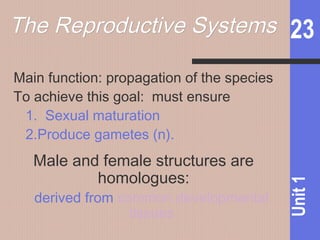

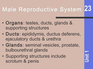


The main functions of the reproductive systems are propagation of the species through sexual maturation and production of gametes. The male and female reproductive structures are homologous, derived from common embryonic tissues. Key parts include testes and ovaries that produce sperm and eggs, duct systems that transport gametes, and accessory glands like the seminal vesicles and prostate in males and mammary glands in females. Hormones regulate development and function of the reproductive systems.


