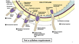
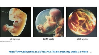
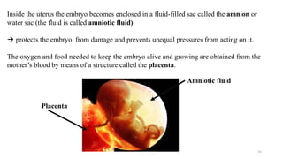
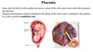
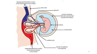
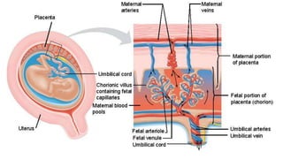
1
16.3 Sexual reproduction in humans
2
16.3 Sexual reproduction in humans
Learning objectives
 Identify the organs, tissues and cells in the male and female human reproductive systems and describe their
functions in reproduction.
 Extended Compare male and female gametes in terms of size, structure, motility and numbers.
 Extended Relate the size, structure, motility and number of sperm and egg cells to their role in
reproduction.
 Describe how the embryo implants into the uterus wall and how structures in the uterus and the placenta
support the developing embryo.
 Extended Describe the functions of the placenta and umbilical cord.
 Outline the growth and development of the human fetus.
 Describe the ante-natal care of pregnant women.
 Describe the processes involved in labour and birth.
 Extended Compare the advantages and disadvantages of breast-feeding and feeding babies with formula
milk.
3
Learning outcomes
 Know that the human male reproductive system includes the testes where sperm are made; the sperm ducts
that carry the sperm to the urethra; the prostate gland and seminal vesicles that produce liquid in which the
sperm swim; the penis, which when erect delivers sperm into the vagina of the woman; and the urethra,
which carries the sperm from the sperm ducts to the outside of the body.
 Know that the human female reproductive system includes the ovaries, where the egg cells are made; the
oviducts, which carry the eggs to the uterus and where fertilisation takes place with sperm cells; the uterus,
where the embryo embeds and develops into a fetus; the cervix, where sperm are deposited at the base of the
uterus; and the vagina, where the penis is inserted during sexual intercourse.
 Extended Know that during development, the embryo (and later the fetus) is protected from mechanical
damage and temperature changes by amniotic fluid that surrounds it in the amniotic sac.
 Know that once the embryo has embedded in the uterus wall, it develops the placenta. This is where nutrients
and waste materials are exchanged between the blood of the mother and of the fetus.
 Know that ante-natal care is the care of the mother before the birth of her baby, and includes checks to make
sure she is healthy and that the baby is developing properly, and advice about how to eat healthily, what to
avoid and how to prepare for the birth.
 Extended Know that breast-feeding of a baby is usually better than bottle-feeding, because of the balance of
nutrients and antibodies that are in the milk.
Human Reproduction
• Sexual Reproduction in humans is the
combination of egg and sperm.
• Sperm are male gametes (haploid)
• ova (ovum singular) are female
gametes. (haploid)
• You get 1/2 your chromosomes from
Dad and the other 1/2 from mom.
• Haploid (1/2) + Haploid (1/2) =
Diploid (1)
4
Female Reproductive Anatomy
• The female external reproductive structures include the clitoris and two sets
of labia
• The internal organs are a pair of gonads and a system of ducts and
chambers that carry gametes and house the embryo and fetus
5
6
State 2 functions of the
ovaries
7
( Fallopian tube)
8
Ovaries
• The female gonads, the ovaries, lie in the abdominal cavity
• Each ovary contains many follicles, which consist of a partially
developed egg, called an oocyte, surrounded by support cells
• Once a month, an oocyte develops into an ovum (egg) by the
process of oogenesis
9
Oviducts and Uterus
• The egg cell travels from the ovary to the uterus via an oviduct, or fallopian
tube
• Cilia in the oviduct convey the egg to the uterus, also called the womb
• The uterus lining, the endometrium, has many blood vessels
• The uterus narrows at the cervix, then opens into the vagina
• normal adult uterus measures approximately
7.2-9.0cm long, 4.5-6.0cm wide and 2.05-3.5 deep
The uterus changes drastically in size during pregnancy to accommodate
the growing baby. When the baby reaches full term, the uterus will be
bigger than five times its original size with a capacity of 500 times.
It'll be 15 times heavier on its own without the baby and the placenta
10
Cilia in the oviduct convey the egg to the uterus
11
12
Vagina and Vulva
• The vagina is a thin-walled chamber
that is the repository for sperm
during copulation and serves as the
birth canal
• The vagina opens to the outside at
the vulva, which consists of the
labia majora, labia minora,
hymen, and clitoris
13
Female Reproductive System
Structure Function
Ovary Produce eggs (ova) and female hormones
Fallopian Tubes • Have funnel-shaped ends that catch the eggs as they are released from
the ovaries
• Carry eggs to the uterus by cilia and peristalsis
• Site of fertilisation
Uterus (Womb) • Site of implantation
• Outer muscular wall which contracts to push baby our during birth
• Inner lining (endometrium) nourishes and protects embryo.
Cervix • a ring of muscles separating the vagina from the uterus.
• it contains a plug of mucus which protects uterus from infection
Vagina • Receives penis and sperm during intercourse
• It is the birth canal - allows the exit of the baby during birth
• Acid produced by normal resident bacteria protect from infection 14
Male Reproductive Anatomy
• The male’s external reproductive organs are the scrotum and penis
• Internal organs are the gonads, which produce sperm and hormones,
and accessory glands
15
16
State 2 functions of the
testes
Cowper’s Gland
17
(Sperm Duct)
Cowper’s Gland
18
Testes
• The male gonads, or testes, consist of highly coiled tubes surrounded by
connective tissue
• Sperm form in these seminiferous tubules
• Production of normal sperm cannot occur at the body temperatures of
most mammals
• The testes of many mammals are held outside the abdominal cavity in
the scrotum, where the temperature (35°C is lower than in the
abdominal cavity) (optimal temp for meiosis)
19
Ducts
• From the seminiferous tubules of a testis, sperm pass
into the coiled tubules of the epididymis
• During ejaculation, sperm are propelled through the
muscular vas deferens and the ejaculatory duct, and
then exit the penis through the urethra
20
21
Accessory Glands
Semen is composed of sperm plus secretions from three sets of accessory
glands
• The two seminal vesicles contribute about 60% of the total volume of
semen
• The prostate gland secretes its products directly into the urethra through
several small ducts
• The bulbourethral glands (Cowper’s gland) secrete a clear mucus
before ejaculation that neutralizes acidic urine remaining in the urethra
22
Penis
• The human penis is composed of three cylinders of spongy erectile tissue
• During sexual arousal, the erectile tissue fills with blood from the arteries,
causing an erection
• The head of the penis has a thinner skin covering than the shaft, and is
more sensitive to stimulation
23
24
Structure Function
Scrotum • Sac of skin in which the testes are held
Testes • Produces sperm and testosterone
Epididymis • Sperm storage and maturation
Sperm Duct (vas
deferens)
• Carries sperm from the epididymis to the urethra
Seminal Vesicles • Produce milky seminal fluid (sugary alkaline fluid in which sperm are
suspended, nourished and protected from the acidic vagina)
Cowper’s Gland • Produces clear sticky fluid which neutralises any urine which remains in
the urethra.
Prostate • Produces another milky nourishing fluid which goes to make up part of
the semen (semen = sperm plus fluid)
Penis • Transfer of sperm into the vagina. Becomes engorged with blood during
sexual arousal. Once hard and erect, it can penetrate the soft tissue of the
vagina.
Urethra • Carries sperm along and out of the penis.
Gametogenesis (Production of gametes)
• Gametogenesis, the production of gametes, differs in male and
female, reflecting the distinct structure and function of their
gametes
• Sperm are small and motile and must pass from male to female
• Eggs are larger, and carry out their function within the female
25
• Spermatogenesis, the development of sperm is continuous and
prolific (millions of sperm are produced per day); each sperm takes
about 74 days to develop.
• Oogenesis, the development of a mature egg, is a prolonged process
• Immature eggs form in the female embryo, but do not complete their
development until years later
26
27
Spermatogenesis
• Stem cells in testes divide mitotically to produce spermatocytes
• Spermatocytes divide by meiosis to produce four equal sized
haploid spermatids that mature into four sperm
.
Spermatogenesis
28
Not a syllabus requirement
Primordial germ cell in embryo
Mitotic divisions
Spermatogonial stem cell
Spermatogonium
Mitotic divisions
Mitotic divisions
Primary spermatocyte
Meiosis I
Meiosis II
Secondary spermatocyte
Early
spermatid
Sperm cell
Differentiation
(Sertoli cells
provide nutrients)
2n
2n
2n
n n
n n n n
n n n n
Meiosis
Mitosis
29
Not a syllabus requirement
30
Describe the structure of
the sperm cell
Plasma
membrane
Tail
Neck
Midpiece Head
Mitochondria
Nucleus
Acrosome
Contain nucleus (n)
Consist of digestive
enzymes
Tail is an extension of the cell
membrane  pushes the sperm
actively forward
Consist of a number of mitochondria 
to produce energy for the movement of
the sperm
Structure of a sperm cell
31
32
Oogenesis: Ovum Formation
• Cells of the ovary divide to form oocytes
• Oocytes divide by meiosis
• Unequal cytoplasmic division
• A discontinuous process
• At birth, oocytes are arrested in meiosis I
• At ovulation, an oocyte continues and stops in meiosis
II
• The four meiotic products produce a functional ovum
and three polar bodies.
33
34
The making of Oocytes
35
Follicular development in the ovary
Ovulation
At birth: two million
primordial follicles with
their primary oocytes in
the ovaries
At puberty: only
about 400,000 primordial
follicles left in the ovaries
36
Compare sperm and egg cells in terms of size
These images are of human
ovulation in progress.
They were captured in 2008
by Dr Jacques Donnez by
chance during a routine
hysterectomy (operation to
remove a woman's uterus).
These were the very first
images ever taken of human
ovulation.
37
Ovulation
Not a syllabus requirement
38
Describe the structure of an
ovum
Structure of an ovum
Eggs are released, one at a
time about every 4 weeks
from alternate ovaries.
The egg released in the
oviduct can live up to 1 day
39
40
Compare male and
female gamete
Comparing male and female gametes
41
Feature Sperm cell Egg cell
size
Movement
Food store
Chromosome
Number
produced
Small Much larger than sperm cell
Swims using tail Moved along the oviduct by cilia and
peristalsis (does not move itself)
Uses sugar in seminal
fluid
Protein and fat in cytoplasm
23 23
Millions constantly
produced (after puberty)
throughout life
One a month after puberty-menopause,
except when pregnant or taking the
contraceptive pill
Fertilization
42
43
Fertilization
• Fertilization is the union of sperm and ovum
• After fertilization, chemical reactions occur preventing
additional sperm from entering the ovum
https://www.babycentre.co.uk/v1049102/inside-pregnancy-fertilisation-video
44
The acrosome at the head of the sperm secretes enzymes which digest part of the egg
membrane.
The sperm then enters the cytoplasm of the ovum and the male nucleus of the sperm
fuses with the female nucleus.
Fertilization
45
Why is the ovum
fertilized only by one
sperm?
Not a syllabus requirement
46
Fertilization
• During intercourse 100 million of sperm are released
into the vagina.
only a few hundred will reach the oviduct and only
one will fertilise the ovum. (The function of the others is not fully understood).
• Sperm swim through the cervix, up through the uterus, and in to the oviducts
• If there is an egg moving down the oviduct it may be fertilized by the sperm.
Ovum can survive for about 24 hours in the oviduct;
Sperm can survive up to 3 days
 So there is about 4 days each month when fertilisation might occur.
It can be used either to achieve or to avoid fertilisation
47
48
Pregnancy
and
development
• The zygote passes along
the oviduct to the uterus
and it begins to divide by
mitosis to form a ball of
cells called embryo.
• The embryo becomes
embedded in the uterine
lining. This is called
implantation.
https://www.babycentre.co.uk/v1049105/inside-pregnancy-early-fetal-development-video
49
Implantation
50
First trimester
Zygote divide by mitosis, 1 cell  2, 4, 8, 16  Morula
In day 6 blastocyst (also called blastula) forms
(fluid –filled cavity ) and implants in the lining
of the uterus
Development of the embryo
https://www.babycentre.co.uk/v1049105/inside-pregnancy-early-fetal-development-video
The inner cell mass of the blastocyst develops
into the embryo
Other cells become the extraembryonic
membranes, important for implantation and
support of embryonic growth
Morula
Blastula
Gastrula
51
Not a syllabus requirement
52
As the embryo grows,
the uterus enlarges to
contain it.
53
(a) 5 weeks (b) 14 weeks (c) 20 weeks
https://www.babycentre.co.uk/v1027475/inside-pregnancy-weeks-1-9-video 54
55
Inside the uterus the embryo becomes enclosed in a fluid-filled sac called the amnion or
water sac (the fluid is called amniotic fluid)
 protects the embryo from damage and prevents unequal pressures from acting on it.
The oxygen and food needed to keep the embryo alive and growing are obtained from the
mother’s blood by means of a structure called the placenta.
Amniotic fluid
Placenta
56
What is the function
of the placenta?
57
Soon after the ball of cells reaches the uterus, some of the cells, grow into a disc-like structure,
the placenta.
The placenta becomes closely attached to the lining of the uterus and is attached to the embryo
by a tube called the umbilical cord
Placenta
58
59
The nervous system (brain, spinal cord and sense organs) start to
develop very quickly.
After a few weeks, the embryo’s heart has developed and is circulating
blood through the umbilical cord and placenta as well as through its own
tissues. Oxygen and nutrients such as glucose and amino acids pass
across the placenta to the embryo’s bloodstream.
Carbon dioxide passes from the embryo’s blood to that of the mother.
Blood entering the placenta from the mother does not mix with the
embryo’s blood.
60
61
62
Functions of the placenta and umbilical cord
Oxygen, glucose, amino acids and salts can pass from the mother’s blood to
the embryo’s blood
Carbon dioxide and urea in the embryo’s blood escape from the vessels in
the placenta and are carried away by the mother’s blood
There is no direct communication between the mother’s blood system and
that of the embryo.
The exchange of substances takes place across the thin walls of the blood
vessels.
 mother’s blood pressure cannot damage the delicate vessels of the
embryo
Placenta selects the substances allowed to pass into the embryo’s blood.
63
Functions of the placenta and umbilical cord
 The placenta can prevent some harmful substances in the mother’s blood from
reaching the embryo.
but It cannot prevent all of them.
Eg. Nicotine and Alcohol, drugs and certain viruses (HIV, rubella virus, zika virus)
If the fetus is infected with Rubella virus
major health problems: deafness, congenital heart disease, diabetes and mental
retardation.
If the fetus is infected with HIV  it can be fatal.
The placenta produces hormones: HCG, oestrogens and progesterone
 play an important part in maintaining the pregnancy and preparing for birth,
but their precise function is not known.
64
65
What are the dos and
don’ts during
pregnancy ?
66
‘Antenatal’ or ‘prenatal’ refers to the period before birth.
Antenatal care is the way a woman should look after herself during pregnancy, so
that the birth will be safe and her baby healthy.
 Eat properly (takes more iron and folic acid to prevent spina bifida)
 No lifting or stooping.
 No drinking or smoking
 No drugs
During pregnancy, a woman should not take any
drugs unless they are strictly necessary and
prescribed by a doctor.
Antenatal care
Not a syllabus requirement
67
Not a syllabus requirement
68
69
Critical Periods of Development
Not a syllabus requirement
70
Fetal alcohol syndrome
Drinking alcohol during pregnancy:
Problems may include an abnormal appearance, short height, low body weight, small
head size, poor coordination, low intelligence, behavior problems, and problems with
hearing or seeing.
Not a syllabus requirement
71
Birth defect due to drug: thalidomide
Phocomelia due to Thalidomide:
A mutation halts limb development
from the third to the fifth week of
the embryonic period, causing
“flippers” to develop in place of
arms and legs
72
Accutane: used for the treatment of severe acne.
Birth defect due to drug: Accutane
Exposure to 13-cis Retinoic
acid (Accutane) during
pregnancy may result in
malformations of the fetus
Not a syllabus requirement
73
How are twins made?
74
Multiple Births
Dizygotic twins
• Form from two different zygotes
• Two ova are fertilized
• Same genetic relationship as any
siblings
Monozygotic twins
• One ovum is fertilized
• Developing embryo splits during
early development
• Genetically identical
75
Not a syllabus requirement
76
• Labour typically has three stages
• Thinning and opening of the cervix, or dilation
• Expulsion or delivery of the baby
• Delivery of the placenta
The period from fertilisation to birth takes about 38 weeks in humans.
 called the gestation period.
When birth starts, the uterus begins to contract rhythmically.
 beginning of ‘labour’.
Birth
77
Dilation of the cervix
2
1
3
Placenta
Umbilical cord
Uterus
Cervix
Expulsion: delivery of the infant
Delivery of the placenta
Uterus
Umbilical cord
Placenta
(detaching)
78
Dilation of the cervix
1
Placenta
Umbilical cord
Uterus
Cervix
the muscular contractions
of the uterus wall and
abdomen push the baby
head-first through the
widened cervix and
vagina
https://www.babycentre.co.uk/v1027490/inside-pregnancy-labour-and-birth-video
79
Cervical effacement and dilation
80
Expulsion: delivery of the infant
2
81
Delivery of the placenta
Uterus
Umbilical
cord
Placenta
(detaching)
3
The umbilical cord, which still
connects the child to the
placenta, is tied and cut.
Later, the placenta breaks away
from the uterus and is pushed
out separately as the ‘afterbirth’.
82
83
The sudden fall in temperature felt by the newly born
baby stimulates it to take its first breath and it usually
cries.
In a few days, the remains of the umbilical cord attached to the baby’s
abdomen shrivel and fall away, leaving a scar in the abdominal wall,
 the navel.
84
85
In certain cases, birth may be induced.
This means that it is started artificially by:
carefully breaking the membrane of the amniotic sac.
 injecting a hormone, oxytocin, into the mother’s veins.
Either of these methods brings on the start of labour.
Sometimes both are used together.
Induced birth
86
During pregnancy: the number of milk-secreting
cells increases  the mammary glands (breasts) enlarge
No milk is secreted during pregnancy.
Baby sucking the nipple  stimulate milk release
The continued production of milk is under the control of hormones.
Feeding and parental care
87
Milk contains the proteins, fats, sugar,
vitamins and salts that babies need for
their energy requirements and tissue-
building,
but there is too little iron present to make
haemoglobin.
All the iron needed for the first weeks or
months is stored in the liver of the foetus
during gestation.
88
How what is the
difference between
breast milk and
formula?
Mammary gland
Not a syllabus requirement 89
90
The liquid produced in the first few days
is called colostrum.
It is sticky and yellow, and contains more
Protein than the milk produced later.
It also contains some of the mother’s antibodies.
 provides passive immunity to infection.
The mother’s milk supply increases with the demands of the baby, up to 1
litre per day.
The milk is gradually reduced and replaced entirely by solid food, a process
known as weaning.
91
Breast milk vs formula milk
vs
92
Human milk Cows’ milk
All nutrients are present at the right
proportion
has more protein, sodium and phosphorus
less sugar, vitamin A and vitamin C
easily digested less easily digested
contains antibodies lack human antibodies
milk free from bacteria  does not require
sterilisation
bottle feeding carries the risk of introducing
bacteria that cause intestinal diseases 
requires sterilisation
no risk of an allergic reaction risk of an allergic reaction
produced at the correct temperature. Need to warm
no additives or preservatives
Free Cost money
does not need to be prepared need to be prepared
Breastfeeding triggers a reduction in the size
of the mother’s uterus
Cows’ milk  obesity and allergy in babies
93
16.4 Sex hormones in humans
95
16.4 Sex hormones in humans
Learning objectives
 Describe the roles of oestrogen and testosterone in the development of secondary sexual characteristics.
 Describe the changes in the uterus and ovaries during the menstrual cycle.
 Extended Explain the role of hormones in controlling the menstrual cycle.
Learning outcomes
 Know that sex hormones, testosterone in men and oestrogen in women, control the development of secondary
sexual characteristics.
 Know that during the menstrual cycle and egg is released from the ovary, and the uterus lining thickens. If
fertilisation does not take place, the uterus lining and egg are shed at the start of the next cycle.
 Extended Know that the hormones oestrogen and progesterone from the ovaries, and luteinising hormone
(LH) and follicle-stimulating hormone (FSH) from the pituitary gland, control the menstrual cycle.
96
What are
sex hormones?
Sex hormones in Human
• Human reproduction is coordinated by hormones from the hypothalamus,
anterior pituitary, and gonads
• hypothalamus secretes: Gonadotropin-releasing hormone (GnRH)
• GnRH stimulate anterior pituitary to secrete Luteinizing hormone (LH) and
follicle-stimulating hormone (FSH)
• FSH and LH stimulate the gonads to secrete sex hormones
Sex hormones are Testosterone and estrogen
testosterone and estrogen have been considered to be male and female sex
hormones, respectively.
Not a syllabus requirement
100
What are the
functions of
sex hormones?
• Sex hormones serve many functions in addition to gamete production,
including sexual behavior and the development of primary and secondary
sex characteristics
• In girls
growth of the breasts, a widening of the hips and
increase in the size of the uterus and vagina….
• In boys
enlargement of the testes and penis, deepening of the voice,
growth of hair in the pubic region, armpits, chest and, later on, the face….
Testosterone
Testosterone is produced in testes in men and by
the ovaries in women, although small quantities
are also produced by the adrenal glands in both
sexes.
Testosterone is a hormone found in men, and less-
so in women.
It stimulates the development of male characteristics:
increased muscle and bone mass, and the growth of body hair.
Estrogen, or oestrogen, is the primary female sex
hormone. (It is also found in men)
Produced by the follicle in the ovaries
It is responsible for the development and regulation of
the female reproductive system and secondary sex
characteristics.
Estrogen
Progesterone
Progesterone is a steroid hormone, involved in
the menstrual cycle, it prepares the uterus for
pregnancy, and to implant the fertilized egg
It is Produced by the corpus luteum in the ovaries
Hormonal Control of the Female Reproductive Cycles
• In females, the secretion of hormones and the reproductive events they
regulate are cyclic
• Prior to ovulation, the endometrium thickens with blood vessels in
preparation for embryo implantation
• If an embryo does not implant in the endometrium, the endometrium is
shed in a process called menstruation ( period)
Hormones closely link the two cycles of female reproduction
• Changes in the uterus define the menstrual cycle (also called the
uterine cycle)
• Changes in the ovaries define the ovarian cycle
107
What is uterine
cycle?
The Uterine (Menstrual) Cycle
• Hormones coordinate the uterine cycle with the ovarian cycle
• Thickening of the endometrium
• Shedding of the endometrium  bleeding : menstruation
• A new cycle begins if no embryo implants in the endometrium
The menstrual cycle
Day 1 to day 5
Day 5 to day 13
Day 14
Day 15 to day 28
111
What is ovarian
cycle?
The Ovarian Cycle
• The release of FSH and LH stimulates follicle growth
• Growing follicle growth releases Oestrogen
• at day 14 of the cycle, the ovum is released  ovulation
• the follicular tissue left behind transforms into the corpus luteum
• The corpus luteum disintegrates, and ovarian steroid (oestrogens
and progesterone) hormones decrease
The Ovarian Cycle
Control by hypothalamus
(a)
Hypothalamus
GnRH
Anterior pituitary
FSH LH
Inhibited by combination of
estradiol and progesterone
Stimulated by high levels
of estradiol
Inhibited by low levels of
estradiol



10
9
8
7
6
5
4
3
2
1
(b) Pituitary gonadotropins
in blood
LH
FSH
FSH and LH stimulate
follicle to grow
6
LH surge triggers
ovulation
Ovarian cycle
Growing follicle Maturing
follicle
Corpus
luteum
Degenerating
corpus luteum
Follicular phase Ovulation Luteal phase
Estradiol secreted
by growing follicle in
increasing amounts
(c)
Progesterone and
estradiol secreted
by corpus luteum
(d) Ovarian hormones
in blood
Estradiol
Peak causes
LH surge
(see )
Progesterone
Progesterone and estra-
diol promote thickening
of endometrium
Estradiol level
very low
(e) Uterine (menstrual) cycle
Endometrium
Menstrual flow phase Proliferative phase Secretory phase
Days
28
25
20
15
14
10
5
0
Hypothalamus
Pituitary gland
ovaries
Uterus
Blood
Blood
Ovarian cycle
Uterine (menstrual) cycle
LH
FSH
FSH and LH stimulate
follicle to grow
LH surge triggers
ovulation
Ovarian cycle
Growing follicle Maturing
follicle
Corpus
luteum
Degenerating
corpus luteum
Ovulation
28
25
20
15
14
11
5
0
Days
3
6
7 8
Copulation could
result in fertilisation
Oestrogen secreted
by growing follicle in
increasing amounts
Progesterone and
Oestrogen secreted
by corpus luteum
(d)
Oestrogen
Peak causes
LH surge
(see )
Progesterone
Progesterone and Oestrogen
promote thickening
of endometrium
Oestrogen level
very low
(e)
Uterine (menstrual) cycle
Endometrium
Days
28
25
20
15
14
10
5
0
4
5
6
9
10
(e)
Ovarian hormones in blood
119
Menopause
•After about 400 to 500 cycles, human females undergo
menopause, the cessation of ovulation and menstruation
•Menopause is very unusual among animals
•Menopause might have evolved to allow a mother to
provide better care for her children and grandchildren
121
Not a syllabus requirement
122
16.5 Methods of birth control in humans
(Family planning)
123
16.5 Methods of birth control in humans
Learning objectives
 Outline how different birth control methods prevent fertilisation.
 Extended Outline the process of artificial insemination (AI) and in vitro fertilisation (IVF).
 Extended Explain the use of hormones in controlling fertility and discuss the social implications of this.
Learning outcomes
 Know that birth control may be by natural methods, chemical methods, mechanical methods or surgical
methods.
 Extended Know that hormones may be used in contraception and fertility treatments, and these have social
implications.
 Extended Know that artificial insemination (AI) or in vitro fertilisation (IVF) may be used to help a couple
have a child.
124
Give one example of
birth control method
125
Methods of birth control in humans
Birth control is used to space out births and limit the size of the family.
• Contraception, the deliberate prevention of pregnancy, can be achieved
in a number of ways
• Contraceptive methods fall into three categories
• Preventing release of eggs and sperm
• Keeping sperm and egg apart
• Preventing implantation of an embryo
A health-care provider should be consulted for complete information on the
choice and risks of contraception methods
Natural methods of family planning
Abstinence couple avoiding sexual intercourse
Monitoring body
temperature
During or soon after ovulation
a woman’s temperature rises by about 0.5°C
Avoid intercourse during fertile period (about 10 days in
mid-cycle)
Cervical mucus As the time for ovulation approaches, the mucus becomes
more fluid  calculate their fertile period
127
Artificial methods of family planning
Barrier methods
Sheath or condom
Femidom (female condom)
 traps the sperm and prevents them from
reaching the uterus
 prevents the transmission of sexually
transmitted infections (STIs).
Diaphragm
thin rubber disc, placed in the vagina
before intercourse
 stops sperm entering the uterus
 But does not prevents the transmission
of sexually transmitted infections
(STIs).
128
Artificial methods of family planning
Chemical methods
Spermicides
cream, gel or foam, is placed in the
vagina
kill or immobilise sperm
Not very effective
Intra-uterine device (IUD)
small T-shaped plastic and copper
device, inserted into the wall of the
uterus
Prevents implantation of a fertilised ovum.
It is about 98% effective
Contraceptive implant • small plastic tube about 4 cm long,
• inserted under the skin of the upper arm  slowly
releases progesterone  prevent pregnancy.
• It lasts for about 3 years  99% effective.
• Does not protect against STIs
129
Femidom
130
Artificial methods of family planning
Chemical methods
Contraceptive pill
oestrogen- and
progesterone-like
chemicals
• suppress ovulation  prevent conception.
• taken everyday for the 21 days between menstrual
periods.
• long-term use  increase risk of breast and cervix cancer.
• Does not protect against STIs.
Contraceptive
injection
• This injection, contains progesterone
• It works by thickening the mucus in the cervix, stopping
sperm reaching an egg.
• thins the lining of the uterus  unsuitable for
implantation of an embryo.
• Effective for 8 up to 12 weeks.
• Does not protect against STIs.
131
Contraceptive pill Contraceptive injection
132
Artificial methods of family planning
Surgical methods
Male
sterilisation –
vasectomy
• the man’s sperm ducts are cut and the ends sealed.
the semen contains the secretions of the prostate gland
and seminal vesicle but no sperm
Operation can be reversed but not always successful
Female
sterilisation –
laparotomy
• oviducts are tied, blocked or cut  sperm can no longer
reach the ova.
Ova are released, but break down in the upper part of the
oviduct.
The operation cannot usually be reversed.
133
Male sterilisation – vasectomy Female sterilisation – laparotomy
134
Infertility
135
Female infertility
Female infertility is
usually caused by a
failure to ovulate or a
blockage or distortion
of the oviducts.
The latter can often be
corrected by surgery.
Not a syllabus requirement
Male infertility
138
The use of hormones in fertility
and contraception treatments
Using hormones to improve fertility
Fertility drugs  to trait cases that fail to produce ova
These drugs  are similar to hormones  act by increasing the levels of
FSH and LH.
Artificial insemination (AI)
When the quantity of sperm in the semen is low or by sperm that are insufficiently mobile
to reach the oviducts  Treatment involves injection of semen through a tube into the top
of the uterus.
In vitro fertilisation (Test tube baby)
In vitro fertilisation
 The woman is given fertility drugs
 cause her ovaries to release several mature ova simultaneously.
 The ova are then collected by laparoscopy
(they are sucked up in a fine tube inserted through the abdominal wall).
 The ova are mixed with the husband’s seminal fluid
 watched under the microscope to see if cell division takes place.
One or more of the dividing zygotes are introduced to the woman’s uterus
Usually, only one (or none) of the zygotes develops (occasionally there are
multiple births).
144
Using hormones for contraception
Oestrogen and progesterone control important events in the menstrual cycle.
Oestrogen
 encourages re-growth of the lining of the uterus wall after a period
 prevents the release of FSH  If FSH is blocked  no further ova are matured.
Progesterone
 maintains the thickness of the uterine lining.
 inhibits the secretion of luteinising hormone (LH)  If LH is suppressed  ovulation
cannot happen  so there are no ova to be fertilised.
Because of the roles of oestrogen and progesterone, they are used, singly or in combination, in
a range of contraceptive methods.
Social implications of contraception and fertility treatments
Some religions are against any artificial forms of contraception and actively
discourage the use of contraceptives such as the sheath and femidom.
Fertility treatments such as in vitro fertilisation are
controversial because of the ‘spare’ embryos that are
created and not returned to the uterus.
Some people believe that since these embryos are
potential human beings, they should not be destroyed or
used for research.
In some cases the ‘spare’ embryos have
been frozen and used later if the first
transplants did not work.
Social implications of contraception and fertility treatments
147
16.5 Sexually transmitted infections (STIs)
149
16.5 Methods of birth control in humans
Learning objectives
 Outline how different birth control methods prevent fertilisation.
 Extended Outline the process of artificial insemination (AI) and in vitro fertilisation (IVF).
 Extended Explain the use of hormones in controlling fertility and discuss the social implications of this.
Learning outcomes
 Know that birth control may be by natural methods, chemical methods, mechanical methods or surgical
methods.
 Extended Know that hormones may be used in contraception and fertility treatments, and these have social
implications.
 Extended Know that artificial insemination (AI) or in vitro fertilisation (IVF) may be used to help a couple
have a child.
150
Give one example of
STD
Sexually transmitted infections (STIs)
A sexually transmitted infection is an infection that is transmitted
via body fluids through sexual contact.
STDs are caused by:
• Bacteria: Chlamydia, Gonorrhea, Treponema pallidum (Syphilis)
• Viruses (Herpes simplex virus (HSV), Hepatitis B virus (HBV),
Human papillomavirus (HPV), HIV
• Parasites (Pubic lice, scabies, trichomoniasis)
Syphilis
Gonorrhea
Chlamydia
Chlamydia
AIDS
Acquired Immune Deficiency Syndrome
A = Acquired, not inherited
I = Weakens the Immune system
D = Creates a Deficiency of CD4+ cells in the immune
system
S = Syndrome, or a group of illnesses taking place at the
same time
AIDS  caused by Human Immunodeficiency Virus (HIV).
HIV
Human Immunodeficiency Virus
H = Infects only Human beings
I = Immunodeficiency virus weakens the immune
system and increases the risk of infection
V = Virus
Methods of transmission:
Sexual Intercourse
Blood-to-blood contact (transfusion, organ transplant)
Sharing needles or other drug-use equipment
Tattoo or body piercing
Infected mother to her baby
A person can be infected for many years before symptoms develop
 They can still infect other people
Sexual Intercourse
with infected person
Infected mother
to her child
Sharing
contaminated
needles
Contaminated blood
Transfusion &
organ transplants
These tests do not detect the virus 
indicate whether antibodies to the virus are
in the blood.
Blood tests to detect HIV infection
If HIV antibodies are present 
the person is HIV positive.
AIDS patients
Journal of the International AIDS
Society, Volume: 22, Issue: S6,
First published: 30 August 2019,
DOI: (10.1002/jia2.25361)
Pompeii was an ancient Roman town-city
in Italy
It was mostly destroyed and buried under 4
to 6 m of volcanic ash and rocks in
the eruption of Mount Vesuvius in AD 79.
Pompeii
Not a syllabus requirement
Prostitution was permitted both socially
and legally in Pompeii.
164
165

Sexual reproduction in Human methods and techniques

  • 1.
  • 2.
    2 16.3 Sexual reproductionin humans Learning objectives  Identify the organs, tissues and cells in the male and female human reproductive systems and describe their functions in reproduction.  Extended Compare male and female gametes in terms of size, structure, motility and numbers.  Extended Relate the size, structure, motility and number of sperm and egg cells to their role in reproduction.  Describe how the embryo implants into the uterus wall and how structures in the uterus and the placenta support the developing embryo.  Extended Describe the functions of the placenta and umbilical cord.  Outline the growth and development of the human fetus.  Describe the ante-natal care of pregnant women.  Describe the processes involved in labour and birth.  Extended Compare the advantages and disadvantages of breast-feeding and feeding babies with formula milk.
  • 3.
    3 Learning outcomes  Knowthat the human male reproductive system includes the testes where sperm are made; the sperm ducts that carry the sperm to the urethra; the prostate gland and seminal vesicles that produce liquid in which the sperm swim; the penis, which when erect delivers sperm into the vagina of the woman; and the urethra, which carries the sperm from the sperm ducts to the outside of the body.  Know that the human female reproductive system includes the ovaries, where the egg cells are made; the oviducts, which carry the eggs to the uterus and where fertilisation takes place with sperm cells; the uterus, where the embryo embeds and develops into a fetus; the cervix, where sperm are deposited at the base of the uterus; and the vagina, where the penis is inserted during sexual intercourse.  Extended Know that during development, the embryo (and later the fetus) is protected from mechanical damage and temperature changes by amniotic fluid that surrounds it in the amniotic sac.  Know that once the embryo has embedded in the uterus wall, it develops the placenta. This is where nutrients and waste materials are exchanged between the blood of the mother and of the fetus.  Know that ante-natal care is the care of the mother before the birth of her baby, and includes checks to make sure she is healthy and that the baby is developing properly, and advice about how to eat healthily, what to avoid and how to prepare for the birth.  Extended Know that breast-feeding of a baby is usually better than bottle-feeding, because of the balance of nutrients and antibodies that are in the milk.
  • 4.
    Human Reproduction • SexualReproduction in humans is the combination of egg and sperm. • Sperm are male gametes (haploid) • ova (ovum singular) are female gametes. (haploid) • You get 1/2 your chromosomes from Dad and the other 1/2 from mom. • Haploid (1/2) + Haploid (1/2) = Diploid (1) 4
  • 5.
    Female Reproductive Anatomy •The female external reproductive structures include the clitoris and two sets of labia • The internal organs are a pair of gonads and a system of ducts and chambers that carry gametes and house the embryo and fetus 5
  • 6.
    6 State 2 functionsof the ovaries
  • 7.
  • 8.
  • 9.
    Ovaries • The femalegonads, the ovaries, lie in the abdominal cavity • Each ovary contains many follicles, which consist of a partially developed egg, called an oocyte, surrounded by support cells • Once a month, an oocyte develops into an ovum (egg) by the process of oogenesis 9
  • 10.
    Oviducts and Uterus •The egg cell travels from the ovary to the uterus via an oviduct, or fallopian tube • Cilia in the oviduct convey the egg to the uterus, also called the womb • The uterus lining, the endometrium, has many blood vessels • The uterus narrows at the cervix, then opens into the vagina • normal adult uterus measures approximately 7.2-9.0cm long, 4.5-6.0cm wide and 2.05-3.5 deep The uterus changes drastically in size during pregnancy to accommodate the growing baby. When the baby reaches full term, the uterus will be bigger than five times its original size with a capacity of 500 times. It'll be 15 times heavier on its own without the baby and the placenta 10
  • 11.
    Cilia in theoviduct convey the egg to the uterus 11
  • 12.
  • 13.
    Vagina and Vulva •The vagina is a thin-walled chamber that is the repository for sperm during copulation and serves as the birth canal • The vagina opens to the outside at the vulva, which consists of the labia majora, labia minora, hymen, and clitoris 13
  • 14.
    Female Reproductive System StructureFunction Ovary Produce eggs (ova) and female hormones Fallopian Tubes • Have funnel-shaped ends that catch the eggs as they are released from the ovaries • Carry eggs to the uterus by cilia and peristalsis • Site of fertilisation Uterus (Womb) • Site of implantation • Outer muscular wall which contracts to push baby our during birth • Inner lining (endometrium) nourishes and protects embryo. Cervix • a ring of muscles separating the vagina from the uterus. • it contains a plug of mucus which protects uterus from infection Vagina • Receives penis and sperm during intercourse • It is the birth canal - allows the exit of the baby during birth • Acid produced by normal resident bacteria protect from infection 14
  • 15.
    Male Reproductive Anatomy •The male’s external reproductive organs are the scrotum and penis • Internal organs are the gonads, which produce sperm and hormones, and accessory glands 15
  • 16.
    16 State 2 functionsof the testes
  • 17.
  • 18.
  • 19.
    Testes • The malegonads, or testes, consist of highly coiled tubes surrounded by connective tissue • Sperm form in these seminiferous tubules • Production of normal sperm cannot occur at the body temperatures of most mammals • The testes of many mammals are held outside the abdominal cavity in the scrotum, where the temperature (35°C is lower than in the abdominal cavity) (optimal temp for meiosis) 19
  • 20.
    Ducts • From theseminiferous tubules of a testis, sperm pass into the coiled tubules of the epididymis • During ejaculation, sperm are propelled through the muscular vas deferens and the ejaculatory duct, and then exit the penis through the urethra 20
  • 21.
  • 22.
    Accessory Glands Semen iscomposed of sperm plus secretions from three sets of accessory glands • The two seminal vesicles contribute about 60% of the total volume of semen • The prostate gland secretes its products directly into the urethra through several small ducts • The bulbourethral glands (Cowper’s gland) secrete a clear mucus before ejaculation that neutralizes acidic urine remaining in the urethra 22
  • 23.
    Penis • The humanpenis is composed of three cylinders of spongy erectile tissue • During sexual arousal, the erectile tissue fills with blood from the arteries, causing an erection • The head of the penis has a thinner skin covering than the shaft, and is more sensitive to stimulation 23
  • 24.
    24 Structure Function Scrotum •Sac of skin in which the testes are held Testes • Produces sperm and testosterone Epididymis • Sperm storage and maturation Sperm Duct (vas deferens) • Carries sperm from the epididymis to the urethra Seminal Vesicles • Produce milky seminal fluid (sugary alkaline fluid in which sperm are suspended, nourished and protected from the acidic vagina) Cowper’s Gland • Produces clear sticky fluid which neutralises any urine which remains in the urethra. Prostate • Produces another milky nourishing fluid which goes to make up part of the semen (semen = sperm plus fluid) Penis • Transfer of sperm into the vagina. Becomes engorged with blood during sexual arousal. Once hard and erect, it can penetrate the soft tissue of the vagina. Urethra • Carries sperm along and out of the penis.
  • 25.
    Gametogenesis (Production ofgametes) • Gametogenesis, the production of gametes, differs in male and female, reflecting the distinct structure and function of their gametes • Sperm are small and motile and must pass from male to female • Eggs are larger, and carry out their function within the female 25
  • 26.
    • Spermatogenesis, thedevelopment of sperm is continuous and prolific (millions of sperm are produced per day); each sperm takes about 74 days to develop. • Oogenesis, the development of a mature egg, is a prolonged process • Immature eggs form in the female embryo, but do not complete their development until years later 26
  • 27.
    27 Spermatogenesis • Stem cellsin testes divide mitotically to produce spermatocytes • Spermatocytes divide by meiosis to produce four equal sized haploid spermatids that mature into four sperm .
  • 28.
  • 29.
    Primordial germ cellin embryo Mitotic divisions Spermatogonial stem cell Spermatogonium Mitotic divisions Mitotic divisions Primary spermatocyte Meiosis I Meiosis II Secondary spermatocyte Early spermatid Sperm cell Differentiation (Sertoli cells provide nutrients) 2n 2n 2n n n n n n n n n n n Meiosis Mitosis 29 Not a syllabus requirement
  • 30.
    30 Describe the structureof the sperm cell
  • 31.
    Plasma membrane Tail Neck Midpiece Head Mitochondria Nucleus Acrosome Contain nucleus(n) Consist of digestive enzymes Tail is an extension of the cell membrane  pushes the sperm actively forward Consist of a number of mitochondria  to produce energy for the movement of the sperm Structure of a sperm cell 31
  • 32.
    32 Oogenesis: Ovum Formation •Cells of the ovary divide to form oocytes • Oocytes divide by meiosis • Unequal cytoplasmic division • A discontinuous process • At birth, oocytes are arrested in meiosis I • At ovulation, an oocyte continues and stops in meiosis II • The four meiotic products produce a functional ovum and three polar bodies.
  • 33.
  • 34.
  • 35.
    35 Follicular development inthe ovary Ovulation At birth: two million primordial follicles with their primary oocytes in the ovaries At puberty: only about 400,000 primordial follicles left in the ovaries
  • 36.
    36 Compare sperm andegg cells in terms of size
  • 37.
    These images areof human ovulation in progress. They were captured in 2008 by Dr Jacques Donnez by chance during a routine hysterectomy (operation to remove a woman's uterus). These were the very first images ever taken of human ovulation. 37 Ovulation Not a syllabus requirement
  • 38.
  • 39.
    Structure of anovum Eggs are released, one at a time about every 4 weeks from alternate ovaries. The egg released in the oviduct can live up to 1 day 39
  • 40.
  • 41.
    Comparing male andfemale gametes 41 Feature Sperm cell Egg cell size Movement Food store Chromosome Number produced Small Much larger than sperm cell Swims using tail Moved along the oviduct by cilia and peristalsis (does not move itself) Uses sugar in seminal fluid Protein and fat in cytoplasm 23 23 Millions constantly produced (after puberty) throughout life One a month after puberty-menopause, except when pregnant or taking the contraceptive pill
  • 42.
  • 43.
    43 Fertilization • Fertilization isthe union of sperm and ovum • After fertilization, chemical reactions occur preventing additional sperm from entering the ovum https://www.babycentre.co.uk/v1049102/inside-pregnancy-fertilisation-video
  • 44.
    44 The acrosome atthe head of the sperm secretes enzymes which digest part of the egg membrane. The sperm then enters the cytoplasm of the ovum and the male nucleus of the sperm fuses with the female nucleus. Fertilization
  • 45.
    45 Why is theovum fertilized only by one sperm?
  • 46.
    Not a syllabusrequirement 46
  • 47.
    Fertilization • During intercourse100 million of sperm are released into the vagina. only a few hundred will reach the oviduct and only one will fertilise the ovum. (The function of the others is not fully understood). • Sperm swim through the cervix, up through the uterus, and in to the oviducts • If there is an egg moving down the oviduct it may be fertilized by the sperm. Ovum can survive for about 24 hours in the oviduct; Sperm can survive up to 3 days  So there is about 4 days each month when fertilisation might occur. It can be used either to achieve or to avoid fertilisation 47
  • 48.
    48 Pregnancy and development • The zygotepasses along the oviduct to the uterus and it begins to divide by mitosis to form a ball of cells called embryo. • The embryo becomes embedded in the uterine lining. This is called implantation. https://www.babycentre.co.uk/v1049105/inside-pregnancy-early-fetal-development-video
  • 49.
  • 50.
    50 First trimester Zygote divideby mitosis, 1 cell  2, 4, 8, 16  Morula In day 6 blastocyst (also called blastula) forms (fluid –filled cavity ) and implants in the lining of the uterus Development of the embryo https://www.babycentre.co.uk/v1049105/inside-pregnancy-early-fetal-development-video The inner cell mass of the blastocyst develops into the embryo Other cells become the extraembryonic membranes, important for implantation and support of embryonic growth Morula Blastula Gastrula
  • 51.
    51 Not a syllabusrequirement
  • 52.
    52 As the embryogrows, the uterus enlarges to contain it.
  • 53.
  • 54.
    (a) 5 weeks(b) 14 weeks (c) 20 weeks https://www.babycentre.co.uk/v1027475/inside-pregnancy-weeks-1-9-video 54
  • 55.
    55 Inside the uterusthe embryo becomes enclosed in a fluid-filled sac called the amnion or water sac (the fluid is called amniotic fluid)  protects the embryo from damage and prevents unequal pressures from acting on it. The oxygen and food needed to keep the embryo alive and growing are obtained from the mother’s blood by means of a structure called the placenta. Amniotic fluid Placenta
  • 56.
    56 What is thefunction of the placenta?
  • 57.
    57 Soon after theball of cells reaches the uterus, some of the cells, grow into a disc-like structure, the placenta. The placenta becomes closely attached to the lining of the uterus and is attached to the embryo by a tube called the umbilical cord Placenta
  • 58.
  • 59.
    59 The nervous system(brain, spinal cord and sense organs) start to develop very quickly. After a few weeks, the embryo’s heart has developed and is circulating blood through the umbilical cord and placenta as well as through its own tissues. Oxygen and nutrients such as glucose and amino acids pass across the placenta to the embryo’s bloodstream. Carbon dioxide passes from the embryo’s blood to that of the mother. Blood entering the placenta from the mother does not mix with the embryo’s blood.
  • 60.
  • 61.
  • 62.
    62 Functions of theplacenta and umbilical cord Oxygen, glucose, amino acids and salts can pass from the mother’s blood to the embryo’s blood Carbon dioxide and urea in the embryo’s blood escape from the vessels in the placenta and are carried away by the mother’s blood There is no direct communication between the mother’s blood system and that of the embryo. The exchange of substances takes place across the thin walls of the blood vessels.  mother’s blood pressure cannot damage the delicate vessels of the embryo Placenta selects the substances allowed to pass into the embryo’s blood.
  • 63.
    63 Functions of theplacenta and umbilical cord  The placenta can prevent some harmful substances in the mother’s blood from reaching the embryo. but It cannot prevent all of them. Eg. Nicotine and Alcohol, drugs and certain viruses (HIV, rubella virus, zika virus) If the fetus is infected with Rubella virus major health problems: deafness, congenital heart disease, diabetes and mental retardation. If the fetus is infected with HIV  it can be fatal. The placenta produces hormones: HCG, oestrogens and progesterone  play an important part in maintaining the pregnancy and preparing for birth, but their precise function is not known.
  • 64.
  • 65.
    65 What are thedos and don’ts during pregnancy ?
  • 66.
    66 ‘Antenatal’ or ‘prenatal’refers to the period before birth. Antenatal care is the way a woman should look after herself during pregnancy, so that the birth will be safe and her baby healthy.  Eat properly (takes more iron and folic acid to prevent spina bifida)  No lifting or stooping.  No drinking or smoking  No drugs During pregnancy, a woman should not take any drugs unless they are strictly necessary and prescribed by a doctor. Antenatal care
  • 67.
    Not a syllabusrequirement 67
  • 68.
    Not a syllabusrequirement 68
  • 69.
    69 Critical Periods ofDevelopment Not a syllabus requirement
  • 70.
    70 Fetal alcohol syndrome Drinkingalcohol during pregnancy: Problems may include an abnormal appearance, short height, low body weight, small head size, poor coordination, low intelligence, behavior problems, and problems with hearing or seeing. Not a syllabus requirement
  • 71.
    71 Birth defect dueto drug: thalidomide Phocomelia due to Thalidomide: A mutation halts limb development from the third to the fifth week of the embryonic period, causing “flippers” to develop in place of arms and legs
  • 72.
    72 Accutane: used forthe treatment of severe acne. Birth defect due to drug: Accutane Exposure to 13-cis Retinoic acid (Accutane) during pregnancy may result in malformations of the fetus Not a syllabus requirement
  • 73.
  • 74.
    74 Multiple Births Dizygotic twins •Form from two different zygotes • Two ova are fertilized • Same genetic relationship as any siblings Monozygotic twins • One ovum is fertilized • Developing embryo splits during early development • Genetically identical
  • 75.
  • 76.
    Not a syllabusrequirement 76
  • 77.
    • Labour typicallyhas three stages • Thinning and opening of the cervix, or dilation • Expulsion or delivery of the baby • Delivery of the placenta The period from fertilisation to birth takes about 38 weeks in humans.  called the gestation period. When birth starts, the uterus begins to contract rhythmically.  beginning of ‘labour’. Birth 77
  • 78.
    Dilation of thecervix 2 1 3 Placenta Umbilical cord Uterus Cervix Expulsion: delivery of the infant Delivery of the placenta Uterus Umbilical cord Placenta (detaching) 78
  • 79.
    Dilation of thecervix 1 Placenta Umbilical cord Uterus Cervix the muscular contractions of the uterus wall and abdomen push the baby head-first through the widened cervix and vagina https://www.babycentre.co.uk/v1027490/inside-pregnancy-labour-and-birth-video 79
  • 80.
  • 81.
    Expulsion: delivery ofthe infant 2 81
  • 82.
    Delivery of theplacenta Uterus Umbilical cord Placenta (detaching) 3 The umbilical cord, which still connects the child to the placenta, is tied and cut. Later, the placenta breaks away from the uterus and is pushed out separately as the ‘afterbirth’. 82
  • 83.
  • 84.
    The sudden fallin temperature felt by the newly born baby stimulates it to take its first breath and it usually cries. In a few days, the remains of the umbilical cord attached to the baby’s abdomen shrivel and fall away, leaving a scar in the abdominal wall,  the navel. 84
  • 85.
    85 In certain cases,birth may be induced. This means that it is started artificially by: carefully breaking the membrane of the amniotic sac.  injecting a hormone, oxytocin, into the mother’s veins. Either of these methods brings on the start of labour. Sometimes both are used together. Induced birth
  • 86.
    86 During pregnancy: thenumber of milk-secreting cells increases  the mammary glands (breasts) enlarge No milk is secreted during pregnancy. Baby sucking the nipple  stimulate milk release The continued production of milk is under the control of hormones. Feeding and parental care
  • 87.
    87 Milk contains theproteins, fats, sugar, vitamins and salts that babies need for their energy requirements and tissue- building, but there is too little iron present to make haemoglobin. All the iron needed for the first weeks or months is stored in the liver of the foetus during gestation.
  • 88.
    88 How what isthe difference between breast milk and formula?
  • 89.
    Mammary gland Not asyllabus requirement 89
  • 90.
    90 The liquid producedin the first few days is called colostrum. It is sticky and yellow, and contains more Protein than the milk produced later. It also contains some of the mother’s antibodies.  provides passive immunity to infection. The mother’s milk supply increases with the demands of the baby, up to 1 litre per day. The milk is gradually reduced and replaced entirely by solid food, a process known as weaning.
  • 91.
    91 Breast milk vsformula milk vs
  • 92.
    92 Human milk Cows’milk All nutrients are present at the right proportion has more protein, sodium and phosphorus less sugar, vitamin A and vitamin C easily digested less easily digested contains antibodies lack human antibodies milk free from bacteria  does not require sterilisation bottle feeding carries the risk of introducing bacteria that cause intestinal diseases  requires sterilisation no risk of an allergic reaction risk of an allergic reaction produced at the correct temperature. Need to warm no additives or preservatives Free Cost money does not need to be prepared need to be prepared Breastfeeding triggers a reduction in the size of the mother’s uterus
  • 93.
    Cows’ milk obesity and allergy in babies 93
  • 94.
  • 95.
    95 16.4 Sex hormonesin humans Learning objectives  Describe the roles of oestrogen and testosterone in the development of secondary sexual characteristics.  Describe the changes in the uterus and ovaries during the menstrual cycle.  Extended Explain the role of hormones in controlling the menstrual cycle. Learning outcomes  Know that sex hormones, testosterone in men and oestrogen in women, control the development of secondary sexual characteristics.  Know that during the menstrual cycle and egg is released from the ovary, and the uterus lining thickens. If fertilisation does not take place, the uterus lining and egg are shed at the start of the next cycle.  Extended Know that the hormones oestrogen and progesterone from the ovaries, and luteinising hormone (LH) and follicle-stimulating hormone (FSH) from the pituitary gland, control the menstrual cycle.
  • 96.
  • 97.
    Sex hormones inHuman • Human reproduction is coordinated by hormones from the hypothalamus, anterior pituitary, and gonads • hypothalamus secretes: Gonadotropin-releasing hormone (GnRH) • GnRH stimulate anterior pituitary to secrete Luteinizing hormone (LH) and follicle-stimulating hormone (FSH) • FSH and LH stimulate the gonads to secrete sex hormones Sex hormones are Testosterone and estrogen testosterone and estrogen have been considered to be male and female sex hormones, respectively.
  • 99.
    Not a syllabusrequirement
  • 100.
  • 101.
    • Sex hormonesserve many functions in addition to gamete production, including sexual behavior and the development of primary and secondary sex characteristics • In girls growth of the breasts, a widening of the hips and increase in the size of the uterus and vagina…. • In boys enlargement of the testes and penis, deepening of the voice, growth of hair in the pubic region, armpits, chest and, later on, the face….
  • 102.
    Testosterone Testosterone is producedin testes in men and by the ovaries in women, although small quantities are also produced by the adrenal glands in both sexes. Testosterone is a hormone found in men, and less- so in women. It stimulates the development of male characteristics: increased muscle and bone mass, and the growth of body hair.
  • 103.
    Estrogen, or oestrogen,is the primary female sex hormone. (It is also found in men) Produced by the follicle in the ovaries It is responsible for the development and regulation of the female reproductive system and secondary sex characteristics. Estrogen
  • 104.
    Progesterone Progesterone is asteroid hormone, involved in the menstrual cycle, it prepares the uterus for pregnancy, and to implant the fertilized egg It is Produced by the corpus luteum in the ovaries
  • 105.
    Hormonal Control ofthe Female Reproductive Cycles • In females, the secretion of hormones and the reproductive events they regulate are cyclic • Prior to ovulation, the endometrium thickens with blood vessels in preparation for embryo implantation • If an embryo does not implant in the endometrium, the endometrium is shed in a process called menstruation ( period)
  • 106.
    Hormones closely linkthe two cycles of female reproduction • Changes in the uterus define the menstrual cycle (also called the uterine cycle) • Changes in the ovaries define the ovarian cycle
  • 107.
  • 108.
    The Uterine (Menstrual)Cycle • Hormones coordinate the uterine cycle with the ovarian cycle • Thickening of the endometrium • Shedding of the endometrium  bleeding : menstruation • A new cycle begins if no embryo implants in the endometrium
  • 109.
    The menstrual cycle Day1 to day 5 Day 5 to day 13 Day 14 Day 15 to day 28
  • 111.
  • 112.
    The Ovarian Cycle •The release of FSH and LH stimulates follicle growth • Growing follicle growth releases Oestrogen • at day 14 of the cycle, the ovum is released  ovulation • the follicular tissue left behind transforms into the corpus luteum • The corpus luteum disintegrates, and ovarian steroid (oestrogens and progesterone) hormones decrease
  • 113.
  • 114.
    Control by hypothalamus (a) Hypothalamus GnRH Anteriorpituitary FSH LH Inhibited by combination of estradiol and progesterone Stimulated by high levels of estradiol Inhibited by low levels of estradiol    10 9 8 7 6 5 4 3 2 1 (b) Pituitary gonadotropins in blood LH FSH FSH and LH stimulate follicle to grow 6 LH surge triggers ovulation Ovarian cycle Growing follicle Maturing follicle Corpus luteum Degenerating corpus luteum Follicular phase Ovulation Luteal phase Estradiol secreted by growing follicle in increasing amounts (c) Progesterone and estradiol secreted by corpus luteum (d) Ovarian hormones in blood Estradiol Peak causes LH surge (see ) Progesterone Progesterone and estra- diol promote thickening of endometrium Estradiol level very low (e) Uterine (menstrual) cycle Endometrium Menstrual flow phase Proliferative phase Secretory phase Days 28 25 20 15 14 10 5 0 Hypothalamus Pituitary gland ovaries Uterus Blood Blood Ovarian cycle Uterine (menstrual) cycle
  • 116.
    LH FSH FSH and LHstimulate follicle to grow LH surge triggers ovulation Ovarian cycle Growing follicle Maturing follicle Corpus luteum Degenerating corpus luteum Ovulation 28 25 20 15 14 11 5 0 Days 3 6 7 8 Copulation could result in fertilisation
  • 118.
    Oestrogen secreted by growingfollicle in increasing amounts Progesterone and Oestrogen secreted by corpus luteum (d) Oestrogen Peak causes LH surge (see ) Progesterone Progesterone and Oestrogen promote thickening of endometrium Oestrogen level very low (e) Uterine (menstrual) cycle Endometrium Days 28 25 20 15 14 10 5 0 4 5 6 9 10 (e) Ovarian hormones in blood
  • 119.
  • 120.
    Menopause •After about 400to 500 cycles, human females undergo menopause, the cessation of ovulation and menstruation •Menopause is very unusual among animals •Menopause might have evolved to allow a mother to provide better care for her children and grandchildren
  • 121.
    121 Not a syllabusrequirement
  • 122.
    122 16.5 Methods ofbirth control in humans (Family planning)
  • 123.
    123 16.5 Methods ofbirth control in humans Learning objectives  Outline how different birth control methods prevent fertilisation.  Extended Outline the process of artificial insemination (AI) and in vitro fertilisation (IVF).  Extended Explain the use of hormones in controlling fertility and discuss the social implications of this. Learning outcomes  Know that birth control may be by natural methods, chemical methods, mechanical methods or surgical methods.  Extended Know that hormones may be used in contraception and fertility treatments, and these have social implications.  Extended Know that artificial insemination (AI) or in vitro fertilisation (IVF) may be used to help a couple have a child.
  • 124.
    124 Give one exampleof birth control method
  • 125.
    125 Methods of birthcontrol in humans Birth control is used to space out births and limit the size of the family. • Contraception, the deliberate prevention of pregnancy, can be achieved in a number of ways • Contraceptive methods fall into three categories • Preventing release of eggs and sperm • Keeping sperm and egg apart • Preventing implantation of an embryo A health-care provider should be consulted for complete information on the choice and risks of contraception methods
  • 126.
    Natural methods offamily planning Abstinence couple avoiding sexual intercourse Monitoring body temperature During or soon after ovulation a woman’s temperature rises by about 0.5°C Avoid intercourse during fertile period (about 10 days in mid-cycle) Cervical mucus As the time for ovulation approaches, the mucus becomes more fluid  calculate their fertile period
  • 127.
    127 Artificial methods offamily planning Barrier methods Sheath or condom Femidom (female condom)  traps the sperm and prevents them from reaching the uterus  prevents the transmission of sexually transmitted infections (STIs). Diaphragm thin rubber disc, placed in the vagina before intercourse  stops sperm entering the uterus  But does not prevents the transmission of sexually transmitted infections (STIs).
  • 128.
    128 Artificial methods offamily planning Chemical methods Spermicides cream, gel or foam, is placed in the vagina kill or immobilise sperm Not very effective Intra-uterine device (IUD) small T-shaped plastic and copper device, inserted into the wall of the uterus Prevents implantation of a fertilised ovum. It is about 98% effective Contraceptive implant • small plastic tube about 4 cm long, • inserted under the skin of the upper arm  slowly releases progesterone  prevent pregnancy. • It lasts for about 3 years  99% effective. • Does not protect against STIs
  • 129.
  • 130.
    130 Artificial methods offamily planning Chemical methods Contraceptive pill oestrogen- and progesterone-like chemicals • suppress ovulation  prevent conception. • taken everyday for the 21 days between menstrual periods. • long-term use  increase risk of breast and cervix cancer. • Does not protect against STIs. Contraceptive injection • This injection, contains progesterone • It works by thickening the mucus in the cervix, stopping sperm reaching an egg. • thins the lining of the uterus  unsuitable for implantation of an embryo. • Effective for 8 up to 12 weeks. • Does not protect against STIs.
  • 131.
  • 132.
    132 Artificial methods offamily planning Surgical methods Male sterilisation – vasectomy • the man’s sperm ducts are cut and the ends sealed. the semen contains the secretions of the prostate gland and seminal vesicle but no sperm Operation can be reversed but not always successful Female sterilisation – laparotomy • oviducts are tied, blocked or cut  sperm can no longer reach the ova. Ova are released, but break down in the upper part of the oviduct. The operation cannot usually be reversed.
  • 133.
    133 Male sterilisation –vasectomy Female sterilisation – laparotomy
  • 134.
  • 135.
    135 Female infertility Female infertilityis usually caused by a failure to ovulate or a blockage or distortion of the oviducts. The latter can often be corrected by surgery.
  • 136.
    Not a syllabusrequirement
  • 137.
  • 138.
    138 The use ofhormones in fertility and contraception treatments Using hormones to improve fertility Fertility drugs  to trait cases that fail to produce ova These drugs  are similar to hormones  act by increasing the levels of FSH and LH.
  • 139.
    Artificial insemination (AI) Whenthe quantity of sperm in the semen is low or by sperm that are insufficiently mobile to reach the oviducts  Treatment involves injection of semen through a tube into the top of the uterus.
  • 140.
    In vitro fertilisation(Test tube baby)
  • 141.
  • 142.
     The womanis given fertility drugs  cause her ovaries to release several mature ova simultaneously.  The ova are then collected by laparoscopy (they are sucked up in a fine tube inserted through the abdominal wall).  The ova are mixed with the husband’s seminal fluid  watched under the microscope to see if cell division takes place. One or more of the dividing zygotes are introduced to the woman’s uterus Usually, only one (or none) of the zygotes develops (occasionally there are multiple births).
  • 144.
    144 Using hormones forcontraception Oestrogen and progesterone control important events in the menstrual cycle. Oestrogen  encourages re-growth of the lining of the uterus wall after a period  prevents the release of FSH  If FSH is blocked  no further ova are matured. Progesterone  maintains the thickness of the uterine lining.  inhibits the secretion of luteinising hormone (LH)  If LH is suppressed  ovulation cannot happen  so there are no ova to be fertilised. Because of the roles of oestrogen and progesterone, they are used, singly or in combination, in a range of contraceptive methods.
  • 145.
    Social implications ofcontraception and fertility treatments Some religions are against any artificial forms of contraception and actively discourage the use of contraceptives such as the sheath and femidom.
  • 146.
    Fertility treatments suchas in vitro fertilisation are controversial because of the ‘spare’ embryos that are created and not returned to the uterus. Some people believe that since these embryos are potential human beings, they should not be destroyed or used for research. In some cases the ‘spare’ embryos have been frozen and used later if the first transplants did not work. Social implications of contraception and fertility treatments
  • 147.
  • 148.
    16.5 Sexually transmittedinfections (STIs)
  • 149.
    149 16.5 Methods ofbirth control in humans Learning objectives  Outline how different birth control methods prevent fertilisation.  Extended Outline the process of artificial insemination (AI) and in vitro fertilisation (IVF).  Extended Explain the use of hormones in controlling fertility and discuss the social implications of this. Learning outcomes  Know that birth control may be by natural methods, chemical methods, mechanical methods or surgical methods.  Extended Know that hormones may be used in contraception and fertility treatments, and these have social implications.  Extended Know that artificial insemination (AI) or in vitro fertilisation (IVF) may be used to help a couple have a child.
  • 150.
  • 151.
    Sexually transmitted infections(STIs) A sexually transmitted infection is an infection that is transmitted via body fluids through sexual contact.
  • 152.
    STDs are causedby: • Bacteria: Chlamydia, Gonorrhea, Treponema pallidum (Syphilis) • Viruses (Herpes simplex virus (HSV), Hepatitis B virus (HBV), Human papillomavirus (HPV), HIV • Parasites (Pubic lice, scabies, trichomoniasis)
  • 153.
  • 154.
    AIDS Acquired Immune DeficiencySyndrome A = Acquired, not inherited I = Weakens the Immune system D = Creates a Deficiency of CD4+ cells in the immune system S = Syndrome, or a group of illnesses taking place at the same time AIDS  caused by Human Immunodeficiency Virus (HIV).
  • 155.
    HIV Human Immunodeficiency Virus H= Infects only Human beings I = Immunodeficiency virus weakens the immune system and increases the risk of infection V = Virus
  • 156.
    Methods of transmission: SexualIntercourse Blood-to-blood contact (transfusion, organ transplant) Sharing needles or other drug-use equipment Tattoo or body piercing Infected mother to her baby A person can be infected for many years before symptoms develop  They can still infect other people
  • 157.
    Sexual Intercourse with infectedperson Infected mother to her child Sharing contaminated needles Contaminated blood Transfusion & organ transplants
  • 159.
    These tests donot detect the virus  indicate whether antibodies to the virus are in the blood. Blood tests to detect HIV infection If HIV antibodies are present  the person is HIV positive.
  • 160.
  • 161.
    Journal of theInternational AIDS Society, Volume: 22, Issue: S6, First published: 30 August 2019, DOI: (10.1002/jia2.25361)
  • 162.
    Pompeii was anancient Roman town-city in Italy It was mostly destroyed and buried under 4 to 6 m of volcanic ash and rocks in the eruption of Mount Vesuvius in AD 79. Pompeii Not a syllabus requirement Prostitution was permitted both socially and legally in Pompeii.
  • 164.
  • 165.