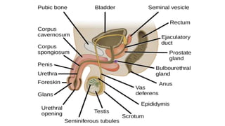
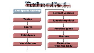
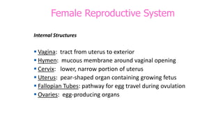
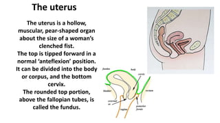
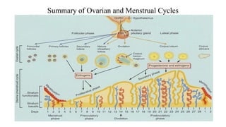
Sexual and reproductive
anatomy and physiology
What is the
reproductive
system?
The reproductive
systems in both the
male and female
consist of primary
and secondary sex
organs and sex
glands.
The primary function of the reproductive systems
is to perpetuate the species through sexual or
germ cell fertilization and reproduction.
Male organs of reproduction
Male Reproductive System
External Structures
• Penis: external male sex organ
- Circumcised: removes some or all of foreskin
- Uncircumcised: foreskin not removed
• Scrotum: sac of skin and muscle containing testicles
Uncircumcised Circumcised
Internal Structures
• Prostate: exocrine gland of male reproductive system
• Vas Deferens: tubes connecting epididymis to ejaculatory ducts
• Epididymis: organ where sperm matures
• Testicles: organ where sperm is created
• Urethra: tube that connects bladder to outside of body
• Cowper’s Gland: secretes lubricating fluid
• Seminal Vesicle: make semen (seminal fluid)
The oval-shaped
testes are located in
the scrotum, and are
each divided into 250
lobules. Coiled
within the lobule are
seminiferous tubes
where the sperm are
formed.
The mature sperm are stored in the epididymis, a
tube 13-20 feet in length, coiled and lying on the
side of each testicle.
Testes and epididymis…
The acrosome
covering the head
of the sperm
contains enzymes
that help it
penetrate the ova.
The head carries
the genetic
material.
The midpiece
supplies energy.
The tail or
flagellum provides
motility.
Sperm carry either an
X/female OR Y/male
chromosome. Since all ova
carry the X/female
chromosome, the male sperm
does influence the baby’s sex.
Sperm
The vas deferens are
muscular tubes
transporting sperm from
the epididymis to the
seminal vesicles.
The seminal vesicles
produce a slightly alkaline
fluid that mixes with the
sperm and becomes part of
the semen ejaculated
during the male orgasm.
It protects the sperm from
the acidity of the urethra.
Vas deferens and seminal vesicles
The Sperm Pathway
Vas deferens Expulsion
from the body
Testes
Sperm
Epididymis
Seminal vesicles
Ejaculatory duct
Prostate gland
Urethra
Female Reproductive system
External Structures
 Mons Pubis: soft mound of flesh above genitals
 Labia: lip-like structures on the outside of the vagina
 Clitoris: sex organ whose sole function is pleasure
External Organs
Female Reproductive System
Internal Structures
 Vagina: tract from uterus to exterior
 Hymen: mucous membrane around vaginal opening
 Cervix: lower, narrow portion of uterus
 Uterus: pear-shaped organ containing growing fetus
 Fallopian Tubes: pathway for egg travel during ovulation
 Ovaries: egg-producing organs
Internal Female Reproductive Organs
The uterus
The uterus is a hollow,
muscular, pear-shaped organ
about the size of a woman’s
clenched fist.
The top is tipped forward in a
normal ‘anteflexion’ position.
It can be divided into the body
or corpus, and the bottom
cervix.
The rounded top portion,
above the fallopian tubes, is
called the fundus.
The uterus is supported and
held in position by a number of
ligaments. Trauma, disease, or
multiple pregnancies can
weaken these ligaments and
result in abnormal positioning.
The uterus wall
has 3 layers: the
outside layer
called the
perimetrium, the
muscular inside
layer called the
myometrium, and
the mucous
membrane lining
the uterus called
the endometrium.
Outer layer
(Perimetrium)
Thick layer of muscle
(Myometrium)
Mucous lining
(Endometrium)
The cervical opening is about
the diameter of a pencil.
The uterus has 3
functions:
1. The endometrium
sheds the lining of the
uterus every 21 to 40
days by menstruation
2. It provides a place for
the protection and
nourishment of the
fetus during
pregnancy
3. It contracts during labor to expel the fetus
The fallopian tubes
The isthmus is
the portion that
connects to
uterus
Tube widens to form the
ampulla
Fimbria are the finger-like
projections around the
opening that trap the egg as
it leaves the ovary
End of tube is
called the
infundibulum
Opening
is called
the
ostium
The fallopian tube is 4-6 inches long. The egg, released
from the ovary, is captured by the fimbria and brought
into the fallopian tube.
The egg is moved along inside the tube by muscular
contractions and the waving action of cilia. It takes an
egg about 3-4 days to travel the length of the tube.
If an egg is fertilized, it occurs here.
The two ovaries are
attached to each
side of the uterus
by a ligament. They
are oval-shaped,
about the size of a
large olive, and lie
close to the fimbria
at the end of the
fallopian tubes.
The ovaries
Each ovary is filled, already at birth, with egg-
containing sacs called follicles.
Each egg is called an ovum.
Once every 21 days,
one follicle in one
ovary ripens. This
mature follicle is a
graafian follicle.
The follicle ruptures in
response to hormones
from the pituitary
gland, releasing the
ovum/egg. A process
called ovulation.
After the follicle ruptures, it
becomes a mass of yellow
cells called the corpus
luteum. This is a temporary,
progesterone-producing
structure.
The vagina extends
from the cervix to the
outside of the body.
It is a 3 ½ inch long
muscular tube that
expands in length and
width during sexual
arousal.
The vagina
The vagina is the female organ for copulation
(sexual intercourse), receiving the seminal fluid
from the male penis. It is also a passageway for
menstruation or the birth of a fetus.
The vulva is 5 organs making up
the external genitalia of the
female:
1. Mons pubis: triangular-shaped
pad of fatty tissue over the
pubis bone, covered with
pubic hair
The vulva
2. Labia majora: 2 large folds of adipose tissue on the sides
of the vaginal opening
3. Labia minora: 2 smaller folds of adipose tissue on the
inside of the labia majora
4. Vestibule: area between labia with openings for the
vagina, urethra, and two excretory ducts for Bartholin’s
glands (provide lubricant)
5. Clitoris: sensitive fold of tissue partially covered by hood
The perineum is the area
located between the vaginal
opening and the anus.
It is a muscular sheet that
can be torn during
childbirth.
Some doctors avoid
uncontrolled tearing of
the perineum by making
a surgical incision called
an episiotomy.
Perineum
The breasts are the
mammary glands,
varying in size according
to age, heredity, and the
amount of fatty tissue
present. Each breast has
15-20 glandular lobes
separated by connective
tissue.
After childbirth, the
pituitary gland stimulates
these lobules with the
hormone prolactin… and
they produce milk.
The breast
The dark-colored circle at
the tip of the breast is
called the areola.
It contains sebaceous
glands to keep the skin
conditioned.
In the center is the nipple,
where ducts from the
lobules open.
The first secretion from
the breast is not a true
milk, but a thin yellowish
substance called
colostrum.
Colostrum contains
nutrients and the
mother’s immunities
that can protect baby.
Reproductive system of both males & females is similar, or sexually
undifferentiated for the 1st 6 weeks of prenatal life.
At 7th week difference between males & female appear in the internal
structures.
Differentiation of the external sexual organs is complete at about 12
weeks.
Female Puberty
▪ Begins anywhere from 8 to 15 years of age
▪ Earlier onset of menstruation may be due
to being overweight.
Puberty lasts about 3 to 5 years
▪ Begins when pituitary gland initiates release of FSH and LH,
which increases the ovaries production of estrogen
▪ Increased size of Fallopian tubes, uterus,
vagina, breasts, buttocks, thighs
Secondary Sex Characteristics
Pubertal changes typically are manifest as:
 Growth spurt
 Increase in the transverse diameter of the pelvis
 Breast development
 Growth of pubic hair
 Onset of menstruation
 Growth of axillary hair
 Vaginal secretions
▪ Menstruation: Is a cycle of regular uterine bleeding that begins with
puberty & ends with menopause, occurs each month except during
pregnancy.
▪ Menstrual cycle: Initiated in response to the interaction between the
reproductive & endocrine system. It is interrupted by pregnancy.
Average every 28 days.
▪ Menarche: 1st cycle normally occurs between the age of 9- 16th.
Heredity, environment, nutritional status & race affect menarche.
▪ Early menstrual periods are often irregular& scant.
▪ Early menstrual cycles are not usually fertile because
ovulation occurs inconsistently.
Female reproductive Cycle:
The female reproductive cycle is often called
Menstrual cycle because menstruation provides
a marker for each cycle’s beginning & end if
pregnancy does not occur
.
▪ The duration of the cycle is about 28 days,
although it may range from 20-45 days.
▪ Significant deviations from the 28days cycle are
associated with reduced fertility.
▪ The female reproductive cycle is divided into two cycles
that reflect changes in the ovaries and uterine
Endometrium.
Inside the ovary
immature
eggs
Day 1-13:
egg developing
inside the
growing follicle
Day 14:
ovulation – egg
released from follicle
Day 15-28:
empty follicle turns
into corpus luteum
(yellow body)
Ovarian Cycle
• In response to GNRH from the hypothalamus ,
the anterior pituitary secretes FSH & LH.
• FSH & LH stimulates the ovaries to mature an
ovum, release it, & secrete other hormones
that will prepare the Endometrium for
implantation of a fertilized ovum.
• It contains 3 phases:
1 Follicular phase.
2 Ovulatory phase.
3 Luteal phase.
1 Follicular phase : Begins after menstruation ends; lasts 6-13
days; ovarian follicles begin to ripen; estrogen promotes
development of endometrium to 2-5 millimeters thick.
2 Ovulatory phase : Around day 14; about 2 days before
ovulation, LH & FSH secretion rises cause a slight fall in
estrogen production & a rise in progesterone secretion,
stimulating final maturation of a single follicle& release of its
mature ovum.
3 Luteal phase:
After ovulation & under the influence of LH , the
remaining cells of the old follicle persist for about 12
days as a corpus luteum. It secretes estrogen & large
amount of progesterone to prepare the
endometerium for a fertilized ovum.
• Level of FSH & LH decrease in response of high
estrogen & progesterone . If ovum is fertilized, it
secretes HCG that causes the corpus luteum to
persist to maintain an early pregnancy.
• If the ovum is not fertilized, FSH& LH falls & the
corpus luteum regresses & decrease in estrogen &
Progesterone results in menstruation.
Ovarian Cycle
Endometerial Cycle:
A- Menstrual phase: From day 1-5, Estrogen &
progesterone at low level, Menses initiated, with a
normal range of 20 to 80 ml it is bright or light red in
color discharged through vagina.
Discharge consist of Endo material sloughed, mucous
and blood.
8/23/2021 Maternity Nursing Mrs samar fadda 16
Uterus lining
The lining of the uterus becomes thicker with blood vessels and
more stable during the menstrual cycle. Why is this important?
period
2 4 6 8 10 12 14 16 18 20 22 24 26 28
days after start of period
A fertilized egg will bury itself (implant) in the uterus lining.
The egg needs a plentiful supply of oxygen and nutrients to
develop.
uterus
lining
thickness
B- Proliferative phase:
Pituitary gland releases FSH  Stimulate
ovary to prepare mature ovum (graafian folloicle)
 release estrogen which help the endometrium to become
more vascular & thicker
.
Release of LH  graafian follicle rupture  mature
ovum  fallopian tube.
C- Secretory phase: Corpus lutueum secrete estrogen &
progesterone to maintain the vascular lining of the uterus until
placental formation.
If no fertilization corpus luteum survives only 8 days then
disintegrates estrogen & progesterone , menstruation occurs.
Hormonal changes causes:
• Basal body temperature  one degree just before the day of
ovulation due to low progesterone level.
• Basal body temperature  one degree following ovulation due
to high progesterone level & temperature remain at this level until
the end of menses.
• If conception occurs Fertilized ovum implants on the endometrium
of the uterus Corpus luteum remains 16-20 wks of pregnancy.
• If conception doesn’t occur Unfertilized ovum atrophies after 4-5
days & corpus luteum regresses.
Summary of Ovarian and Menstrual Cycles
Ovulation:
❑Each month Anterior pituitary release FSH which
stimulate Primordial follicle to grow & mature.
❑The cell of Primordial follicle produce clear fluid high in
estrogen (Estridol) & some progesterone.
❑Estrogen: Produced by the ovaries, encourages the
uterus to start building up a thick lining in preparation
for pregnancy.
❑As the primordial follicle grows it become visible as a
clear water blister on the surface of the ovaries.
❑Then it becomes as Graafian follicle small ovum has
follicle membrane & fluid.
Ovum mature & set on the surface of the
ovaries.
Ovulation period
FSH LH
FSH Enhance rupture graafian follicle then go to the
fallopian tube.
LH Act on the follicle cell to produce follicular fluid
yellow, rich in progesterone, (Corpus luteum)
Changes in cervical mucous
❑During most of the female reproductive cycle. The
mucous of the cervix is scant & thick.
❑Just before ovulation cervical mucous becomes thin,
clear & elastic to promote passage of sperm into the
uterus & fallopian tube, where they can fertilize the
ovum.
Menstrual Problems
1- Premenstrual syndrome (PMS): is a combination of
emotional, physical, psychological, and mood disturbances that
occur after a woman's ovulation and typically ending with the
onset of her menstrual flow.
The most common mood-related symptoms are irritability,
depression, crying, oversensitivity, and mood swings with
alternating sadness and anger
.
The most common physical symptoms are fatigue, bloating,
breast tenderness (mastalgia), acne, and appetite changes with
food cravings
General management includes a healthy lifestyle
including:
• Exercise.
• Family and friends can provide emotional support
during the time of a woman's cycle.
• Avoid salt before the menstrual period.
• Reduce caffeine intake.
• Quite smoking.
• Reduce alcohol& refined sugars.
• some studies suggest that vitamin B6, vitamin E, calcium, and
magnesium supplements may have some benefit.
• In severe cases Non steroidal anti inflammatory
medications.
• Oral contraceptive pills (OCPs).
2- Dysmenorrhea: Menstrual cramps are pains in the belly
and pelvic areas that are experienced by a woman as a result
of her menstrual period
There are two types of dysmenorrhea, primary and
secondary.
Primary dysmenorrhea: there is no underlying gynecologic
problem causing the pain. This type of cramping may begin
within six months to a year following menarche (the
beginning of menstruation), the time when a girl starts
having menstrual periods. Menstrual cramps typically do not
begin until ovulatory menstrual cycles (when an egg is
released from the ovaries) occur, and actual menstrual
bleeding usually begins before the onset of ovulation.
Secondary dysmenorrhea: some underlying
abnormal condition contributes to the
menstrual pain.
Secondary dysmenorrhea may be evident at
menarche but, more often, the condition
develops later
.
Treatment for primary dysmenorrhea:
• Every woman needs to find a treatment that works
for her. There are a number of possible remedies for
menstrual cramps.
1- include not only adequate rest and sleep, but also
regular exercise (especially walking). Some women find
that abdominal massage, yoga, or orgasmic sexual
activity may bring relief.
2-A heating pad applied to the abdominal area may
relieve the pain and congestion and decrease
symptoms.
3-The main agents for treating moderate menstrual
cramps are the nonsteroidal antiinflammatory drugs
For mild cramps, aspirin or acetaminophen
• For severe low doses of birth control pills (oral
contraceptives) containing estrogen and
progestin in a regular or extended cycle.
• This type of approach can prevent ovulation
(the monthly release of an egg) and reduce
the production of prostaglandins which, in
turn, reduces the severity of cramping and
causes a light menstrual flow.
3- Amenorrhea:
• Amenorrhea is the medical term for the
absence of menstrual periods, either on a
permanent or temporary basis. Amenorrhea
can be classified as primary or secondary.
Primary amenorrhea: menstrual periods have
never begun (by age 16),
Secondary amenorrhea : absence of
menstrual periods for three consecutive cycles
or a time period of more than six months in a
woman who was previously menstruating.
What causes amenorrhea?
The normal menstrual cycle occurs because of
changing levels of hormones made and secreted
by the ovaries.
The ovaries respond to hormonal signals from
the pituitary gland located at the base of the
brain, which is, in turn, controlled by hormones
produced in the hypothalamus of the brain.
Disorders that affect any component of this
regulatory cycle can lead to amenorrhea.
Treatment: According to the causative Factor,
mainly hormonal therapy.
4- Oligomenorrhea: Infrequent menses may result
from hormonal imbalances.
5 - Menorrhagia: Excessive menstrual flow.
6 -Metrorrhagia: Bleeding at various time during
7 - Menopause: Cessation of menses & the cycle.
Reproductive capability in older adult women ~at 50
50 yrs. Occurs due to in ovarian activity & in
circulating estrogen.
Conception and ovum development
Gametogenesis:
• Gametes (ovum and spermatazon) are formed by meiosis ; each
has one set of 23 chromosomes; other body cells have two sets (46
chromosomes, 23 pairs)
• Oogenesis the process of egg ( ovum/oocyte) formation in female.
• Spermatogenesis the process of sperm
(spermatocyte)formation in male.
Oögenesis – Oögonia to Oöcytes
• Germ cells from yolk sac migrate to ovary and
become potential egg cells called oögonia
• In fetus, millions of oögonia produced by mitosis
but most of them degenerate .
• Some develop into immature egg cells called
primary oöcytes during fetal development
– 200,000 to 2 millions present at birth
– 40,000 remain at puberty but only 400 mature during a
woman’s reproductive life
• Each month about 20 primary oöcytes become
secondary oöcytes but usually only one survives to
be ovulated from Graffian follicle
Oögenesis
Egg forming cells (oöcytes) go through
two divisions
1º = primary
2º = secondary
•Starts with a 2n=46 1ºoöcyte that
divides, resulting in two n=23 cells,
but one is a large 2º oöcyte and one
is a small 1st polar body that may
itself divide
•Second division only occurs if
2º oöcyte is fertilized. Results in
one large n=23 ovum (egg) and one
small n=23 2nd polar body
•Thus oögenesis results in one large
fertilized egg (zygote) and possibly
three small polar bodies
Spermatogenesis
•Begins during puberty in the male.
•Primitive sperm cells spermatogonia develop
during fetal life & begin multiplying by mitosis
during puberty.
•Male produces new spermatogonia that can
mature into sperm throughout his life time.
❑The halves form a new cell & restore the
chromosome number 46.
❑Mother’s ovum carries a single X-
chromosome.
❑Father’s spermatozoa carries either an X or Y
chromosome.
❑If an X – bearing spermatozoon fertilizes the
ovum the off spring’s genetic cell is female.
❑If a Y- bearing spermatozoon fertilizes the
ovum a male off spring results.
Ejaculation
❑Male ejaculates ~ 200- 500 million sperm, are
deposited in the upper vagina & over the cervix.
❑Sperm are suspended in seminal fluid which nourishes
& protects them from the acidic vaginal environment.
❑To hold the semen deeply in the vagina the seminal
fluid coagulates some what after ejaculation.
❑Sperm are relatively immobile for about 15-30 min until
other seminal enzymes dissolve the coagulated fluid &
allow the sperm to begin moving upward throughout
the cervix.
Conception:
Timing between release of a mature ovum at
ovulation & ejaculation of enough healthy,
mature, motile sperm into the vagina.
Ovum lives waiting for fertilization 24-72 hrs.
Mature sperm live 24-48 hrs although a few
may remain fertile in the woman’s
reproductive tract as long as 5 days.
• Fertilization usually occurs in the ampulla of the
fallopian tube.
• Estrogen levels increases during ovulation,
aiding fertilization and easing transit of the
ovum down the fallopian tube.
The ovum membrane is surrounded by two layers
of tissues:
a. Zona pellucida : an inner layer
b. Corona radiate : outer layer
.
Conception
Preparation for conception in the female
❑Before ovulation , several oocytes begin to
mature under the influence of FSH & LH,
from the anterior pituitary gland.
❑Maturing oocytes are contained in a sac
called Graafian follicle which produce
estrogen + progestron to prepare the
endometrium for possible pregnancy.
Fertilization and tubal transport of the zygote. From fertilization to implantation,
the zygote travels through the fallopian tube, experiencing rapid mitotic division
(cleavage). During the journey toward the uterus the zygote evolves through
several stages, including morula and blastocyst.
Zygote Development: Cell
Division & Implantation
Preparation of sperm for fertilization
Sperm undergoes two processes before it is able to
penetrate the ovum.
1- Capacitation: Structural changes occur once in the
female genital tract.
2- Acrosomal reaction: the sperm releases enzymes.
•Hyaluronidase: causes separation of the corona radiate.
•Acrosin and neuraminidase allow the sperm enter the
zona pellucida.
• Fertilization:
The joining of the egg and sperm is
fertilization, usually in the outer 1/3 of the fallopian
tube.
• It occurs about 24 hours after ovulation when sperm
enters the ovum.
• The new cell contains 23 chromosomes from the sperm
and 23 chromosomes from the ova.
• The fertilized ovum is termed zygote; the chromosome
number is thus restored to two sets (46 chromosomes)
• After fertilization changes in the zona pellucida
surrounding the ovum prevent the sperm from entering.
• Cleavage:
• Rapid mitotic division of zygot
produces a morula( a ball of 16 cells) that divides
to form a blastocyst.
• Morula divides into trophoblast( which become
the placenta and chorion) and embryoblast which
becomes the embryon.
• The total structure of the developing embryo is
called blastocyst
8/23/2021 Maternity Nursing 77
A- Pre-embryonic (zygotic)stage.
Fertilized ovum  Fallopian tube  Uterus takes ~ 3 days.
• Zygote undergoes Mitotic Division (Cleavage)

Plastomers ( Further Multiplication)

Morula (floats in uterus for 3-4 days)under going changes

Mass cell

Blastocyst( Inner solid mass)
(Amnion )

Trophoblast
(Chorion )
Inner cell mass
Destined to
become fetus
Fertilization
Trophoblast
Accomplishes
implantation
and develops
into fetal
portions of
placenta
Blastocyst
(cross
section)
Blastocoele Becomes amniotic sac
Morula
Cleavage
Spermatozoa
Ovum
(cross
section)
Secondary
oocyte
(ovum)
Ovulation
Ovary
Implantation
Endometrium
of uterus
B- Implantation:
Trophoblast outer layer of mass cell secrete
enzyme that help the Blastocyst embed in
the uterine lining.
Time from conception to implantation ~ 7
days.
Endometrium is very thick and vascular
.
Blastocyst: Implant into the rich vascular
endometrium until completely covered &
implant it self.
The site of implantation is important because it is
the place were the placenta develops.
Implantation occurs in the upper uterus
posterior portion.
After implantation Endometrium called Decidua
.
Decidua continues its thicking under the
implanted blastocyst.
Trophoblast develops finger like projections called
chorionic villa contain blood of the developing
zygot & gives O2 & nutrient from mother’s blood
vessels walls by osmosis & diffusion.
Endometrium has 3 separate areas:
Decidua basalis: Part of endometrium lying
directly under the embryo or where the
Trophoblast cells are establishing communication
with maternal blood vessels.
Decidua Capsularis: The portion of
endometrium that stretches or encapsulates the
surface of the Trophoblast.
Decidua vera: Remaining portion of the uterine
lining.
Thank you

human reproduction.pptx

  • 1.
  • 2.
    What is the reproductive system? Thereproductive systems in both the male and female consist of primary and secondary sex organs and sex glands. The primary function of the reproductive systems is to perpetuate the species through sexual or germ cell fertilization and reproduction.
  • 3.
    Male organs ofreproduction
  • 4.
    Male Reproductive System ExternalStructures • Penis: external male sex organ - Circumcised: removes some or all of foreskin - Uncircumcised: foreskin not removed • Scrotum: sac of skin and muscle containing testicles Uncircumcised Circumcised
  • 6.
    Internal Structures • Prostate:exocrine gland of male reproductive system • Vas Deferens: tubes connecting epididymis to ejaculatory ducts • Epididymis: organ where sperm matures • Testicles: organ where sperm is created • Urethra: tube that connects bladder to outside of body • Cowper’s Gland: secretes lubricating fluid • Seminal Vesicle: make semen (seminal fluid)
  • 7.
    The oval-shaped testes arelocated in the scrotum, and are each divided into 250 lobules. Coiled within the lobule are seminiferous tubes where the sperm are formed. The mature sperm are stored in the epididymis, a tube 13-20 feet in length, coiled and lying on the side of each testicle. Testes and epididymis…
  • 8.
    The acrosome covering thehead of the sperm contains enzymes that help it penetrate the ova. The head carries the genetic material. The midpiece supplies energy. The tail or flagellum provides motility. Sperm carry either an X/female OR Y/male chromosome. Since all ova carry the X/female chromosome, the male sperm does influence the baby’s sex. Sperm
  • 9.
    The vas deferensare muscular tubes transporting sperm from the epididymis to the seminal vesicles. The seminal vesicles produce a slightly alkaline fluid that mixes with the sperm and becomes part of the semen ejaculated during the male orgasm. It protects the sperm from the acidity of the urethra. Vas deferens and seminal vesicles
  • 10.
    The Sperm Pathway Vasdeferens Expulsion from the body Testes Sperm Epididymis Seminal vesicles Ejaculatory duct Prostate gland Urethra
  • 11.
  • 12.
    External Structures  MonsPubis: soft mound of flesh above genitals  Labia: lip-like structures on the outside of the vagina  Clitoris: sex organ whose sole function is pleasure
  • 13.
  • 14.
    Female Reproductive System InternalStructures  Vagina: tract from uterus to exterior  Hymen: mucous membrane around vaginal opening  Cervix: lower, narrow portion of uterus  Uterus: pear-shaped organ containing growing fetus  Fallopian Tubes: pathway for egg travel during ovulation  Ovaries: egg-producing organs
  • 15.
  • 16.
    The uterus The uterusis a hollow, muscular, pear-shaped organ about the size of a woman’s clenched fist. The top is tipped forward in a normal ‘anteflexion’ position. It can be divided into the body or corpus, and the bottom cervix. The rounded top portion, above the fallopian tubes, is called the fundus.
  • 17.
    The uterus issupported and held in position by a number of ligaments. Trauma, disease, or multiple pregnancies can weaken these ligaments and result in abnormal positioning.
  • 18.
    The uterus wall has3 layers: the outside layer called the perimetrium, the muscular inside layer called the myometrium, and the mucous membrane lining the uterus called the endometrium. Outer layer (Perimetrium) Thick layer of muscle (Myometrium) Mucous lining (Endometrium) The cervical opening is about the diameter of a pencil.
  • 19.
    The uterus has3 functions: 1. The endometrium sheds the lining of the uterus every 21 to 40 days by menstruation 2. It provides a place for the protection and nourishment of the fetus during pregnancy 3. It contracts during labor to expel the fetus
  • 20.
    The fallopian tubes Theisthmus is the portion that connects to uterus Tube widens to form the ampulla Fimbria are the finger-like projections around the opening that trap the egg as it leaves the ovary End of tube is called the infundibulum Opening is called the ostium
  • 21.
    The fallopian tubeis 4-6 inches long. The egg, released from the ovary, is captured by the fimbria and brought into the fallopian tube. The egg is moved along inside the tube by muscular contractions and the waving action of cilia. It takes an egg about 3-4 days to travel the length of the tube. If an egg is fertilized, it occurs here.
  • 22.
    The two ovariesare attached to each side of the uterus by a ligament. They are oval-shaped, about the size of a large olive, and lie close to the fimbria at the end of the fallopian tubes. The ovaries Each ovary is filled, already at birth, with egg- containing sacs called follicles. Each egg is called an ovum.
  • 23.
    Once every 21days, one follicle in one ovary ripens. This mature follicle is a graafian follicle. The follicle ruptures in response to hormones from the pituitary gland, releasing the ovum/egg. A process called ovulation. After the follicle ruptures, it becomes a mass of yellow cells called the corpus luteum. This is a temporary, progesterone-producing structure.
  • 24.
    The vagina extends fromthe cervix to the outside of the body. It is a 3 ½ inch long muscular tube that expands in length and width during sexual arousal. The vagina The vagina is the female organ for copulation (sexual intercourse), receiving the seminal fluid from the male penis. It is also a passageway for menstruation or the birth of a fetus.
  • 25.
    The vulva is5 organs making up the external genitalia of the female: 1. Mons pubis: triangular-shaped pad of fatty tissue over the pubis bone, covered with pubic hair The vulva 2. Labia majora: 2 large folds of adipose tissue on the sides of the vaginal opening 3. Labia minora: 2 smaller folds of adipose tissue on the inside of the labia majora 4. Vestibule: area between labia with openings for the vagina, urethra, and two excretory ducts for Bartholin’s glands (provide lubricant) 5. Clitoris: sensitive fold of tissue partially covered by hood
  • 26.
    The perineum isthe area located between the vaginal opening and the anus. It is a muscular sheet that can be torn during childbirth. Some doctors avoid uncontrolled tearing of the perineum by making a surgical incision called an episiotomy. Perineum
  • 27.
    The breasts arethe mammary glands, varying in size according to age, heredity, and the amount of fatty tissue present. Each breast has 15-20 glandular lobes separated by connective tissue. After childbirth, the pituitary gland stimulates these lobules with the hormone prolactin… and they produce milk. The breast
  • 28.
    The dark-colored circleat the tip of the breast is called the areola. It contains sebaceous glands to keep the skin conditioned. In the center is the nipple, where ducts from the lobules open. The first secretion from the breast is not a true milk, but a thin yellowish substance called colostrum. Colostrum contains nutrients and the mother’s immunities that can protect baby.
  • 29.
    Reproductive system ofboth males & females is similar, or sexually undifferentiated for the 1st 6 weeks of prenatal life. At 7th week difference between males & female appear in the internal structures. Differentiation of the external sexual organs is complete at about 12 weeks.
  • 30.
    Female Puberty ▪ Beginsanywhere from 8 to 15 years of age ▪ Earlier onset of menstruation may be due to being overweight. Puberty lasts about 3 to 5 years ▪ Begins when pituitary gland initiates release of FSH and LH, which increases the ovaries production of estrogen ▪ Increased size of Fallopian tubes, uterus, vagina, breasts, buttocks, thighs
  • 31.
    Secondary Sex Characteristics Pubertalchanges typically are manifest as:  Growth spurt  Increase in the transverse diameter of the pelvis  Breast development  Growth of pubic hair  Onset of menstruation  Growth of axillary hair  Vaginal secretions
  • 32.
    ▪ Menstruation: Isa cycle of regular uterine bleeding that begins with puberty & ends with menopause, occurs each month except during pregnancy. ▪ Menstrual cycle: Initiated in response to the interaction between the reproductive & endocrine system. It is interrupted by pregnancy. Average every 28 days. ▪ Menarche: 1st cycle normally occurs between the age of 9- 16th. Heredity, environment, nutritional status & race affect menarche. ▪ Early menstrual periods are often irregular& scant. ▪ Early menstrual cycles are not usually fertile because ovulation occurs inconsistently.
  • 33.
    Female reproductive Cycle: Thefemale reproductive cycle is often called Menstrual cycle because menstruation provides a marker for each cycle’s beginning & end if pregnancy does not occur .
  • 34.
    ▪ The durationof the cycle is about 28 days, although it may range from 20-45 days. ▪ Significant deviations from the 28days cycle are associated with reduced fertility. ▪ The female reproductive cycle is divided into two cycles that reflect changes in the ovaries and uterine Endometrium.
  • 35.
    Inside the ovary immature eggs Day1-13: egg developing inside the growing follicle Day 14: ovulation – egg released from follicle Day 15-28: empty follicle turns into corpus luteum (yellow body)
  • 36.
    Ovarian Cycle • Inresponse to GNRH from the hypothalamus , the anterior pituitary secretes FSH & LH. • FSH & LH stimulates the ovaries to mature an ovum, release it, & secrete other hormones that will prepare the Endometrium for implantation of a fertilized ovum. • It contains 3 phases: 1 Follicular phase. 2 Ovulatory phase. 3 Luteal phase.
  • 37.
    1 Follicular phase: Begins after menstruation ends; lasts 6-13 days; ovarian follicles begin to ripen; estrogen promotes development of endometrium to 2-5 millimeters thick. 2 Ovulatory phase : Around day 14; about 2 days before ovulation, LH & FSH secretion rises cause a slight fall in estrogen production & a rise in progesterone secretion, stimulating final maturation of a single follicle& release of its mature ovum.
  • 38.
    3 Luteal phase: Afterovulation & under the influence of LH , the remaining cells of the old follicle persist for about 12 days as a corpus luteum. It secretes estrogen & large amount of progesterone to prepare the endometerium for a fertilized ovum. • Level of FSH & LH decrease in response of high estrogen & progesterone . If ovum is fertilized, it secretes HCG that causes the corpus luteum to persist to maintain an early pregnancy. • If the ovum is not fertilized, FSH& LH falls & the corpus luteum regresses & decrease in estrogen & Progesterone results in menstruation.
  • 39.
  • 40.
    Endometerial Cycle: A- Menstrualphase: From day 1-5, Estrogen & progesterone at low level, Menses initiated, with a normal range of 20 to 80 ml it is bright or light red in color discharged through vagina. Discharge consist of Endo material sloughed, mucous and blood. 8/23/2021 Maternity Nursing Mrs samar fadda 16
  • 41.
    Uterus lining The liningof the uterus becomes thicker with blood vessels and more stable during the menstrual cycle. Why is this important? period 2 4 6 8 10 12 14 16 18 20 22 24 26 28 days after start of period A fertilized egg will bury itself (implant) in the uterus lining. The egg needs a plentiful supply of oxygen and nutrients to develop. uterus lining thickness
  • 42.
    B- Proliferative phase: Pituitarygland releases FSH  Stimulate ovary to prepare mature ovum (graafian folloicle)  release estrogen which help the endometrium to become more vascular & thicker . Release of LH  graafian follicle rupture  mature ovum  fallopian tube. C- Secretory phase: Corpus lutueum secrete estrogen & progesterone to maintain the vascular lining of the uterus until placental formation. If no fertilization corpus luteum survives only 8 days then disintegrates estrogen & progesterone , menstruation occurs.
  • 43.
    Hormonal changes causes: •Basal body temperature  one degree just before the day of ovulation due to low progesterone level. • Basal body temperature  one degree following ovulation due to high progesterone level & temperature remain at this level until the end of menses. • If conception occurs Fertilized ovum implants on the endometrium of the uterus Corpus luteum remains 16-20 wks of pregnancy. • If conception doesn’t occur Unfertilized ovum atrophies after 4-5 days & corpus luteum regresses.
  • 44.
    Summary of Ovarianand Menstrual Cycles
  • 45.
    Ovulation: ❑Each month Anteriorpituitary release FSH which stimulate Primordial follicle to grow & mature. ❑The cell of Primordial follicle produce clear fluid high in estrogen (Estridol) & some progesterone. ❑Estrogen: Produced by the ovaries, encourages the uterus to start building up a thick lining in preparation for pregnancy. ❑As the primordial follicle grows it become visible as a clear water blister on the surface of the ovaries. ❑Then it becomes as Graafian follicle small ovum has follicle membrane & fluid.
  • 46.
    Ovum mature &set on the surface of the ovaries. Ovulation period FSH LH FSH Enhance rupture graafian follicle then go to the fallopian tube. LH Act on the follicle cell to produce follicular fluid yellow, rich in progesterone, (Corpus luteum)
  • 48.
    Changes in cervicalmucous ❑During most of the female reproductive cycle. The mucous of the cervix is scant & thick. ❑Just before ovulation cervical mucous becomes thin, clear & elastic to promote passage of sperm into the uterus & fallopian tube, where they can fertilize the ovum.
  • 49.
    Menstrual Problems 1- Premenstrualsyndrome (PMS): is a combination of emotional, physical, psychological, and mood disturbances that occur after a woman's ovulation and typically ending with the onset of her menstrual flow. The most common mood-related symptoms are irritability, depression, crying, oversensitivity, and mood swings with alternating sadness and anger . The most common physical symptoms are fatigue, bloating, breast tenderness (mastalgia), acne, and appetite changes with food cravings
  • 50.
    General management includesa healthy lifestyle including: • Exercise. • Family and friends can provide emotional support during the time of a woman's cycle. • Avoid salt before the menstrual period. • Reduce caffeine intake. • Quite smoking. • Reduce alcohol& refined sugars. • some studies suggest that vitamin B6, vitamin E, calcium, and magnesium supplements may have some benefit.
  • 51.
    • In severecases Non steroidal anti inflammatory medications. • Oral contraceptive pills (OCPs).
  • 52.
    2- Dysmenorrhea: Menstrualcramps are pains in the belly and pelvic areas that are experienced by a woman as a result of her menstrual period There are two types of dysmenorrhea, primary and secondary. Primary dysmenorrhea: there is no underlying gynecologic problem causing the pain. This type of cramping may begin within six months to a year following menarche (the beginning of menstruation), the time when a girl starts having menstrual periods. Menstrual cramps typically do not begin until ovulatory menstrual cycles (when an egg is released from the ovaries) occur, and actual menstrual bleeding usually begins before the onset of ovulation.
  • 53.
    Secondary dysmenorrhea: someunderlying abnormal condition contributes to the menstrual pain. Secondary dysmenorrhea may be evident at menarche but, more often, the condition develops later .
  • 54.
    Treatment for primarydysmenorrhea: • Every woman needs to find a treatment that works for her. There are a number of possible remedies for menstrual cramps. 1- include not only adequate rest and sleep, but also regular exercise (especially walking). Some women find that abdominal massage, yoga, or orgasmic sexual activity may bring relief. 2-A heating pad applied to the abdominal area may relieve the pain and congestion and decrease symptoms. 3-The main agents for treating moderate menstrual cramps are the nonsteroidal antiinflammatory drugs For mild cramps, aspirin or acetaminophen
  • 55.
    • For severelow doses of birth control pills (oral contraceptives) containing estrogen and progestin in a regular or extended cycle. • This type of approach can prevent ovulation (the monthly release of an egg) and reduce the production of prostaglandins which, in turn, reduces the severity of cramping and causes a light menstrual flow.
  • 56.
    3- Amenorrhea: • Amenorrheais the medical term for the absence of menstrual periods, either on a permanent or temporary basis. Amenorrhea can be classified as primary or secondary. Primary amenorrhea: menstrual periods have never begun (by age 16), Secondary amenorrhea : absence of menstrual periods for three consecutive cycles or a time period of more than six months in a woman who was previously menstruating.
  • 57.
    What causes amenorrhea? Thenormal menstrual cycle occurs because of changing levels of hormones made and secreted by the ovaries. The ovaries respond to hormonal signals from the pituitary gland located at the base of the brain, which is, in turn, controlled by hormones produced in the hypothalamus of the brain. Disorders that affect any component of this regulatory cycle can lead to amenorrhea. Treatment: According to the causative Factor, mainly hormonal therapy.
  • 58.
    4- Oligomenorrhea: Infrequentmenses may result from hormonal imbalances. 5 - Menorrhagia: Excessive menstrual flow. 6 -Metrorrhagia: Bleeding at various time during 7 - Menopause: Cessation of menses & the cycle. Reproductive capability in older adult women ~at 50 50 yrs. Occurs due to in ovarian activity & in circulating estrogen.
  • 59.
  • 60.
    Gametogenesis: • Gametes (ovumand spermatazon) are formed by meiosis ; each has one set of 23 chromosomes; other body cells have two sets (46 chromosomes, 23 pairs) • Oogenesis the process of egg ( ovum/oocyte) formation in female. • Spermatogenesis the process of sperm (spermatocyte)formation in male.
  • 61.
    Oögenesis – Oögoniato Oöcytes • Germ cells from yolk sac migrate to ovary and become potential egg cells called oögonia • In fetus, millions of oögonia produced by mitosis but most of them degenerate . • Some develop into immature egg cells called primary oöcytes during fetal development – 200,000 to 2 millions present at birth – 40,000 remain at puberty but only 400 mature during a woman’s reproductive life • Each month about 20 primary oöcytes become secondary oöcytes but usually only one survives to be ovulated from Graffian follicle
  • 62.
    Oögenesis Egg forming cells(oöcytes) go through two divisions 1º = primary 2º = secondary •Starts with a 2n=46 1ºoöcyte that divides, resulting in two n=23 cells, but one is a large 2º oöcyte and one is a small 1st polar body that may itself divide •Second division only occurs if 2º oöcyte is fertilized. Results in one large n=23 ovum (egg) and one small n=23 2nd polar body •Thus oögenesis results in one large fertilized egg (zygote) and possibly three small polar bodies
  • 63.
    Spermatogenesis •Begins during pubertyin the male. •Primitive sperm cells spermatogonia develop during fetal life & begin multiplying by mitosis during puberty. •Male produces new spermatogonia that can mature into sperm throughout his life time.
  • 64.
    ❑The halves forma new cell & restore the chromosome number 46. ❑Mother’s ovum carries a single X- chromosome. ❑Father’s spermatozoa carries either an X or Y chromosome. ❑If an X – bearing spermatozoon fertilizes the ovum the off spring’s genetic cell is female. ❑If a Y- bearing spermatozoon fertilizes the ovum a male off spring results.
  • 65.
    Ejaculation ❑Male ejaculates ~200- 500 million sperm, are deposited in the upper vagina & over the cervix. ❑Sperm are suspended in seminal fluid which nourishes & protects them from the acidic vaginal environment. ❑To hold the semen deeply in the vagina the seminal fluid coagulates some what after ejaculation. ❑Sperm are relatively immobile for about 15-30 min until other seminal enzymes dissolve the coagulated fluid & allow the sperm to begin moving upward throughout the cervix.
  • 66.
    Conception: Timing between releaseof a mature ovum at ovulation & ejaculation of enough healthy, mature, motile sperm into the vagina. Ovum lives waiting for fertilization 24-72 hrs. Mature sperm live 24-48 hrs although a few may remain fertile in the woman’s reproductive tract as long as 5 days.
  • 67.
    • Fertilization usuallyoccurs in the ampulla of the fallopian tube. • Estrogen levels increases during ovulation, aiding fertilization and easing transit of the ovum down the fallopian tube. The ovum membrane is surrounded by two layers of tissues: a. Zona pellucida : an inner layer b. Corona radiate : outer layer .
  • 68.
  • 69.
    Preparation for conceptionin the female ❑Before ovulation , several oocytes begin to mature under the influence of FSH & LH, from the anterior pituitary gland. ❑Maturing oocytes are contained in a sac called Graafian follicle which produce estrogen + progestron to prepare the endometrium for possible pregnancy.
  • 71.
    Fertilization and tubaltransport of the zygote. From fertilization to implantation, the zygote travels through the fallopian tube, experiencing rapid mitotic division (cleavage). During the journey toward the uterus the zygote evolves through several stages, including morula and blastocyst.
  • 72.
  • 73.
    Preparation of spermfor fertilization Sperm undergoes two processes before it is able to penetrate the ovum. 1- Capacitation: Structural changes occur once in the female genital tract. 2- Acrosomal reaction: the sperm releases enzymes. •Hyaluronidase: causes separation of the corona radiate. •Acrosin and neuraminidase allow the sperm enter the zona pellucida.
  • 74.
    • Fertilization: The joiningof the egg and sperm is fertilization, usually in the outer 1/3 of the fallopian tube. • It occurs about 24 hours after ovulation when sperm enters the ovum. • The new cell contains 23 chromosomes from the sperm and 23 chromosomes from the ova. • The fertilized ovum is termed zygote; the chromosome number is thus restored to two sets (46 chromosomes) • After fertilization changes in the zona pellucida surrounding the ovum prevent the sperm from entering.
  • 76.
    • Cleavage: • Rapidmitotic division of zygot produces a morula( a ball of 16 cells) that divides to form a blastocyst. • Morula divides into trophoblast( which become the placenta and chorion) and embryoblast which becomes the embryon. • The total structure of the developing embryo is called blastocyst
  • 77.
  • 78.
    A- Pre-embryonic (zygotic)stage. Fertilizedovum  Fallopian tube  Uterus takes ~ 3 days. • Zygote undergoes Mitotic Division (Cleavage)  Plastomers ( Further Multiplication)  Morula (floats in uterus for 3-4 days)under going changes  Mass cell  Blastocyst( Inner solid mass) (Amnion )  Trophoblast (Chorion )
  • 79.
    Inner cell mass Destinedto become fetus Fertilization Trophoblast Accomplishes implantation and develops into fetal portions of placenta Blastocyst (cross section) Blastocoele Becomes amniotic sac Morula Cleavage Spermatozoa Ovum (cross section) Secondary oocyte (ovum) Ovulation Ovary Implantation Endometrium of uterus
  • 80.
    B- Implantation: Trophoblast outerlayer of mass cell secrete enzyme that help the Blastocyst embed in the uterine lining. Time from conception to implantation ~ 7 days. Endometrium is very thick and vascular . Blastocyst: Implant into the rich vascular endometrium until completely covered & implant it self.
  • 81.
    The site ofimplantation is important because it is the place were the placenta develops. Implantation occurs in the upper uterus posterior portion. After implantation Endometrium called Decidua . Decidua continues its thicking under the implanted blastocyst. Trophoblast develops finger like projections called chorionic villa contain blood of the developing zygot & gives O2 & nutrient from mother’s blood vessels walls by osmosis & diffusion.
  • 82.
    Endometrium has 3separate areas: Decidua basalis: Part of endometrium lying directly under the embryo or where the Trophoblast cells are establishing communication with maternal blood vessels. Decidua Capsularis: The portion of endometrium that stretches or encapsulates the surface of the Trophoblast. Decidua vera: Remaining portion of the uterine lining.
  • 83.