Downloaded 84 times


![DIPTHERIA
Management :
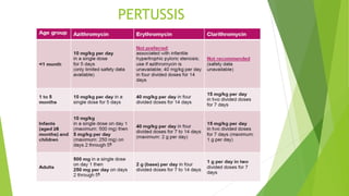
Antimicrobial therapy :
Erythromycin (40-50 mg/kg/day divided every 6 hr
by mouth [PO] or intravenously [IV]; maximum 2
g/day).
Aqueous crystalline penicillin G (100,000-150,000
units/kg/day divided every 6 hr IV or intramuscularly
[IM]).
Procaine penicillin (300,000units every 12 hr IM for
those ≤10 kg in weight; 600,000 units every 12 hr IM
for those >10 kg in weight) for14 days.
Once oral medications are tolerated, oral
penicillin V (250 mg four times daily)may be used.](https://image.slidesharecdn.com/diptheria-converted-200726175940/85/Diptheria-Pertusis-Tetanus-14-320.jpg)


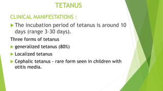


1) Diphtheria, pertussis, and tetanus are acute infectious diseases caused by Corynebacterium diphtheriae, Bordetella pertussis/parapertussis, and Clostridium tetani respectively. 2) They present with respiratory symptoms like sore throat and cough and neurological symptoms like muscle spasms. Diagnosis involves identification of bacteria and supportive lab tests. 3) Treatment involves antitoxins, antibiotics, wound care and supportive measures. Immunization provides effective prevention against these diseases.


![Bordetella pertusis ppt [Autosaved].pptx](https://cdn.slidesharecdn.com/ss_thumbnails/bordetellapertusispptautosaved-250119054438-6c19987e-thumbnail.jpg?width=640&height=640&fit=bounds)
![Bordetella pertusis ppt [Autosaved].pptx](https://cdn.slidesharecdn.com/ss_thumbnails/bordetellapertusispptautosaved-250119055602-ce139642-thumbnail.jpg?width=640&height=640&fit=bounds)
![Bordetella pertusis ppt [Autosaved].pptx](https://cdn.slidesharecdn.com/ss_thumbnails/bordetellapertusispptautosaved-250119061234-b158cb40-thumbnail.jpg?width=640&height=640&fit=bounds)
![Bordetella pertusis ppt [Autosaved].pptx](https://cdn.slidesharecdn.com/ss_thumbnails/bordetellapertusispptautosaved-250119062118-946806e0-thumbnail.jpg?width=640&height=640&fit=bounds)


