SlideShare a Scribd company logo
White Paper | DRX-Evolution Plus Digital Radiography System
Technical Brief Series
Digital Tomosynthesis – Theory of Operation
Pending FDA 510(k) clearance
Author: Xiahui (Ed) Wang
Digital Tomosynthesis (DT) is a new radiographic imaging
technique that is revived from the nearly century-old traditional
film-screen tomography. This rejuvenation is all made possible
by the recent advances in high frame-rate, high-sensitivity flat-
panel digital radiographic detector, rapid pulsed-exposure
sequence-capable high-frequency x-ray generator, the widely
available and low-cost computer GPU processing power, and
the precision motion controls built in the digital radiography
system hardware.
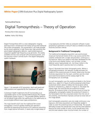
Figure 1: An example of DT acquisition. Both wall stand and
table positions are supported by the Carestream DT option.
Carestream Health implements the DT functionality as an
upgradable option on the standard DRX-Evolution Plus Digital
Radiography System (Figure 1). The portable DRXPlus detector
can be placed inside either the wall bucky or the table bucky to
accommodate different exam types and positions.
The major benefit of DT over the traditional film-screen
tomography is that DT greatly simplifies the operator’s
workflow by separating the process of DT exposure acquisition
from image volume formation; it needs only a single sweep of
x-ray exposures and then relies on computer software in post-
processing to generate as many DT slices as needed at any slice
thickness and slice interval.
Background in Traditional Tomography
The traditional tomography acquisition uses synchronized,
reciprocal mechanical motion of the x-ray source and the film
cassette combined with a single, long-duration, continuous x-
ray exposure. Many scan patterns have been developed for the
x-ray source and cassette motion, such as linear, circular,
elliptical, spiral, hypocycloidal, etc., but linear motion is the
most frequently used in practice due to its simplicity.
Figure 2 illustrates how linear tomography works. Before a
tomography procedure, the operator visually estimates the
midline of the anatomical area of interest, determines the slice
thickness requirement for the exam, and then sets the initial
pivot level (fulcral plane) and the sweep angle for the
tomography scan accordingly.
For each tomographic scan the anatomical details in the fulcral
plane (e.g., object O in Figure 2) will be preserved while those
anatomical clutters above or below the fulcral plane will be
blurred or smeared (e.g., object X in Figure 2). The larger the
sweep angle, the more blurred the anatomical clutters
become. As anatomy clutter is the primary source of noise for
radiography, tomography isolates the anatomy of interest in
the fulcral plane from the unwanted clutters from the rest of
the anatomy, and therefore improves contrast-to-noise ratio of
the image details.
Each tomography acquisition uses one single continuous x-ray
exposure, and many acquisitions are frequently needed in
order to cover the anatomical area of interest fully. This would
definitely increase the overall exam time and the total radiation
dose by the total number of scans. As the overall exam time
and the total radiation dose must be limited, the number of
White Paper | DRX-Evolution Plus Digital Radiography System
2
tomographic scans is limited, which may limit the smallest
tomographic slice interval (slice pitch) and the spatial resolution
in the depth direction of the tomography volume.
Digital Tomosynthesis
Figure 3(a) illustrated how DT acquisition works. DT operates
based on the same principle as tomography except the
detector can either be mechanically motorized in
synchronization with the x-ray source or simply be stationary
during the exam. The DT implemented by Carestream chooses
to use stationary x-ray detector for the purpose of reducing
motion blur and improving spatial resolution. In addition, DT
uses multiple, low-power, pulsed x-ray exposures instead of a
single continuous exposure. Correspondingly the x-ray detector
in a DT exam will capture each short x-ray pulse as individual
images from different projection angles of the x-ray source.
These projections are used to reconstruct a stack of DT slices
for viewing, of which each slice is equivalent to a traditional
tomogram with fulcral plane set at the center of the DT slice.
Because the image acquisition is separated from the DT image
volume formation process, sophisticated reconstruction
processes are used to maximize the anatomy contrast-to-noise
ratio in the DT slices. Figure 3(b) and (c) shows the “Shift-and-
Add” method used in some of the early DT systems.
Individually captured images are shifted differently and added
Figure 3: Schematic view of DT acquisition (a) and reconstructions of slices at different depth (b) (c).
O
X
X-Ray Source
Motion
O X
X O
X
O
X X X
O
O
O
O X
X O
X
O
O O
X
X
X
O
(a) (b) (c)
Stationary
Detector
Plane A
Plane B
Plane A
Reconstruction
Plane B
Reconstruction
Figure 2: In traditional tomographic imaging, object structure
details (e.g., Object O) in the fulcral plane stays focused
(stationary in the film plane) while objects outside the focal
plane (e.g., object X) become blurred or smeared due to
motion.
O
X
O X X O
O
X
Focal Plane
X-Ray Source
Motion
Synchronized
Detector Motion
White Paper | DRX-Evolution Plus Digital Radiography System
3
together in the software to generate DT slices focused at
different depths of the anatomy. The Carestream DT
reconstruction software leverages the state-of-the-art
algorithms that are the same in principle to those applied in
the computed tomography (CT), such as filtered back
projection or iterative reconstruction. As a consequence, the
anatomical details are greatly improved with DT when
compared to tomogram. Figure 4 shows two DT slices of the
pelvis region at different depth locations.
The exam time is greatly reduced with DT than with traditional
tomography, and the operator’s trial-and-error process in
locating the anatomical region of interest is eliminated.
Multiple projections are acquired during a single DT acquisition
sweep. Afterward the anatomical features at different depth
locations are reconstructed mathematically. The total number
of slices and the slice interval can be made arbitrarily large or
small in the software as needed; and they are limited by only
the computer memory capacity, network bandwidth, PACS
storage space, and most importantly the diagnostic needs on
the image resolution.
DT Scout
Each DT acquisition sequence starts with a scout view. The
scout serves two purposes for DT: 1) collimation and anatomy
positioning, and 2) exposure control. The scout view is
captured at the same technique settings (kVp, mAs, etc.) as
with a standard radiography except the projection angle is
always straight on to the detector center and the SID is fixed
(110 cm for table and 180 cm for wall stand). The scout is
processed as a radiographic image and then presented to the
operator to verify the collimation for anatomy coverage as well
as the anatomy positioning. The scout image can be captured
using either automatic exposure control (AEC) or manual
technique settings. The operator would decide on the
appropriateness of the image quality of the scout view for
diagnosis or retake the exposure if adjustment is needed.
DT Sequence Exposure Control
Once the DT scout is captured and accepted by the operator,
its exposure technique will be used as the basis to derive the
total exposure for the DT sequence. This is an important
Figure 4: DT slice examples of the pelvis region at different depth locations.
White Paper | DRX-Evolution Plus Digital Radiography System
4
operation step for automatic exposure control of the DT
sequence.
The total exposure level, mAs in particular, of a DT sequence is
set as the multiplication of the DT scout exposure level (mAs)
and the DT Technique Scale Factor that is preconfigured in the
Preference Editor. This is regardless of the total number of
projections and the x-ray tube sweep angle specified. The total
DT mAs is allocated equally to all the DT projections. The other
exposure technique settings between the scout and the DT
sequence, such as kVp and beam filtration, are kept the same.
DT Acquisition Modes and Reconstruction
Resolution
DT projection images can be acquired in either high-resolution
(HR) mode or high-speed (HS) mode. In the HR acquisition
mode, the detector works in its native resolution (i.e., no pixel
binning) and acquires images at 4.5 frames per second (fps); in
the HS acquisition mode the detector uses 2x1 pixel binning
and acquires images at 9.0 fps due to 50% reduced data rate.
The primary usage of the HR mode is for musculoskeletal
exams (hand, foot, knee, etc.), when the patient motion
artifact due to the longer acquisition time is limited and it
would not be a major concern to the DT image quality. The
improved frame rate in the HS acquisition mode is directly
translated into 50% reduction in total DT image acquisition
time. Because the HS acquisition mode is 2X faster, it will be
helpful to reduce the patient motion artifact. There is a
potential image resolution loss due to 2x1 pixel binning;
however, the impact is minimized in the system design by
binning the pixels in the x-ray tube motion direction as well as
applying sharpness enhancement filtering operation to
compensate for the minor resolution loss. A comparative
image quality example is shown in Figure 5.
Regardless of the acquisition mode, DT slices can be
reconstructed and presented in two spatial resolutions during
post processing: the native 1x1 pixel resolution (HR-res) or the
2x2 pixel average resolution (STD-res). The size of an STD-res
DT volumetric image will be 1/8 (½ x ½ x ½) of that of the
corresponding HR-res image.
In summary, the DT volumetric images can be generated in
four combinations (see Table 1 for the recommended
applications). In theory, the HR/HR-res combination will offer
the best spatial resolution in the reconstructed DT slice image
because both the detector and the reconstruction work in the
native 1x1 resolution.
Figure 5: Image quality comparison of a hand phantom acquired in HR mode (left) and HS mode (right). Both images are
reconstructed in HR-res using 0.3 cm slice thickness and 0.05 cm slice interval. Sharpness enhancement is applied automatically in
the software to bring up the anatomical details in the image acquired in HS mode.
White Paper | DRX-Evolution Plus Digital Radiography System
5
HR Std. Res
Acquisition
HR (1x1)
Hand/Wrist
Foot/Ankle
Knee
Shoulder
Acquisition
HS (1x2)
Hand/Wrist
Foot/Ankle
Knee
Shoulder
Chest
Abdomen
Pelvis
C-Spine
Table 1: Recommended DT operation for different exam types.
HR-res reconstruction is optimal for musculoskeletal exams,
and the HS acquisition helps to reduce patient motion artifact.
DT Section Thickness and Sweep Angle α
As mentioned before, DT imaging follows the same principles
as traditional tomography. For any anatomical details that are
outside the fulcral plane, the greater the sweep angle, the
greater the blur effect. Because all the details in the fulcral
plane are unchanged, increasing the sweep angle effectively
reduces the clutter caused by anything that is outside the
fulcral plane, which makes the tomographic slice appear
“thinner.” In practice, because the anatomical details gradually
become blurrier as their distance from the fulcral plane
increases, Section Thickness is used to define the region of the
anatomy within which all the anatomical details are under a
permissible blur level. Section Thickness is a value that is
determined solely by the acquisition geometry and therefore is
inherent to the acquisition. It is different from and should not
be confused with the Slice Thickness used in the DT
reconstruction. Table 2 shows the pre-calculated section
thickness at different sweep angles α and 0.25 mm permissible
blur values.
Sweep Angle α
(Degrees)
Section
Thickness (mm)
10 2.86
15 1.90
20 1.42
25 1.13
30 0.93
35 0.79
40 0.69
Table 2: Look up table for selecting DT sweep angle to achieve
desired slice thickness, assuming the maximum permissible blur
is 0.25 mm.
DT Slice Thickness t and Slice Interval δ
The DT image is presented as a stack of reconstructed 2D
coronal slices. The center location of each slice corresponds to
the fulcral plane at which anatomical details are maximized.
The distance between the reconstruction slices is the slice
interval (δ). The smaller the δ value, the finer the DT
reconstruction resolution in the depth direction. The slice
interval can be set accordingly by the operator, depending on
the requirements on the anatomical features to be visualized
for diagnosis. For example, for small musculoskeletal exams
such as wrist, etc., the δ value can be set between 0.5 mm to
1.0 mm, and for chest exams between 1.0 mm to 2.0 mm.
Each DT slice covers a slab of anatomical region and its
thickness (t) can be made arbitrarily by the DT reconstruction
algorithm, which is independent of (i.e., decoupled from) the
Section Thickness or the slice interval (δ). In practice the
reconstruction slice thickness (t) should always be greater than
or equal to the slice interval such that no anatomical void is
created (see Figure 6), causing the slices to overlap each other
partially. A smaller t value more likely helps to resolve the
anatomical details better but at the potential expense of
greater noise appearance. On the other hand, greater t value
helps to reduce the image noise but potentially blends more
White Paper | DRX-Evolution Plus Digital Radiography System
6
anatomical details from the region outside the fulcral plane
therefore defeating the purpose of DT. A default slice
thickness of 20.0 mm is a good starting point for all exam
types.
Figure 6: Thick slices can overlap with each other yet slice
interval can be much smaller than slice thickness.
DT Projection Image Number N
The number of discrete DT projection images acquired across
the sweep angle dictates the reconstruction rippling artifact.
Figure 7 shows a DT slice of an ankle in the arterial-posterior
view. The rippling artifact at the bottom of the image is caused
by the sharp transitions between the open-air region and the
foot, with each ripple corresponding to a DT projection. When
the sweep angle is fixed, more projections will help to reduce
the artifact but at the expense of increased exam time. The
ripple artifact will manifest itself more with thicker anatomy, in
which case increasing the DT projection number will help.
Figure 7: An example of ripple artifact in DT foot exam.
Summary
Compared to traditional tomography, DT significantly improves
the workflow, reduces the overall exam time, minimizes
exposure retakes, and offers improved image quality. Good
radiographic practice in anatomy positioning and x-ray
exposure technique selection should be followed for a DT
exam, just as in standard radiography.
Patient motion during a DT exam degrades the image quality
and therefore should be minimized as much as possible. In
addition to taking measures to restrain patient motion, a
better timing to start the DT sequence with respect to the
patient motion pattern would help. Finally, patient motion
artifact can be reduced by reducing the total exam time using
the HS acquisition mode.
Slice A Slice B
Slice
Increment
White Paper | DRX-Evolution Plus Digital Radiography System
carestream.com
©Carestream Health, Inc., 2019. CARESTREAM is a trademark of
Carestream Health. CAT 2000 265 5/19

More Related Content

What's hot

MR reconstruction 101
MR reconstruction 101MR reconstruction 101
MR reconstruction 101
Sairam Geethanath
 
An Ultrasound Image Despeckling Approach Based on Principle Component Analysis
An Ultrasound Image Despeckling Approach Based on Principle Component AnalysisAn Ultrasound Image Despeckling Approach Based on Principle Component Analysis
An Ultrasound Image Despeckling Approach Based on Principle Component Analysis
CSCJournals
 
Image Quality, Artifacts and it's Remedies in CT-Avinesh Shrestha
Image Quality, Artifacts and it's Remedies in CT-Avinesh ShresthaImage Quality, Artifacts and it's Remedies in CT-Avinesh Shrestha
Image Quality, Artifacts and it's Remedies in CT-Avinesh Shrestha
Avinesh Shrestha
 
Session 6 image quality ct and session 8 dose ct
Session 6  image quality ct and  session 8 dose ctSession 6  image quality ct and  session 8 dose ct
Session 6 image quality ct and session 8 dose ct
Centro Nacional de Radioterapia Nora Astorga
 
CT Scan Image reconstruction
CT Scan Image reconstructionCT Scan Image reconstruction
CT Scan Image reconstructionGunjan Patel
 
Dose reduction in MDCT . Daniel J.P , Khorfakhan hospital . UAE
Dose reduction in MDCT . Daniel J.P , Khorfakhan hospital . UAEDose reduction in MDCT . Daniel J.P , Khorfakhan hospital . UAE
Dose reduction in MDCT . Daniel J.P , Khorfakhan hospital . UAE
haijaypee_dan
 
automatic detection of pulmonary nodules in lung ct images
automatic detection of pulmonary nodules in lung ct imagesautomatic detection of pulmonary nodules in lung ct images
automatic detection of pulmonary nodules in lung ct imagesWookjin Choi
 
Chapter 4 image display
Chapter 4 image displayChapter 4 image display
Chapter 4 image display
Muntaser S.Ahmad
 
Implementation of an audit and dose reduction program for ct matyagin
Implementation of an audit and dose reduction program for ct matyaginImplementation of an audit and dose reduction program for ct matyagin
Implementation of an audit and dose reduction program for ct matyaginLeishman Associates
 
CT Image reconstruction
CT Image reconstructionCT Image reconstruction
CT Image reconstruction
Santosh Ojha
 
Digital X-ray Problems..
Digital X-ray Problems..Digital X-ray Problems..
Digital X-ray Problems..
Ganesan Yogananthem
 
Basics of ct lecture 1
Basics of ct  lecture 1Basics of ct  lecture 1
Basics of ct lecture 1
Gamal Mahdaly
 
Chapter 6 image quality in ct
Chapter 6 image quality in ct Chapter 6 image quality in ct
Chapter 6 image quality in ct
Muntaser S.Ahmad
 
Nihms 619230
Nihms 619230Nihms 619230
Nihms 619230
Maria Vergakh
 
Image reconstrsuction in ct pdf
Image reconstrsuction in ct pdfImage reconstrsuction in ct pdf
Image reconstrsuction in ct pdf
mitians
 
Flat panel ---nihms864608
Flat panel ---nihms864608Flat panel ---nihms864608
Flat panel ---nihms864608
Maria Vergakh
 

What's hot (20)

MR reconstruction 101
MR reconstruction 101MR reconstruction 101
MR reconstruction 101
 
An Ultrasound Image Despeckling Approach Based on Principle Component Analysis
An Ultrasound Image Despeckling Approach Based on Principle Component AnalysisAn Ultrasound Image Despeckling Approach Based on Principle Component Analysis
An Ultrasound Image Despeckling Approach Based on Principle Component Analysis
 
Image Quality, Artifacts and it's Remedies in CT-Avinesh Shrestha
Image Quality, Artifacts and it's Remedies in CT-Avinesh ShresthaImage Quality, Artifacts and it's Remedies in CT-Avinesh Shrestha
Image Quality, Artifacts and it's Remedies in CT-Avinesh Shrestha
 
Session 6 image quality ct and session 8 dose ct
Session 6  image quality ct and  session 8 dose ctSession 6  image quality ct and  session 8 dose ct
Session 6 image quality ct and session 8 dose ct
 
CT Scan Image reconstruction
CT Scan Image reconstructionCT Scan Image reconstruction
CT Scan Image reconstruction
 
Dose reduction in MDCT . Daniel J.P , Khorfakhan hospital . UAE
Dose reduction in MDCT . Daniel J.P , Khorfakhan hospital . UAEDose reduction in MDCT . Daniel J.P , Khorfakhan hospital . UAE
Dose reduction in MDCT . Daniel J.P , Khorfakhan hospital . UAE
 
Ct i patient_whitepaper
Ct i patient_whitepaperCt i patient_whitepaper
Ct i patient_whitepaper
 
automatic detection of pulmonary nodules in lung ct images
automatic detection of pulmonary nodules in lung ct imagesautomatic detection of pulmonary nodules in lung ct images
automatic detection of pulmonary nodules in lung ct images
 
Chapter 4 image display
Chapter 4 image displayChapter 4 image display
Chapter 4 image display
 
Implementation of an audit and dose reduction program for ct matyagin
Implementation of an audit and dose reduction program for ct matyaginImplementation of an audit and dose reduction program for ct matyagin
Implementation of an audit and dose reduction program for ct matyagin
 
CT Image reconstruction
CT Image reconstructionCT Image reconstruction
CT Image reconstruction
 
Digital X-ray Problems..
Digital X-ray Problems..Digital X-ray Problems..
Digital X-ray Problems..
 
Basics of ct lecture 1
Basics of ct  lecture 1Basics of ct  lecture 1
Basics of ct lecture 1
 
Dose reduction
Dose reductionDose reduction
Dose reduction
 
Chapter 6 image quality in ct
Chapter 6 image quality in ct Chapter 6 image quality in ct
Chapter 6 image quality in ct
 
Medphysics Planning
Medphysics PlanningMedphysics Planning
Medphysics Planning
 
Nihms 619230
Nihms 619230Nihms 619230
Nihms 619230
 
Radiotherapy Treatment Simulation
Radiotherapy Treatment SimulationRadiotherapy Treatment Simulation
Radiotherapy Treatment Simulation
 
Image reconstrsuction in ct pdf
Image reconstrsuction in ct pdfImage reconstrsuction in ct pdf
Image reconstrsuction in ct pdf
 
Flat panel ---nihms864608
Flat panel ---nihms864608Flat panel ---nihms864608
Flat panel ---nihms864608
 

Similar to Digital Tomosynthesis: Theory of Operation

whitepaper-advanced-ultrasound-imaging-201507
whitepaper-advanced-ultrasound-imaging-201507whitepaper-advanced-ultrasound-imaging-201507
whitepaper-advanced-ultrasound-imaging-201507Ajay Anand
 
Image processing techniques in nm 08,09
Image processing techniques in nm 08,09Image processing techniques in nm 08,09
Image processing techniques in nm 08,09
Rutuja Solkar
 
Robust Algorithm for Discrete Tomography with Gray Value Estimation
Robust Algorithm for Discrete Tomography with Gray Value EstimationRobust Algorithm for Discrete Tomography with Gray Value Estimation
Robust Algorithm for Discrete Tomography with Gray Value Estimation
Association of Scientists, Developers and Faculties
 
Tube and Grid Alignment System
Tube and Grid Alignment System Tube and Grid Alignment System
Tube and Grid Alignment System
Carestream
 
High Speed and Area Efficient 2D DWT Processor Based Image Compression
High Speed and Area Efficient 2D DWT Processor Based Image CompressionHigh Speed and Area Efficient 2D DWT Processor Based Image Compression
High Speed and Area Efficient 2D DWT Processor Based Image Compression
sipij
 
A NOVEL ALGORITHM FOR IMAGE DENOISING USING DT-CWT
A NOVEL ALGORITHM FOR IMAGE DENOISING USING DT-CWT A NOVEL ALGORITHM FOR IMAGE DENOISING USING DT-CWT
A NOVEL ALGORITHM FOR IMAGE DENOISING USING DT-CWT
sipij
 
Tomosurgery Presentation
Tomosurgery PresentationTomosurgery Presentation
Tomosurgery Presentationngowdar
 
Contourlet Transform Based Method For Medical Image Denoising
Contourlet Transform Based Method For Medical Image DenoisingContourlet Transform Based Method For Medical Image Denoising
Contourlet Transform Based Method For Medical Image Denoising
CSCJournals
 
CBCT in dental practice
CBCT in dental practiceCBCT in dental practice
CBCT in dental practiceZana Hussein
 
3-D WAVELET CODEC (COMPRESSION/DECOMPRESSION) FOR 3-D MEDICAL IMAGES
3-D WAVELET CODEC (COMPRESSION/DECOMPRESSION) FOR 3-D MEDICAL IMAGES3-D WAVELET CODEC (COMPRESSION/DECOMPRESSION) FOR 3-D MEDICAL IMAGES
3-D WAVELET CODEC (COMPRESSION/DECOMPRESSION) FOR 3-D MEDICAL IMAGES
ijitcs
 
A Novel Blind SR Method to Improve the Spatial Resolution of Real Life Video ...
A Novel Blind SR Method to Improve the Spatial Resolution of Real Life Video ...A Novel Blind SR Method to Improve the Spatial Resolution of Real Life Video ...
A Novel Blind SR Method to Improve the Spatial Resolution of Real Life Video ...
IRJET Journal
 
G0352039045
G0352039045G0352039045
G0352039045
inventionjournals
 
Review on Medical Image Fusion using Shearlet Transform
Review on Medical Image Fusion using Shearlet TransformReview on Medical Image Fusion using Shearlet Transform
Review on Medical Image Fusion using Shearlet Transform
IRJET Journal
 
- Lecture 2.ppt
- Lecture 2.ppt- Lecture 2.ppt
- Lecture 2.ppt
Magde Gad
 
5-IMAGE RECONSTRUCTION in CT Scan-Rohit.pptx
5-IMAGE RECONSTRUCTION in CT Scan-Rohit.pptx5-IMAGE RECONSTRUCTION in CT Scan-Rohit.pptx
5-IMAGE RECONSTRUCTION in CT Scan-Rohit.pptx
Rohit Bansal
 
Cb34474478
Cb34474478Cb34474478
Cb34474478
IJERA Editor
 
7200 35328
7200 353287200 35328
Basic principle of CT and generation of CT.pptx
Basic principle of CT and generation of CT.pptxBasic principle of CT and generation of CT.pptx
Basic principle of CT and generation of CT.pptx
sarita gaire
 

Similar to Digital Tomosynthesis: Theory of Operation (20)

whitepaper-advanced-ultrasound-imaging-201507
whitepaper-advanced-ultrasound-imaging-201507whitepaper-advanced-ultrasound-imaging-201507
whitepaper-advanced-ultrasound-imaging-201507
 
Image processing techniques in nm 08,09
Image processing techniques in nm 08,09Image processing techniques in nm 08,09
Image processing techniques in nm 08,09
 
Robust Algorithm for Discrete Tomography with Gray Value Estimation
Robust Algorithm for Discrete Tomography with Gray Value EstimationRobust Algorithm for Discrete Tomography with Gray Value Estimation
Robust Algorithm for Discrete Tomography with Gray Value Estimation
 
Tube and Grid Alignment System
Tube and Grid Alignment System Tube and Grid Alignment System
Tube and Grid Alignment System
 
High Speed and Area Efficient 2D DWT Processor Based Image Compression
High Speed and Area Efficient 2D DWT Processor Based Image CompressionHigh Speed and Area Efficient 2D DWT Processor Based Image Compression
High Speed and Area Efficient 2D DWT Processor Based Image Compression
 
A NOVEL ALGORITHM FOR IMAGE DENOISING USING DT-CWT
A NOVEL ALGORITHM FOR IMAGE DENOISING USING DT-CWT A NOVEL ALGORITHM FOR IMAGE DENOISING USING DT-CWT
A NOVEL ALGORITHM FOR IMAGE DENOISING USING DT-CWT
 
Tomosurgery Presentation
Tomosurgery PresentationTomosurgery Presentation
Tomosurgery Presentation
 
Contourlet Transform Based Method For Medical Image Denoising
Contourlet Transform Based Method For Medical Image DenoisingContourlet Transform Based Method For Medical Image Denoising
Contourlet Transform Based Method For Medical Image Denoising
 
CBCT in dental practice
CBCT in dental practiceCBCT in dental practice
CBCT in dental practice
 
3-D WAVELET CODEC (COMPRESSION/DECOMPRESSION) FOR 3-D MEDICAL IMAGES
3-D WAVELET CODEC (COMPRESSION/DECOMPRESSION) FOR 3-D MEDICAL IMAGES3-D WAVELET CODEC (COMPRESSION/DECOMPRESSION) FOR 3-D MEDICAL IMAGES
3-D WAVELET CODEC (COMPRESSION/DECOMPRESSION) FOR 3-D MEDICAL IMAGES
 
A Novel Blind SR Method to Improve the Spatial Resolution of Real Life Video ...
A Novel Blind SR Method to Improve the Spatial Resolution of Real Life Video ...A Novel Blind SR Method to Improve the Spatial Resolution of Real Life Video ...
A Novel Blind SR Method to Improve the Spatial Resolution of Real Life Video ...
 
G0352039045
G0352039045G0352039045
G0352039045
 
Review on Medical Image Fusion using Shearlet Transform
Review on Medical Image Fusion using Shearlet TransformReview on Medical Image Fusion using Shearlet Transform
Review on Medical Image Fusion using Shearlet Transform
 
CT History
CT HistoryCT History
CT History
 
- Lecture 2.ppt
- Lecture 2.ppt- Lecture 2.ppt
- Lecture 2.ppt
 
5-IMAGE RECONSTRUCTION in CT Scan-Rohit.pptx
5-IMAGE RECONSTRUCTION in CT Scan-Rohit.pptx5-IMAGE RECONSTRUCTION in CT Scan-Rohit.pptx
5-IMAGE RECONSTRUCTION in CT Scan-Rohit.pptx
 
Cb34474478
Cb34474478Cb34474478
Cb34474478
 
7200 35328
7200 353287200 35328
7200 35328
 
W6P3622650776P65
W6P3622650776P65W6P3622650776P65
W6P3622650776P65
 
Basic principle of CT and generation of CT.pptx
Basic principle of CT and generation of CT.pptxBasic principle of CT and generation of CT.pptx
Basic principle of CT and generation of CT.pptx
 

More from Carestream

Affordable Digital Upgrade for Medical Imaging - the Benefits and the Return ...
Affordable Digital Upgrade for Medical Imaging - the Benefits and the Return ...Affordable Digital Upgrade for Medical Imaging - the Benefits and the Return ...
Affordable Digital Upgrade for Medical Imaging - the Benefits and the Return ...
Carestream
 
Dose Efficient Dual Energy Subtraction Radiography - Theory of Operations
Dose Efficient Dual Energy Subtraction Radiography - Theory of OperationsDose Efficient Dual Energy Subtraction Radiography - Theory of Operations
Dose Efficient Dual Energy Subtraction Radiography - Theory of Operations
Carestream
 
Special Report: Challenges and Solutions in Pediatric X-ray
Special Report: Challenges and Solutions in Pediatric X-raySpecial Report: Challenges and Solutions in Pediatric X-ray
Special Report: Challenges and Solutions in Pediatric X-ray
Carestream
 
Special Report: Getting the Optimal Return on X-ray Equipment
Special Report: Getting the Optimal Return on X-ray EquipmentSpecial Report: Getting the Optimal Return on X-ray Equipment
Special Report: Getting the Optimal Return on X-ray Equipment
Carestream
 
The Pursuit of Excellence in Image Quality
The Pursuit of Excellence in Image QualityThe Pursuit of Excellence in Image Quality
The Pursuit of Excellence in Image Quality
Carestream
 
Whitepaper: Healthcare Data Migration - Top 10 Questions
Whitepaper: Healthcare Data Migration - Top 10 Questions Whitepaper: Healthcare Data Migration - Top 10 Questions
Whitepaper: Healthcare Data Migration - Top 10 Questions
Carestream
 
Whitepaper: Image Quality Impact of SmartGrid Processing in Bedside Chest Ima...
Whitepaper: Image Quality Impact of SmartGrid Processing in Bedside Chest Ima...Whitepaper: Image Quality Impact of SmartGrid Processing in Bedside Chest Ima...
Whitepaper: Image Quality Impact of SmartGrid Processing in Bedside Chest Ima...
Carestream
 
Using Carbon Nano-Tube Field Emitters to Miniaturize X-Ray Tubes
Using Carbon Nano-Tube Field Emitters to Miniaturize X-Ray TubesUsing Carbon Nano-Tube Field Emitters to Miniaturize X-Ray Tubes
Using Carbon Nano-Tube Field Emitters to Miniaturize X-Ray Tubes
Carestream
 
Guia obsolescencia seram
Guia obsolescencia seramGuia obsolescencia seram
Guia obsolescencia seram
Carestream
 
Cloud Security
Cloud Security Cloud Security
Cloud Security
Carestream
 
Sunway Medical Centre Installs CARESTREAM Vue PACS to Streamline Imaging Proc...
Sunway Medical Centre Installs CARESTREAM Vue PACS to Streamline Imaging Proc...Sunway Medical Centre Installs CARESTREAM Vue PACS to Streamline Imaging Proc...
Sunway Medical Centre Installs CARESTREAM Vue PACS to Streamline Imaging Proc...
Carestream
 
White Paper: The Benefits of Mobile X-rays in Thoracic and Cardiac Care
White Paper: The Benefits of Mobile X-rays in Thoracic and Cardiac CareWhite Paper: The Benefits of Mobile X-rays in Thoracic and Cardiac Care
White Paper: The Benefits of Mobile X-rays in Thoracic and Cardiac Care
Carestream
 
Study: University Clinic, Regensburg, Evaluates Smart Flow in Ultrasound
Study: University Clinic, Regensburg, Evaluates Smart Flow in UltrasoundStudy: University Clinic, Regensburg, Evaluates Smart Flow in Ultrasound
Study: University Clinic, Regensburg, Evaluates Smart Flow in Ultrasound
Carestream
 
Financial Implications for Integrating Carestream OnSight 3D Extremity System...
Financial Implications for Integrating Carestream OnSight 3D Extremity System...Financial Implications for Integrating Carestream OnSight 3D Extremity System...
Financial Implications for Integrating Carestream OnSight 3D Extremity System...
Carestream
 
Whitepaper: Enterprise Access Viewer Connecting technologies, physicians, and...
Whitepaper: Enterprise Access Viewer Connecting technologies, physicians, and...Whitepaper: Enterprise Access Viewer Connecting technologies, physicians, and...
Whitepaper: Enterprise Access Viewer Connecting technologies, physicians, and...
Carestream
 
Evaluating Enterprise Clinical Data-Management Systems at RSNA 2016
Evaluating Enterprise Clinical Data-Management Systems at RSNA 2016Evaluating Enterprise Clinical Data-Management Systems at RSNA 2016
Evaluating Enterprise Clinical Data-Management Systems at RSNA 2016
Carestream
 
3 Reasons to Visit Carestream at RSNA2016 #RNSA16
3 Reasons to Visit Carestream at RSNA2016 #RNSA163 Reasons to Visit Carestream at RSNA2016 #RNSA16
3 Reasons to Visit Carestream at RSNA2016 #RNSA16
Carestream
 
Casa di Cura Sileno Hospital Italy
Casa di Cura Sileno Hospital ItalyCasa di Cura Sileno Hospital Italy
Casa di Cura Sileno Hospital Italy
Carestream
 
The Power of Together
The Power of TogetherThe Power of Together
The Power of Together
Carestream
 
Whitepaper: Leveraging the Cloud to Enhance an Enterprise Imaging Strategy
Whitepaper: Leveraging the Cloud to Enhance an Enterprise Imaging StrategyWhitepaper: Leveraging the Cloud to Enhance an Enterprise Imaging Strategy
Whitepaper: Leveraging the Cloud to Enhance an Enterprise Imaging Strategy
Carestream
 

More from Carestream (20)

Affordable Digital Upgrade for Medical Imaging - the Benefits and the Return ...
Affordable Digital Upgrade for Medical Imaging - the Benefits and the Return ...Affordable Digital Upgrade for Medical Imaging - the Benefits and the Return ...
Affordable Digital Upgrade for Medical Imaging - the Benefits and the Return ...
 
Dose Efficient Dual Energy Subtraction Radiography - Theory of Operations
Dose Efficient Dual Energy Subtraction Radiography - Theory of OperationsDose Efficient Dual Energy Subtraction Radiography - Theory of Operations
Dose Efficient Dual Energy Subtraction Radiography - Theory of Operations
 
Special Report: Challenges and Solutions in Pediatric X-ray
Special Report: Challenges and Solutions in Pediatric X-raySpecial Report: Challenges and Solutions in Pediatric X-ray
Special Report: Challenges and Solutions in Pediatric X-ray
 
Special Report: Getting the Optimal Return on X-ray Equipment
Special Report: Getting the Optimal Return on X-ray EquipmentSpecial Report: Getting the Optimal Return on X-ray Equipment
Special Report: Getting the Optimal Return on X-ray Equipment
 
The Pursuit of Excellence in Image Quality
The Pursuit of Excellence in Image QualityThe Pursuit of Excellence in Image Quality
The Pursuit of Excellence in Image Quality
 
Whitepaper: Healthcare Data Migration - Top 10 Questions
Whitepaper: Healthcare Data Migration - Top 10 Questions Whitepaper: Healthcare Data Migration - Top 10 Questions
Whitepaper: Healthcare Data Migration - Top 10 Questions
 
Whitepaper: Image Quality Impact of SmartGrid Processing in Bedside Chest Ima...
Whitepaper: Image Quality Impact of SmartGrid Processing in Bedside Chest Ima...Whitepaper: Image Quality Impact of SmartGrid Processing in Bedside Chest Ima...
Whitepaper: Image Quality Impact of SmartGrid Processing in Bedside Chest Ima...
 
Using Carbon Nano-Tube Field Emitters to Miniaturize X-Ray Tubes
Using Carbon Nano-Tube Field Emitters to Miniaturize X-Ray TubesUsing Carbon Nano-Tube Field Emitters to Miniaturize X-Ray Tubes
Using Carbon Nano-Tube Field Emitters to Miniaturize X-Ray Tubes
 
Guia obsolescencia seram
Guia obsolescencia seramGuia obsolescencia seram
Guia obsolescencia seram
 
Cloud Security
Cloud Security Cloud Security
Cloud Security
 
Sunway Medical Centre Installs CARESTREAM Vue PACS to Streamline Imaging Proc...
Sunway Medical Centre Installs CARESTREAM Vue PACS to Streamline Imaging Proc...Sunway Medical Centre Installs CARESTREAM Vue PACS to Streamline Imaging Proc...
Sunway Medical Centre Installs CARESTREAM Vue PACS to Streamline Imaging Proc...
 
White Paper: The Benefits of Mobile X-rays in Thoracic and Cardiac Care
White Paper: The Benefits of Mobile X-rays in Thoracic and Cardiac CareWhite Paper: The Benefits of Mobile X-rays in Thoracic and Cardiac Care
White Paper: The Benefits of Mobile X-rays in Thoracic and Cardiac Care
 
Study: University Clinic, Regensburg, Evaluates Smart Flow in Ultrasound
Study: University Clinic, Regensburg, Evaluates Smart Flow in UltrasoundStudy: University Clinic, Regensburg, Evaluates Smart Flow in Ultrasound
Study: University Clinic, Regensburg, Evaluates Smart Flow in Ultrasound
 
Financial Implications for Integrating Carestream OnSight 3D Extremity System...
Financial Implications for Integrating Carestream OnSight 3D Extremity System...Financial Implications for Integrating Carestream OnSight 3D Extremity System...
Financial Implications for Integrating Carestream OnSight 3D Extremity System...
 
Whitepaper: Enterprise Access Viewer Connecting technologies, physicians, and...
Whitepaper: Enterprise Access Viewer Connecting technologies, physicians, and...Whitepaper: Enterprise Access Viewer Connecting technologies, physicians, and...
Whitepaper: Enterprise Access Viewer Connecting technologies, physicians, and...
 
Evaluating Enterprise Clinical Data-Management Systems at RSNA 2016
Evaluating Enterprise Clinical Data-Management Systems at RSNA 2016Evaluating Enterprise Clinical Data-Management Systems at RSNA 2016
Evaluating Enterprise Clinical Data-Management Systems at RSNA 2016
 
3 Reasons to Visit Carestream at RSNA2016 #RNSA16
3 Reasons to Visit Carestream at RSNA2016 #RNSA163 Reasons to Visit Carestream at RSNA2016 #RNSA16
3 Reasons to Visit Carestream at RSNA2016 #RNSA16
 
Casa di Cura Sileno Hospital Italy
Casa di Cura Sileno Hospital ItalyCasa di Cura Sileno Hospital Italy
Casa di Cura Sileno Hospital Italy
 
The Power of Together
The Power of TogetherThe Power of Together
The Power of Together
 
Whitepaper: Leveraging the Cloud to Enhance an Enterprise Imaging Strategy
Whitepaper: Leveraging the Cloud to Enhance an Enterprise Imaging StrategyWhitepaper: Leveraging the Cloud to Enhance an Enterprise Imaging Strategy
Whitepaper: Leveraging the Cloud to Enhance an Enterprise Imaging Strategy
 

Recently uploaded

Empowering ACOs: Leveraging Quality Management Tools for MIPS and Beyond
Empowering ACOs: Leveraging Quality Management Tools for MIPS and BeyondEmpowering ACOs: Leveraging Quality Management Tools for MIPS and Beyond
Empowering ACOs: Leveraging Quality Management Tools for MIPS and Beyond
Health Catalyst
 
CHAPTER 1 SEMESTER V - ROLE OF PEADIATRIC NURSE.pdf
CHAPTER 1 SEMESTER V - ROLE OF PEADIATRIC NURSE.pdfCHAPTER 1 SEMESTER V - ROLE OF PEADIATRIC NURSE.pdf
CHAPTER 1 SEMESTER V - ROLE OF PEADIATRIC NURSE.pdf
Sachin Sharma
 
Global launch of the Healthy Ageing and Prevention Index 2nd wave – alongside...
Global launch of the Healthy Ageing and Prevention Index 2nd wave – alongside...Global launch of the Healthy Ageing and Prevention Index 2nd wave – alongside...
Global launch of the Healthy Ageing and Prevention Index 2nd wave – alongside...
ILC- UK
 
ventilator, child on ventilator, newborn
ventilator, child on ventilator, newbornventilator, child on ventilator, newborn
ventilator, child on ventilator, newborn
Pooja Rani
 
CANCER CANCER CANCER CANCER CANCER CANCER
CANCER  CANCER  CANCER  CANCER  CANCER CANCERCANCER  CANCER  CANCER  CANCER  CANCER CANCER
CANCER CANCER CANCER CANCER CANCER CANCER
KRISTELLEGAMBOA2
 
ABDOMINAL COMPARTMENT SYSNDROME
ABDOMINAL COMPARTMENT SYSNDROMEABDOMINAL COMPARTMENT SYSNDROME
ABDOMINAL COMPARTMENT SYSNDROME
Rommel Luis III Israel
 
GLOBAL WARMING BY PRIYA BHOJWANI @..pptx
GLOBAL WARMING BY PRIYA BHOJWANI @..pptxGLOBAL WARMING BY PRIYA BHOJWANI @..pptx
GLOBAL WARMING BY PRIYA BHOJWANI @..pptx
priyabhojwani1200
 
💘Ludhiana ℂall Girls 📞]][89011★83002][[ 📱 ❤ESCORTS service in Ludhiana💃💦Ludhi...
💘Ludhiana ℂall Girls 📞]][89011★83002][[ 📱 ❤ESCORTS service in Ludhiana💃💦Ludhi...💘Ludhiana ℂall Girls 📞]][89011★83002][[ 📱 ❤ESCORTS service in Ludhiana💃💦Ludhi...
💘Ludhiana ℂall Girls 📞]][89011★83002][[ 📱 ❤ESCORTS service in Ludhiana💃💦Ludhi...
ranishasharma67
 
CHAPTER 1 SEMESTER V PREVENTIVE-PEDIATRICS.pdf
CHAPTER 1 SEMESTER V PREVENTIVE-PEDIATRICS.pdfCHAPTER 1 SEMESTER V PREVENTIVE-PEDIATRICS.pdf
CHAPTER 1 SEMESTER V PREVENTIVE-PEDIATRICS.pdf
Sachin Sharma
 
一比一原版纽约大学毕业证(NYU毕业证)成绩单留信认证
一比一原版纽约大学毕业证(NYU毕业证)成绩单留信认证一比一原版纽约大学毕业证(NYU毕业证)成绩单留信认证
一比一原版纽约大学毕业证(NYU毕业证)成绩单留信认证
o6ov5dqmf
 
Medical Technology Tackles New Health Care Demand - Research Report - March 2...
Medical Technology Tackles New Health Care Demand - Research Report - March 2...Medical Technology Tackles New Health Care Demand - Research Report - March 2...
Medical Technology Tackles New Health Care Demand - Research Report - March 2...
pchutichetpong
 
TOP AND BEST GLUTE BUILDER A 606 | Fitking Fitness
TOP AND BEST GLUTE BUILDER A 606 | Fitking FitnessTOP AND BEST GLUTE BUILDER A 606 | Fitking Fitness
TOP AND BEST GLUTE BUILDER A 606 | Fitking Fitness
Fitking Fitness
 
10 Ideas for Enhancing Your Meeting Experience
10 Ideas for Enhancing Your Meeting Experience10 Ideas for Enhancing Your Meeting Experience
10 Ideas for Enhancing Your Meeting Experience
ranishasharma67
 
ICH Guidelines for Pharmacovigilance.pdf
ICH Guidelines for Pharmacovigilance.pdfICH Guidelines for Pharmacovigilance.pdf
ICH Guidelines for Pharmacovigilance.pdf
NEHA GUPTA
 
India Diagnostic Labs Market: Dynamics, Key Players, and Industry Projections...
India Diagnostic Labs Market: Dynamics, Key Players, and Industry Projections...India Diagnostic Labs Market: Dynamics, Key Players, and Industry Projections...
India Diagnostic Labs Market: Dynamics, Key Players, and Industry Projections...
Kumar Satyam
 
How many patients does case series should have In comparison to case reports.pdf
How many patients does case series should have In comparison to case reports.pdfHow many patients does case series should have In comparison to case reports.pdf
How many patients does case series should have In comparison to case reports.pdf
pubrica101
 
Haridwar ❤CALL Girls 🔝 89011★83002 🔝 ❤ℂall Girls IN Haridwar ESCORT SERVICE❤
Haridwar ❤CALL Girls 🔝 89011★83002 🔝 ❤ℂall Girls IN Haridwar ESCORT SERVICE❤Haridwar ❤CALL Girls 🔝 89011★83002 🔝 ❤ℂall Girls IN Haridwar ESCORT SERVICE❤
Haridwar ❤CALL Girls 🔝 89011★83002 🔝 ❤ℂall Girls IN Haridwar ESCORT SERVICE❤
ranishasharma67
 
Navigating Challenges: Mental Health, Legislation, and the Prison System in B...
Navigating Challenges: Mental Health, Legislation, and the Prison System in B...Navigating Challenges: Mental Health, Legislation, and the Prison System in B...
Navigating Challenges: Mental Health, Legislation, and the Prison System in B...
Guillermo Rivera
 
The Importance of Community Nursing Care.pdf
The Importance of Community Nursing Care.pdfThe Importance of Community Nursing Care.pdf
The Importance of Community Nursing Care.pdf
AD Healthcare
 
Contact ME {89011**83002} Haridwar ℂall Girls By Full Service Call Girl In Ha...
Contact ME {89011**83002} Haridwar ℂall Girls By Full Service Call Girl In Ha...Contact ME {89011**83002} Haridwar ℂall Girls By Full Service Call Girl In Ha...
Contact ME {89011**83002} Haridwar ℂall Girls By Full Service Call Girl In Ha...
ranishasharma67
 

Recently uploaded (20)

Empowering ACOs: Leveraging Quality Management Tools for MIPS and Beyond
Empowering ACOs: Leveraging Quality Management Tools for MIPS and BeyondEmpowering ACOs: Leveraging Quality Management Tools for MIPS and Beyond
Empowering ACOs: Leveraging Quality Management Tools for MIPS and Beyond
 
CHAPTER 1 SEMESTER V - ROLE OF PEADIATRIC NURSE.pdf
CHAPTER 1 SEMESTER V - ROLE OF PEADIATRIC NURSE.pdfCHAPTER 1 SEMESTER V - ROLE OF PEADIATRIC NURSE.pdf
CHAPTER 1 SEMESTER V - ROLE OF PEADIATRIC NURSE.pdf
 
Global launch of the Healthy Ageing and Prevention Index 2nd wave – alongside...
Global launch of the Healthy Ageing and Prevention Index 2nd wave – alongside...Global launch of the Healthy Ageing and Prevention Index 2nd wave – alongside...
Global launch of the Healthy Ageing and Prevention Index 2nd wave – alongside...
 
ventilator, child on ventilator, newborn
ventilator, child on ventilator, newbornventilator, child on ventilator, newborn
ventilator, child on ventilator, newborn
 
CANCER CANCER CANCER CANCER CANCER CANCER
CANCER  CANCER  CANCER  CANCER  CANCER CANCERCANCER  CANCER  CANCER  CANCER  CANCER CANCER
CANCER CANCER CANCER CANCER CANCER CANCER
 
ABDOMINAL COMPARTMENT SYSNDROME
ABDOMINAL COMPARTMENT SYSNDROMEABDOMINAL COMPARTMENT SYSNDROME
ABDOMINAL COMPARTMENT SYSNDROME
 
GLOBAL WARMING BY PRIYA BHOJWANI @..pptx
GLOBAL WARMING BY PRIYA BHOJWANI @..pptxGLOBAL WARMING BY PRIYA BHOJWANI @..pptx
GLOBAL WARMING BY PRIYA BHOJWANI @..pptx
 
💘Ludhiana ℂall Girls 📞]][89011★83002][[ 📱 ❤ESCORTS service in Ludhiana💃💦Ludhi...
💘Ludhiana ℂall Girls 📞]][89011★83002][[ 📱 ❤ESCORTS service in Ludhiana💃💦Ludhi...💘Ludhiana ℂall Girls 📞]][89011★83002][[ 📱 ❤ESCORTS service in Ludhiana💃💦Ludhi...
💘Ludhiana ℂall Girls 📞]][89011★83002][[ 📱 ❤ESCORTS service in Ludhiana💃💦Ludhi...
 
CHAPTER 1 SEMESTER V PREVENTIVE-PEDIATRICS.pdf
CHAPTER 1 SEMESTER V PREVENTIVE-PEDIATRICS.pdfCHAPTER 1 SEMESTER V PREVENTIVE-PEDIATRICS.pdf
CHAPTER 1 SEMESTER V PREVENTIVE-PEDIATRICS.pdf
 
一比一原版纽约大学毕业证(NYU毕业证)成绩单留信认证
一比一原版纽约大学毕业证(NYU毕业证)成绩单留信认证一比一原版纽约大学毕业证(NYU毕业证)成绩单留信认证
一比一原版纽约大学毕业证(NYU毕业证)成绩单留信认证
 
Medical Technology Tackles New Health Care Demand - Research Report - March 2...
Medical Technology Tackles New Health Care Demand - Research Report - March 2...Medical Technology Tackles New Health Care Demand - Research Report - March 2...
Medical Technology Tackles New Health Care Demand - Research Report - March 2...
 
TOP AND BEST GLUTE BUILDER A 606 | Fitking Fitness
TOP AND BEST GLUTE BUILDER A 606 | Fitking FitnessTOP AND BEST GLUTE BUILDER A 606 | Fitking Fitness
TOP AND BEST GLUTE BUILDER A 606 | Fitking Fitness
 
10 Ideas for Enhancing Your Meeting Experience
10 Ideas for Enhancing Your Meeting Experience10 Ideas for Enhancing Your Meeting Experience
10 Ideas for Enhancing Your Meeting Experience
 
ICH Guidelines for Pharmacovigilance.pdf
ICH Guidelines for Pharmacovigilance.pdfICH Guidelines for Pharmacovigilance.pdf
ICH Guidelines for Pharmacovigilance.pdf
 
India Diagnostic Labs Market: Dynamics, Key Players, and Industry Projections...
India Diagnostic Labs Market: Dynamics, Key Players, and Industry Projections...India Diagnostic Labs Market: Dynamics, Key Players, and Industry Projections...
India Diagnostic Labs Market: Dynamics, Key Players, and Industry Projections...
 
How many patients does case series should have In comparison to case reports.pdf
How many patients does case series should have In comparison to case reports.pdfHow many patients does case series should have In comparison to case reports.pdf
How many patients does case series should have In comparison to case reports.pdf
 
Haridwar ❤CALL Girls 🔝 89011★83002 🔝 ❤ℂall Girls IN Haridwar ESCORT SERVICE❤
Haridwar ❤CALL Girls 🔝 89011★83002 🔝 ❤ℂall Girls IN Haridwar ESCORT SERVICE❤Haridwar ❤CALL Girls 🔝 89011★83002 🔝 ❤ℂall Girls IN Haridwar ESCORT SERVICE❤
Haridwar ❤CALL Girls 🔝 89011★83002 🔝 ❤ℂall Girls IN Haridwar ESCORT SERVICE❤
 
Navigating Challenges: Mental Health, Legislation, and the Prison System in B...
Navigating Challenges: Mental Health, Legislation, and the Prison System in B...Navigating Challenges: Mental Health, Legislation, and the Prison System in B...
Navigating Challenges: Mental Health, Legislation, and the Prison System in B...
 
The Importance of Community Nursing Care.pdf
The Importance of Community Nursing Care.pdfThe Importance of Community Nursing Care.pdf
The Importance of Community Nursing Care.pdf
 
Contact ME {89011**83002} Haridwar ℂall Girls By Full Service Call Girl In Ha...
Contact ME {89011**83002} Haridwar ℂall Girls By Full Service Call Girl In Ha...Contact ME {89011**83002} Haridwar ℂall Girls By Full Service Call Girl In Ha...
Contact ME {89011**83002} Haridwar ℂall Girls By Full Service Call Girl In Ha...
 

Digital Tomosynthesis: Theory of Operation

  • 1. White Paper | DRX-Evolution Plus Digital Radiography System Technical Brief Series Digital Tomosynthesis – Theory of Operation Pending FDA 510(k) clearance Author: Xiahui (Ed) Wang Digital Tomosynthesis (DT) is a new radiographic imaging technique that is revived from the nearly century-old traditional film-screen tomography. This rejuvenation is all made possible by the recent advances in high frame-rate, high-sensitivity flat- panel digital radiographic detector, rapid pulsed-exposure sequence-capable high-frequency x-ray generator, the widely available and low-cost computer GPU processing power, and the precision motion controls built in the digital radiography system hardware. Figure 1: An example of DT acquisition. Both wall stand and table positions are supported by the Carestream DT option. Carestream Health implements the DT functionality as an upgradable option on the standard DRX-Evolution Plus Digital Radiography System (Figure 1). The portable DRXPlus detector can be placed inside either the wall bucky or the table bucky to accommodate different exam types and positions. The major benefit of DT over the traditional film-screen tomography is that DT greatly simplifies the operator’s workflow by separating the process of DT exposure acquisition from image volume formation; it needs only a single sweep of x-ray exposures and then relies on computer software in post- processing to generate as many DT slices as needed at any slice thickness and slice interval. Background in Traditional Tomography The traditional tomography acquisition uses synchronized, reciprocal mechanical motion of the x-ray source and the film cassette combined with a single, long-duration, continuous x- ray exposure. Many scan patterns have been developed for the x-ray source and cassette motion, such as linear, circular, elliptical, spiral, hypocycloidal, etc., but linear motion is the most frequently used in practice due to its simplicity. Figure 2 illustrates how linear tomography works. Before a tomography procedure, the operator visually estimates the midline of the anatomical area of interest, determines the slice thickness requirement for the exam, and then sets the initial pivot level (fulcral plane) and the sweep angle for the tomography scan accordingly. For each tomographic scan the anatomical details in the fulcral plane (e.g., object O in Figure 2) will be preserved while those anatomical clutters above or below the fulcral plane will be blurred or smeared (e.g., object X in Figure 2). The larger the sweep angle, the more blurred the anatomical clutters become. As anatomy clutter is the primary source of noise for radiography, tomography isolates the anatomy of interest in the fulcral plane from the unwanted clutters from the rest of the anatomy, and therefore improves contrast-to-noise ratio of the image details. Each tomography acquisition uses one single continuous x-ray exposure, and many acquisitions are frequently needed in order to cover the anatomical area of interest fully. This would definitely increase the overall exam time and the total radiation dose by the total number of scans. As the overall exam time and the total radiation dose must be limited, the number of
  • 2. White Paper | DRX-Evolution Plus Digital Radiography System 2 tomographic scans is limited, which may limit the smallest tomographic slice interval (slice pitch) and the spatial resolution in the depth direction of the tomography volume. Digital Tomosynthesis Figure 3(a) illustrated how DT acquisition works. DT operates based on the same principle as tomography except the detector can either be mechanically motorized in synchronization with the x-ray source or simply be stationary during the exam. The DT implemented by Carestream chooses to use stationary x-ray detector for the purpose of reducing motion blur and improving spatial resolution. In addition, DT uses multiple, low-power, pulsed x-ray exposures instead of a single continuous exposure. Correspondingly the x-ray detector in a DT exam will capture each short x-ray pulse as individual images from different projection angles of the x-ray source. These projections are used to reconstruct a stack of DT slices for viewing, of which each slice is equivalent to a traditional tomogram with fulcral plane set at the center of the DT slice. Because the image acquisition is separated from the DT image volume formation process, sophisticated reconstruction processes are used to maximize the anatomy contrast-to-noise ratio in the DT slices. Figure 3(b) and (c) shows the “Shift-and- Add” method used in some of the early DT systems. Individually captured images are shifted differently and added Figure 3: Schematic view of DT acquisition (a) and reconstructions of slices at different depth (b) (c). O X X-Ray Source Motion O X X O X O X X X O O O O X X O X O O O X X X O (a) (b) (c) Stationary Detector Plane A Plane B Plane A Reconstruction Plane B Reconstruction Figure 2: In traditional tomographic imaging, object structure details (e.g., Object O) in the fulcral plane stays focused (stationary in the film plane) while objects outside the focal plane (e.g., object X) become blurred or smeared due to motion. O X O X X O O X Focal Plane X-Ray Source Motion Synchronized Detector Motion
  • 3. White Paper | DRX-Evolution Plus Digital Radiography System 3 together in the software to generate DT slices focused at different depths of the anatomy. The Carestream DT reconstruction software leverages the state-of-the-art algorithms that are the same in principle to those applied in the computed tomography (CT), such as filtered back projection or iterative reconstruction. As a consequence, the anatomical details are greatly improved with DT when compared to tomogram. Figure 4 shows two DT slices of the pelvis region at different depth locations. The exam time is greatly reduced with DT than with traditional tomography, and the operator’s trial-and-error process in locating the anatomical region of interest is eliminated. Multiple projections are acquired during a single DT acquisition sweep. Afterward the anatomical features at different depth locations are reconstructed mathematically. The total number of slices and the slice interval can be made arbitrarily large or small in the software as needed; and they are limited by only the computer memory capacity, network bandwidth, PACS storage space, and most importantly the diagnostic needs on the image resolution. DT Scout Each DT acquisition sequence starts with a scout view. The scout serves two purposes for DT: 1) collimation and anatomy positioning, and 2) exposure control. The scout view is captured at the same technique settings (kVp, mAs, etc.) as with a standard radiography except the projection angle is always straight on to the detector center and the SID is fixed (110 cm for table and 180 cm for wall stand). The scout is processed as a radiographic image and then presented to the operator to verify the collimation for anatomy coverage as well as the anatomy positioning. The scout image can be captured using either automatic exposure control (AEC) or manual technique settings. The operator would decide on the appropriateness of the image quality of the scout view for diagnosis or retake the exposure if adjustment is needed. DT Sequence Exposure Control Once the DT scout is captured and accepted by the operator, its exposure technique will be used as the basis to derive the total exposure for the DT sequence. This is an important Figure 4: DT slice examples of the pelvis region at different depth locations.
  • 4. White Paper | DRX-Evolution Plus Digital Radiography System 4 operation step for automatic exposure control of the DT sequence. The total exposure level, mAs in particular, of a DT sequence is set as the multiplication of the DT scout exposure level (mAs) and the DT Technique Scale Factor that is preconfigured in the Preference Editor. This is regardless of the total number of projections and the x-ray tube sweep angle specified. The total DT mAs is allocated equally to all the DT projections. The other exposure technique settings between the scout and the DT sequence, such as kVp and beam filtration, are kept the same. DT Acquisition Modes and Reconstruction Resolution DT projection images can be acquired in either high-resolution (HR) mode or high-speed (HS) mode. In the HR acquisition mode, the detector works in its native resolution (i.e., no pixel binning) and acquires images at 4.5 frames per second (fps); in the HS acquisition mode the detector uses 2x1 pixel binning and acquires images at 9.0 fps due to 50% reduced data rate. The primary usage of the HR mode is for musculoskeletal exams (hand, foot, knee, etc.), when the patient motion artifact due to the longer acquisition time is limited and it would not be a major concern to the DT image quality. The improved frame rate in the HS acquisition mode is directly translated into 50% reduction in total DT image acquisition time. Because the HS acquisition mode is 2X faster, it will be helpful to reduce the patient motion artifact. There is a potential image resolution loss due to 2x1 pixel binning; however, the impact is minimized in the system design by binning the pixels in the x-ray tube motion direction as well as applying sharpness enhancement filtering operation to compensate for the minor resolution loss. A comparative image quality example is shown in Figure 5. Regardless of the acquisition mode, DT slices can be reconstructed and presented in two spatial resolutions during post processing: the native 1x1 pixel resolution (HR-res) or the 2x2 pixel average resolution (STD-res). The size of an STD-res DT volumetric image will be 1/8 (½ x ½ x ½) of that of the corresponding HR-res image. In summary, the DT volumetric images can be generated in four combinations (see Table 1 for the recommended applications). In theory, the HR/HR-res combination will offer the best spatial resolution in the reconstructed DT slice image because both the detector and the reconstruction work in the native 1x1 resolution. Figure 5: Image quality comparison of a hand phantom acquired in HR mode (left) and HS mode (right). Both images are reconstructed in HR-res using 0.3 cm slice thickness and 0.05 cm slice interval. Sharpness enhancement is applied automatically in the software to bring up the anatomical details in the image acquired in HS mode.
  • 5. White Paper | DRX-Evolution Plus Digital Radiography System 5 HR Std. Res Acquisition HR (1x1) Hand/Wrist Foot/Ankle Knee Shoulder Acquisition HS (1x2) Hand/Wrist Foot/Ankle Knee Shoulder Chest Abdomen Pelvis C-Spine Table 1: Recommended DT operation for different exam types. HR-res reconstruction is optimal for musculoskeletal exams, and the HS acquisition helps to reduce patient motion artifact. DT Section Thickness and Sweep Angle α As mentioned before, DT imaging follows the same principles as traditional tomography. For any anatomical details that are outside the fulcral plane, the greater the sweep angle, the greater the blur effect. Because all the details in the fulcral plane are unchanged, increasing the sweep angle effectively reduces the clutter caused by anything that is outside the fulcral plane, which makes the tomographic slice appear “thinner.” In practice, because the anatomical details gradually become blurrier as their distance from the fulcral plane increases, Section Thickness is used to define the region of the anatomy within which all the anatomical details are under a permissible blur level. Section Thickness is a value that is determined solely by the acquisition geometry and therefore is inherent to the acquisition. It is different from and should not be confused with the Slice Thickness used in the DT reconstruction. Table 2 shows the pre-calculated section thickness at different sweep angles α and 0.25 mm permissible blur values. Sweep Angle α (Degrees) Section Thickness (mm) 10 2.86 15 1.90 20 1.42 25 1.13 30 0.93 35 0.79 40 0.69 Table 2: Look up table for selecting DT sweep angle to achieve desired slice thickness, assuming the maximum permissible blur is 0.25 mm. DT Slice Thickness t and Slice Interval δ The DT image is presented as a stack of reconstructed 2D coronal slices. The center location of each slice corresponds to the fulcral plane at which anatomical details are maximized. The distance between the reconstruction slices is the slice interval (δ). The smaller the δ value, the finer the DT reconstruction resolution in the depth direction. The slice interval can be set accordingly by the operator, depending on the requirements on the anatomical features to be visualized for diagnosis. For example, for small musculoskeletal exams such as wrist, etc., the δ value can be set between 0.5 mm to 1.0 mm, and for chest exams between 1.0 mm to 2.0 mm. Each DT slice covers a slab of anatomical region and its thickness (t) can be made arbitrarily by the DT reconstruction algorithm, which is independent of (i.e., decoupled from) the Section Thickness or the slice interval (δ). In practice the reconstruction slice thickness (t) should always be greater than or equal to the slice interval such that no anatomical void is created (see Figure 6), causing the slices to overlap each other partially. A smaller t value more likely helps to resolve the anatomical details better but at the potential expense of greater noise appearance. On the other hand, greater t value helps to reduce the image noise but potentially blends more
  • 6. White Paper | DRX-Evolution Plus Digital Radiography System 6 anatomical details from the region outside the fulcral plane therefore defeating the purpose of DT. A default slice thickness of 20.0 mm is a good starting point for all exam types. Figure 6: Thick slices can overlap with each other yet slice interval can be much smaller than slice thickness. DT Projection Image Number N The number of discrete DT projection images acquired across the sweep angle dictates the reconstruction rippling artifact. Figure 7 shows a DT slice of an ankle in the arterial-posterior view. The rippling artifact at the bottom of the image is caused by the sharp transitions between the open-air region and the foot, with each ripple corresponding to a DT projection. When the sweep angle is fixed, more projections will help to reduce the artifact but at the expense of increased exam time. The ripple artifact will manifest itself more with thicker anatomy, in which case increasing the DT projection number will help. Figure 7: An example of ripple artifact in DT foot exam. Summary Compared to traditional tomography, DT significantly improves the workflow, reduces the overall exam time, minimizes exposure retakes, and offers improved image quality. Good radiographic practice in anatomy positioning and x-ray exposure technique selection should be followed for a DT exam, just as in standard radiography. Patient motion during a DT exam degrades the image quality and therefore should be minimized as much as possible. In addition to taking measures to restrain patient motion, a better timing to start the DT sequence with respect to the patient motion pattern would help. Finally, patient motion artifact can be reduced by reducing the total exam time using the HS acquisition mode. Slice A Slice B Slice Increment
  • 7. White Paper | DRX-Evolution Plus Digital Radiography System carestream.com ©Carestream Health, Inc., 2019. CARESTREAM is a trademark of Carestream Health. CAT 2000 265 5/19