Digital images in nuclear medicine consist of grids of pixels that represent discrete picture elements. Image processing techniques are used to analyze these images. Key techniques include:
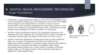
1. Visualizing images by adjusting grayscale, color scale, and windowing.
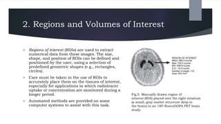
2. Defining regions and volumes of interest to extract numerical data from tissues.
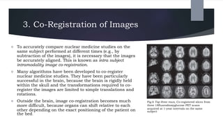
3. Co-registering images acquired at different times to compare changes in the same subject.
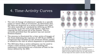
4. Creating time-activity curves from series of frames to analyze radiotracer uptake over time in regions of interest.
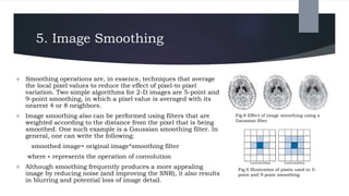
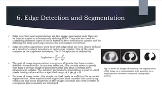
5. Smoothing images reduces noise but blurs details, while edge detection and segmentation identify boundaries and classify tissues.