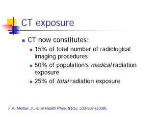
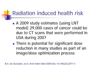
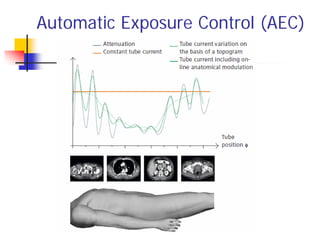
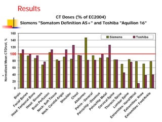
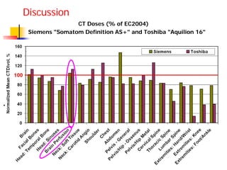
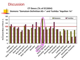
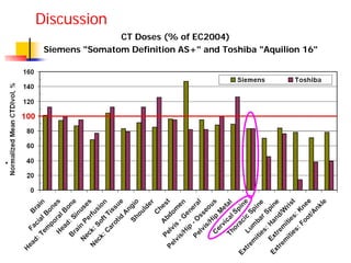
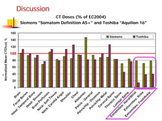
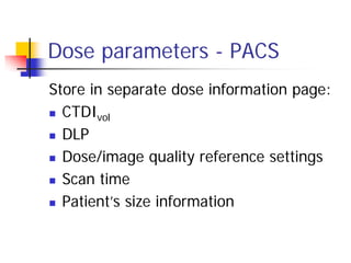
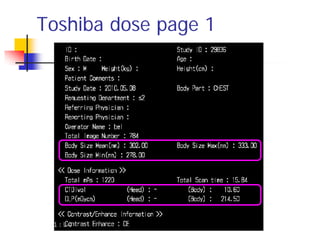
Implementation of an Audit and
Dose Reduction Program for CT
Y. Matyagin and P.J. Collins
Department of Nuclear Medicine, PET and Bone
Densitometry
Royal Adelaide Hospital
CT exposure
 Increasing concern worldwide regarding the
relatively high level of CT radiation exposure
 National Council on Radiation Protection and
Measurements: US average dose from medical
procedures increased 6x between 1980 and
2007
 60% due to CT
F.A Mettler Jr., et al. Radiology 2009 Nov;253(2):520-31.
CT exposure
 CT now constitutes:
 15% of total number of radiological
imaging procedures
 50% of population’s medical radiation
exposure
 25% of total radiation exposure
F.A. Mettler Jr., et al Health Phys. 95(5), 502-507 (2008).
Radiation induced health risk
 A 2009 study estimates (using LNT
model) 29,000 cases of cancer could be
due to CT scans that were performed in
USA during 2007
 There is potential for significant dose
reduction in many studies as part of an
image/dose optimization process
B.A. de Gonzales, et al. Arch Intern Med 2009 Dec 14;169(22):2071-7.
Spiral CT
CT machine
Motorized table
Rotating
X-ray
detectors
X-ray beam
Rotating
X-ray
source
Weighted Computed Tomography
Dose Index (CTDIw)
http://www.impactscan.org/slides/impactcourse/principles%20of%20ct%20dosimetry/index.html
• Weighted CTDI represents the average dose in scan
plane of Perspex phantom
• CTDIW = 1/3 CTDICentre + 2/3 CTDIPeriphery
• Phantom diameter:
32 cm or 16 cm
• Units: mGy
Volume CTDI (CTDIvol)
 CTDIvol: average dose over scanned volume
 CTDIvol: accounts for non-contiguous exposure along z-axis
 CTDIvol = CTDIw / pitch
Pitch 1.5
Pitch 1.0
Pitch 0.75
Dose-length product (DLP)
 Total dose: CTDIvol integrated along the scan
length, ie.
 DLP = CTDIvol • L (where L = scan length)
 Units: mGy • cm
 Different tissues of the body have
different radiosensitivity
 Unit of Effective dose (E) is the Sievert
where wT = weighting factor for organ, or tissue T
HT = equivalent dose to organ/tissue
Effective dose
E w HT T
T
 
Automatic Exposure Control (AEC)
Aim
 Compare CT doses (adults) at RAH with
an International benchmark
 Modify protocol settings to optimize
dose/image quality
Dose reference levels
 RANZCR have adopted EC1999 values
 European Guidelines (EC 2004) were
used in this study
 Address multi-slice scanners
 Includes data from 8 countries
EC 2004. G. Bongartz, et al. European Guidelines for Multislice Computed
Tomography. FIGM-CT2000-20078-CT-TIP. March 2004
Methods
 CT configured to display relevant dose
information (CTDIvol, DLP etc.) in PACS
 Baseline audit performed
 CT scanner exposure settings were
reduced in step-wise fashion so as not
to compromise image quality
Results
CT Doses (% of EC2004)
Siemens "Somatom Definition AS+" and Toshiba "Aquilion 16"
Brain perfusion
 Major International concern regarding
brain perfusion CT
 Cedars-Sinai Medical Center in USA:
 excessive CT doses in 250 patients
 Some studies 8 times expected level
 FDA reported significant CT
overexposure at several other medical
centers in 2009
Brain perfusion overdose
Hair loss in patients who
received radiation overdoses
W. Bogdanich, New York Times, July 31, 2010
Discussion
CT Doses (% of EC2004)
Siemens "Somatom Definition AS+" and Toshiba "Aquilion 16"
CT Doses (% of EC2004)
Siemens "Somatom Definition AS+" and Toshiba "Aquilion 16"
Discussion
CT Doses (% of EC2004)
Siemens "Somatom Definition AS+" and Toshiba "Aquilion 16"
Discussion
CT Doses (% of EC2004)
Siemens "Somatom Definition AS+" and Toshiba "Aquilion 16"
Discussion
Dose parameters - PACS
Store in separate dose information page:
 CTDIvol
 DLP
 Dose/image quality reference settings
 Scan time
 Patient’s size information
Toshiba dose page 1
Toshiba dose page 2
Siemens dose page
Automatic exposure control (AEC)
 AEC switched on
 Dose references level set
 Effect of other CT parameters on
functioning of AEC evaluated
 Min and max tube current
 Tube voltage
 Beam collimation and reconstructed slice
thickness
 Image reconstruction filters and algorithms
Large/small patients
A separate protocol should be employed:
 Higher tube voltages for large and
(possibly) lower tube voltages for small
patients
 Rotation time and pitch adjusted to
ensure tube current is within acceptable
limits
 Somewhat higher image noise is
acceptable for large patients
Future work
 More detailed analysis of image quality
 eg. using phantoms
 Investigate other key parameters
 Slice thickness
 Inter-slice intervals
 Streak artefacts
Acknowledgement
 Radiology Department
 George Kourlis (CT senior)

Implementation of an audit and dose reduction program for ct matyagin

  • 1.
    Implementation of anAudit and Dose Reduction Program for CT Y. Matyagin and P.J. Collins Department of Nuclear Medicine, PET and Bone Densitometry Royal Adelaide Hospital
  • 2.
    CT exposure  Increasingconcern worldwide regarding the relatively high level of CT radiation exposure  National Council on Radiation Protection and Measurements: US average dose from medical procedures increased 6x between 1980 and 2007  60% due to CT F.A Mettler Jr., et al. Radiology 2009 Nov;253(2):520-31.
  • 3.
    CT exposure  CTnow constitutes:  15% of total number of radiological imaging procedures  50% of population’s medical radiation exposure  25% of total radiation exposure F.A. Mettler Jr., et al Health Phys. 95(5), 502-507 (2008).
  • 4.
    Radiation induced healthrisk  A 2009 study estimates (using LNT model) 29,000 cases of cancer could be due to CT scans that were performed in USA during 2007  There is potential for significant dose reduction in many studies as part of an image/dose optimization process B.A. de Gonzales, et al. Arch Intern Med 2009 Dec 14;169(22):2071-7.
  • 5.
    Spiral CT CT machine Motorizedtable Rotating X-ray detectors X-ray beam Rotating X-ray source
  • 6.
    Weighted Computed Tomography DoseIndex (CTDIw) http://www.impactscan.org/slides/impactcourse/principles%20of%20ct%20dosimetry/index.html • Weighted CTDI represents the average dose in scan plane of Perspex phantom • CTDIW = 1/3 CTDICentre + 2/3 CTDIPeriphery • Phantom diameter: 32 cm or 16 cm • Units: mGy
  • 7.
    Volume CTDI (CTDIvol) CTDIvol: average dose over scanned volume  CTDIvol: accounts for non-contiguous exposure along z-axis  CTDIvol = CTDIw / pitch Pitch 1.5 Pitch 1.0 Pitch 0.75
  • 8.
    Dose-length product (DLP) Total dose: CTDIvol integrated along the scan length, ie.  DLP = CTDIvol • L (where L = scan length)  Units: mGy • cm
  • 9.
     Different tissuesof the body have different radiosensitivity  Unit of Effective dose (E) is the Sievert where wT = weighting factor for organ, or tissue T HT = equivalent dose to organ/tissue Effective dose E w HT T T  
  • 10.
  • 11.
    Aim  Compare CTdoses (adults) at RAH with an International benchmark  Modify protocol settings to optimize dose/image quality
  • 12.
    Dose reference levels RANZCR have adopted EC1999 values  European Guidelines (EC 2004) were used in this study  Address multi-slice scanners  Includes data from 8 countries EC 2004. G. Bongartz, et al. European Guidelines for Multislice Computed Tomography. FIGM-CT2000-20078-CT-TIP. March 2004
  • 13.
    Methods  CT configuredto display relevant dose information (CTDIvol, DLP etc.) in PACS  Baseline audit performed  CT scanner exposure settings were reduced in step-wise fashion so as not to compromise image quality
  • 14.
    Results CT Doses (%of EC2004) Siemens "Somatom Definition AS+" and Toshiba "Aquilion 16"
  • 15.
    Brain perfusion  MajorInternational concern regarding brain perfusion CT  Cedars-Sinai Medical Center in USA:  excessive CT doses in 250 patients  Some studies 8 times expected level  FDA reported significant CT overexposure at several other medical centers in 2009
  • 16.
    Brain perfusion overdose Hairloss in patients who received radiation overdoses W. Bogdanich, New York Times, July 31, 2010
  • 17.
    Discussion CT Doses (%of EC2004) Siemens "Somatom Definition AS+" and Toshiba "Aquilion 16"
  • 18.
    CT Doses (%of EC2004) Siemens "Somatom Definition AS+" and Toshiba "Aquilion 16" Discussion
  • 19.
    CT Doses (%of EC2004) Siemens "Somatom Definition AS+" and Toshiba "Aquilion 16" Discussion
  • 20.
    CT Doses (%of EC2004) Siemens "Somatom Definition AS+" and Toshiba "Aquilion 16" Discussion
  • 21.
    Dose parameters -PACS Store in separate dose information page:  CTDIvol  DLP  Dose/image quality reference settings  Scan time  Patient’s size information
  • 22.
  • 23.
  • 24.
  • 25.
    Automatic exposure control(AEC)  AEC switched on  Dose references level set  Effect of other CT parameters on functioning of AEC evaluated  Min and max tube current  Tube voltage  Beam collimation and reconstructed slice thickness  Image reconstruction filters and algorithms
  • 26.
    Large/small patients A separateprotocol should be employed:  Higher tube voltages for large and (possibly) lower tube voltages for small patients  Rotation time and pitch adjusted to ensure tube current is within acceptable limits  Somewhat higher image noise is acceptable for large patients
  • 27.
    Future work  Moredetailed analysis of image quality  eg. using phantoms  Investigate other key parameters  Slice thickness  Inter-slice intervals  Streak artefacts
  • 28.