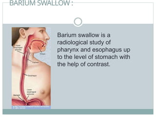
BARIUM SWALLOW :
Bariumswallow is a
radiological study of
pharynx and esophagus up
to the level of stomach with
the help of contrast.
4.
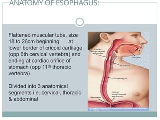
ANATOMY OF ESOPHAGUS:
Flattenedmuscular tube, size
18 to 26cm beginning at
lower border of cricoid cartilage
(opp 6th cervical vertebra) and
ending at cardiac orifice of
stomach (opp 11 thoracic
ᵗʰ
vertebra)
Divided into 3 anatomical
segments i.e. cervical, thoracic
& abdominal
5.
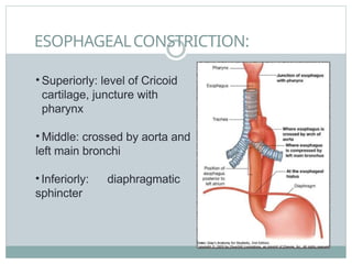
ESOPHAGEALCONSTRICTION:
• Superiorly: levelof Cricoid
cartilage, juncture with
pharynx
• Middle: crossed by aorta and
left main bronchi
• Inferiorly: diaphragmatic
sphincter
6.
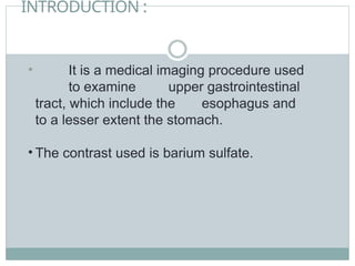
INTRODUCTION :
• Itis a medical imaging procedure used
to examine upper gastrointestinal
tract, which include the esophagus and
to a lesser extent the stomach.
• The contrast used is barium sulfate.
7.
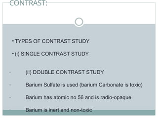
CONTRAST:
• TYPES OFCONTRAST STUDY
• (i) SINGLE CONTRAST STUDY
• (ii) DOUBLE CONTRAST STUDY
• Barium Sulfate is used (barium Carbonate is toxic)
• Barium has atomic no 56 and is radio-opaque
• Barium is inert and non-toxic
8.
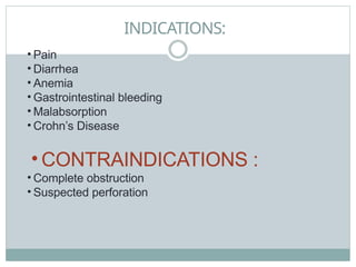
INDICATIONS:
• Dysphagia
• Heartburn, retrosternal pain, regurgitation &
odynophagia.
• Hiatus hernia
• Reflux esophagitis
• Stricture formation.
• Esophageal carcinoma.
• Motility disorder like
• Achalasia
• diffuse esophageal spasms.
• Pressure or invasion from extrinsic lesions.
• Assessment of abnormality of
• i. pharyngo esophageal junction including zenkers
diverticulum
• ii
.
• ii
cricoid webs
cricopharyngeal Achalasia.
9.
CONTRAINDICATIONS :
• Suspectedleakage from esophagus into the
mediastinum or pleura and peritoneal cavities
(Diatrazole Meglumine - 66% to be used)
• Tracheo-esophageal fistula (Diatrazole Meglumine -66%
to be used)
• Recent Biopsy
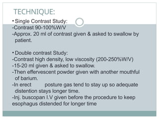
TECHNIQUE:
• Single ContrastStudy:
-Contrast 90-100%W/V
-Approx. 20 ml of contrast given & asked to swallow by
patient.
• Double contrast Study:
-Contrast high density, low viscosity (200-250%W/V)
-15-20 ml given & asked to swallow.
-Then effervescent powder given with another mouthful
of barium.
-In erect posture gas tend to stay up so adequate
distention stays longer time.
-Inj. buscopan I.V given before the procedure to keep
esophagus distended for longer time
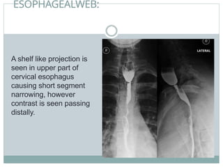
ESOPHAGEALWEB:
A shelf likeprojection is
seen in upper part of
cervical esophagus
causing short segment
narrowing, however
contrast is seen passing
distally.
14.
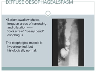
DIFFUSE OESOPHAGEALSPASM
• Bariumswallow shows
irregular areas of narrowing
and dilatation -----
“corkscrew” “rosary bead"
esophagus.
The esophageal muscle is
hypertrophied, but
histologically normal.
15.
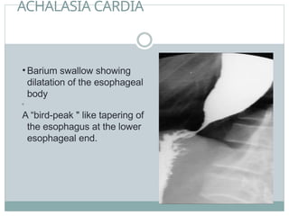
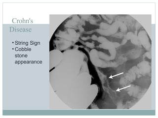
ACHALASIA CARDIA
• Bariumswallow showing
dilatation of the esophageal
body
•
A “bird-peak " like tapering of
the esophagus at the lower
esophageal end.
16.
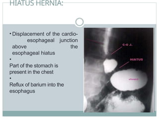
HIATUS HERNIA:
• Displacementof the cardio-
esophageal junction
above the
esophageal hiatus
•
Part of the stomach is
present in the chest
•
Reflux of barium into the
esophagus
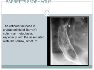
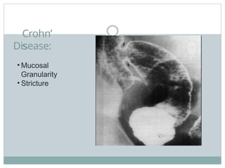
BARRETT’S ESOPHAGUS:
The reticularmucosa is
characteristic of Barrett's
columnar metaplasia,
especially with the associated
web-like (arrow) stricture.
19.
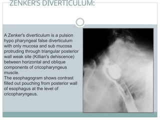
ZENKER’S DIVERTICULUM:
A Zenker'sdiverticulum is a pulsion
hypo pharyngeal false diverticulum
with only mucosa and sub mucosa
protruding through triangular posterior
wall weak site (Killian's dehiscence)
between horizontal and oblique
components of cricopharyngeus
muscle.
The esophagogram shows contrast
filled out pouching from posterior wall
of esophagus at the level of
cricopharyngeus.
20.
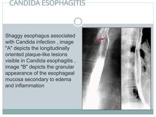
CANDIDA ESOPHAGITIS
Shaggy esophagusassociated
with Candida infection , image
"A" depicts the longitudinally
oriented plaque-like lesions
visible in Candida esophagitis ,
image "B" depicts the granular
appearance of the esophageal
mucosa secondary to edema
and inflammation
21.
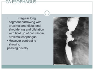
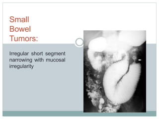
CA ESOPHAGUS
• Irregularlong
segment narrowing with
proximal and distal end
shouldering and dilatation
with hold up of contrast in
proximal esophagus
• However contrast is
showing
passing distally
BARIUM MEAL:
• Bariummeal is radiological study of lower
esophagus, stomach and duodenum.
• Done by oral administration of contrast media barium
sulphate.
24.
INDICATIONS:
• 1.Dyspepsia
• 2.Weightloss
• 3.Upper abdominal mass
• 4.Gastrointestinal hemorrhage or unexplained iron
deficiency anemia
• 5. Partial obstruction
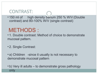
CONTRAST:
• 150 mlof high density barium 250 % W/V (Double
contrast) and 80-100% W/V (single contrast)
METHODS :
• 1. Double contrast: Method of choice to demonstrate
mucosal pattern.
• 2. Single Contrast:
• a) Children -since it usually is not necessary to
demonstrate mucosal pattern
• b) Very ill adults – to demonstrate gross pathology
only
27.
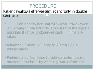
PROCEDURE
Patient swallows effervescentagent (only in double
contrast)
• High density barium(250% w/v) is swallowed
while lyingon the left side. Then turn to the supine
position. If reflux is observed spot films are
taken
• A hypotonic agent –Buscopan(20 mg I.V ) is
administered
• Patient rolled from side to side so barium coats
mucosal surfaces by washing mucus from the
gastric mucosa
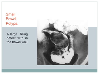
GASTRIC POLYP
• Multiplewell defined
filling defects with a
surrounding ring of
barium are noted along
the dependent wall of
stomach suggesting
multiple gastric polyps
34.
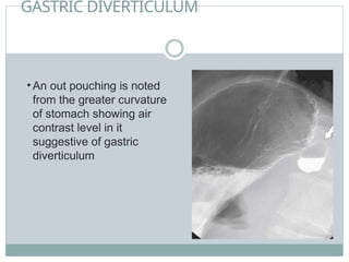
GASTRIC DIVERTICULUM
• Anout pouching is noted
from the greater curvature
of stomach showing air
contrast level in it
suggestive of gastric
diverticulum
35.
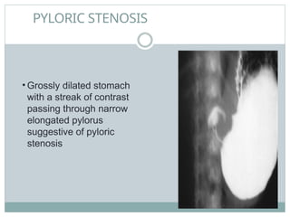
PYLORIC STENOSIS
• Grosslydilated stomach
with a streak of contrast
passing through narrow
elongated pylorus
suggestive of pyloric
stenosis
36.
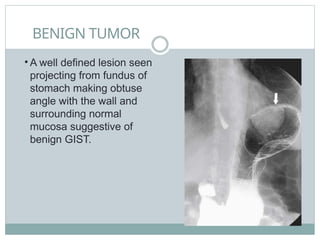
BENIGN TUMOR
• Awell defined lesion seen
projecting from fundus of
stomach making obtuse
angle with the wall and
surrounding normal
mucosa suggestive of
benign GIST.
37.
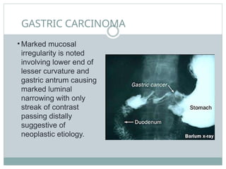
GASTRIC CARCINOMA
• Markedmucosal
irregularity is noted
involving lower end of
lesser curvature and
gastric antrum causing
marked luminal
narrowing with only
streak of contrast
passing distally
suggestive of
neoplastic etiology.
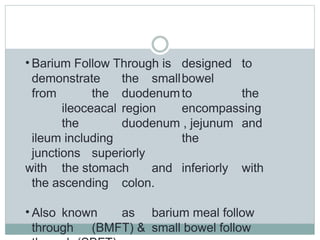
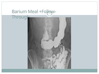
• Barium FollowThrough is designed to
demonstrate the smallbowel
from the duodenumto the
ileoceacal region encompassing
the duodenum , jejunum and
ileum including the
junctions superiorly
with the stomach and inferiorly with
the ascending colon.
• Also known as barium meal follow
through (BMFT) & small bowel follow
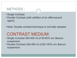
METHODS :
• SingleContrast
• Double Contrast (with addition of an effervescent
agent)
• Note: Double contrast technique is normally adopted
CONTRAST MEDIUM:
• Single Contrast 300-400 ml of 50-60% w/v Barium
suspension
• Double Contrast 300-400 ml of 80-100% w/v Barium
suspension
42.
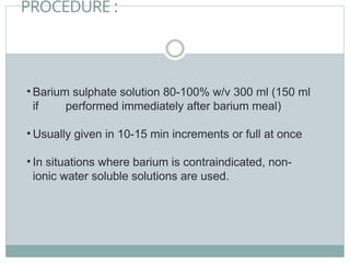
PROCEDURE :
• Bariumsulphate solution 80-100% w/v 300 ml (150 ml
if performed immediately after barium meal)
• Usually given in 10-15 min increments or full at once
• In situations where barium is contraindicated, non-
ionic water soluble solutions are used.
43.
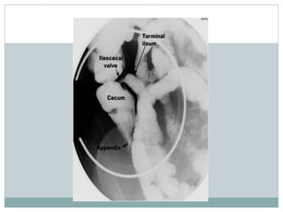
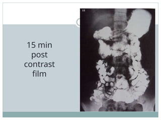
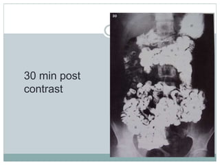
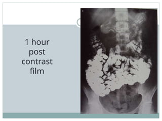
FILMING:
• Prone PAfilms of the abdomen are taken.
• The first radiograph is taken 10 min following the drink,
with the second image at 30 min stage. Then the
radiographs are taken at 30 min intervals until the
barium has reached terminal ileum.
• Pressure on the abdomen helps to compress abdominal
contents so that the loops of small bowel are separated.
Thus for better radiographic quality, prone position is
used.
• Spot films of the terminal ileum are taken supine.
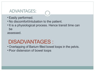
ADVANTAGES:
• Easily performed.
•No discomfort/intubation to the patient.
• It is a physiological process. Hence transit time can
be
assessed.
DISADVANTAGES :
• Overlapping of Barium filled bowel loops in the pelvis.
• Poor distension of bowel loops
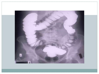
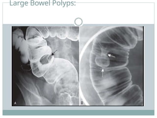
BARIUM ENEMA:
• Abarium enema is a test used to help visualize the
colon (large bowel).
• A barium enema is used to look for problems in the
colon, such as polyps, inflammation (colitis), narrowing
of the colon, tumors, diverticulitis.
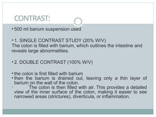
CONTRAST:
• 500 mlbarium suspension used
• 1. SINGLE CONTRAST STUDY (20% W/V)
The colon is filled with barium, which outlines the intestine and
reveals large abnormalities.
• 2. DOUBLE CONTRAST (100% W/V)
• the colon is first filled with barium
• then the barium is drained out, leaving only a thin layer of
barium on the wall of the colon.
• The colon is then filled with air. This provides a detailed
view of the inner surface of the colon, making it easier to see
narrowed areas (strictures), diverticula, or inflammation.


![COMPLETE BARIUM STUDIES Of GIT NAD [Adrian Dungu Niyimpa].pdf](https://cdn.slidesharecdn.com/ss_thumbnails/bastudies-gitnad-220902221043-d535eaa3-thumbnail.jpg?width=640&height=640&fit=bounds)


