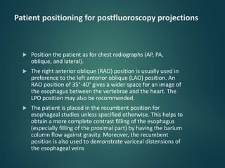
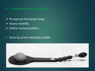
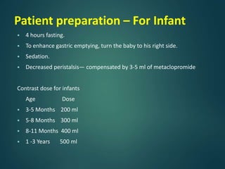
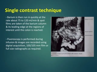
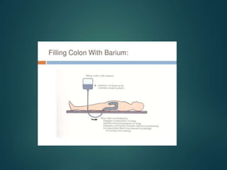
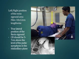
This document provides information about barium procedures, including barium swallow, barium meal, and barium follow through examinations. It defines barium as a radioopaque contrast agent used to provide a roadmap of GI tract pathologies in x-ray exams. Barium sulphate is commonly used because it is non-toxic, non-absorbable, and coats the mucosa, allowing double contrast studies. The document describes the techniques, positions, and views used in various barium exams to visualize the esophagus, stomach, and small intestine. It also lists common indications, contraindications, and complications.
Learn Barium Meal & Follow Through for the beginners from a Radiology Resident.For some image description please go through the text book "David Sutton" because i have described these image during my presentation Verbally..There are many animations used inside this presentation so to see all the pictures which are placed layer by layer with the help of animations you simple need to download this presentation first.... Thanx.
Barium meal ppt presentation is very important for radiology resident , radiologist and radiographers. this slide contents lots of barium image and technique, position, indication and modification and lots of information. this presentation help alot thanks .
Achalasia cardia is a primary oesophageal motility disorder. Barium swallow demonstrates a classic bird beak appearance. This presentation briefly explains the barium swallow procedure and findings in achalasia cardia.
Title: Sense of Taste
Presenter: Dr. Faiza, Assistant Professor of Physiology
Qualifications:
MBBS (Best Graduate, AIMC Lahore)
FCPS Physiology
ICMT, CHPE, DHPE (STMU)
MPH (GC University, Faisalabad)
MBA (Virtual University of Pakistan)
Learning Objectives:
Describe the structure and function of taste buds.
Describe the relationship between the taste threshold and taste index of common substances.
Explain the chemical basis and signal transduction of taste perception for each type of primary taste sensation.
Recognize different abnormalities of taste perception and their causes.
Key Topics:
Significance of Taste Sensation:
Differentiation between pleasant and harmful food
Influence on behavior
Selection of food based on metabolic needs
Receptors of Taste:
Taste buds on the tongue
Influence of sense of smell, texture of food, and pain stimulation (e.g., by pepper)
Primary and Secondary Taste Sensations:
Primary taste sensations: Sweet, Sour, Salty, Bitter, Umami
Chemical basis and signal transduction mechanisms for each taste
Taste Threshold and Index:
Taste threshold values for Sweet (sucrose), Salty (NaCl), Sour (HCl), and Bitter (Quinine)
Taste index relationship: Inversely proportional to taste threshold
Taste Blindness:
Inability to taste certain substances, particularly thiourea compounds
Example: Phenylthiocarbamide
Structure and Function of Taste Buds:
Composition: Epithelial cells, Sustentacular/Supporting cells, Taste cells, Basal cells
Features: Taste pores, Taste hairs/microvilli, and Taste nerve fibers
Location of Taste Buds:
Found in papillae of the tongue (Fungiform, Circumvallate, Foliate)
Also present on the palate, tonsillar pillars, epiglottis, and proximal esophagus
Mechanism of Taste Stimulation:
Interaction of taste substances with receptors on microvilli
Signal transduction pathways for Umami, Sweet, Bitter, Sour, and Salty tastes
Taste Sensitivity and Adaptation:
Decrease in sensitivity with age
Rapid adaptation of taste sensation
Role of Saliva in Taste:
Dissolution of tastants to reach receptors
Washing away the stimulus
Taste Preferences and Aversions:
Mechanisms behind taste preference and aversion
Influence of receptors and neural pathways
Impact of Sensory Nerve Damage:
Degeneration of taste buds if the sensory nerve fiber is cut
Abnormalities of Taste Detection:
Conditions: Ageusia, Hypogeusia, Dysgeusia (parageusia)
Causes: Nerve damage, neurological disorders, infections, poor oral hygiene, adverse drug effects, deficiencies, aging, tobacco use, altered neurotransmitter levels
Neurotransmitters and Taste Threshold:
Effects of serotonin (5-HT) and norepinephrine (NE) on taste sensitivity
Supertasters:
25% of the population with heightened sensitivity to taste, especially bitterness
Increased number of fungiform papillae
HOT NEW PRODUCT! BIG SALES FAST SHIPPING NOW FROM CHINA!! EU KU DB BK substit...GL Anaacs
Contact us if you are interested:
Email / Skype : kefaya1771@gmail.com
Threema: PXHY5PDH
New BATCH Ku !!! MUCH IN DEMAND FAST SALE EVERY BATCH HAPPY GOOD EFFECT BIG BATCH !
Contact me on Threema or skype to start big business!!
Hot-sale products:
NEW HOT EUTYLONE WHITE CRYSTAL!!
5cl-adba precursor (semi finished )
5cl-adba raw materials
ADBB precursor (semi finished )
ADBB raw materials
APVP powder
5fadb/4f-adb
Jwh018 / Jwh210
Eutylone crystal
Protonitazene (hydrochloride) CAS: 119276-01-6
Flubrotizolam CAS: 57801-95-3
Metonitazene CAS: 14680-51-4
Payment terms: Western Union,MoneyGram,Bitcoin or USDT.
Deliver Time: Usually 7-15days
Shipping method: FedEx, TNT, DHL,UPS etc.Our deliveries are 100% safe, fast, reliable and discreet.
Samples will be sent for your evaluation!If you are interested in, please contact me, let's talk details.
We specializes in exporting high quality Research chemical, medical intermediate, Pharmaceutical chemicals and so on. Products are exported to USA, Canada, France, Korea, Japan,Russia, Southeast Asia and other countries.
Ethanol (CH3CH2OH), or beverage alcohol, is a two-carbon alcohol
that is rapidly distributed in the body and brain. Ethanol alters many
neurochemical systems and has rewarding and addictive properties. It
is the oldest recreational drug and likely contributes to more morbidity,
mortality, and public health costs than all illicit drugs combined. The
5th edition of the Diagnostic and Statistical Manual of Mental Disorders
(DSM-5) integrates alcohol abuse and alcohol dependence into a single
disorder called alcohol use disorder (AUD), with mild, moderate,
and severe subclassifications (American Psychiatric Association, 2013).
In the DSM-5, all types of substance abuse and dependence have been
combined into a single substance use disorder (SUD) on a continuum
from mild to severe. A diagnosis of AUD requires that at least two of
the 11 DSM-5 behaviors be present within a 12-month period (mild
AUD: 2–3 criteria; moderate AUD: 4–5 criteria; severe AUD: 6–11 criteria).
The four main behavioral effects of AUD are impaired control over
drinking, negative social consequences, risky use, and altered physiological
effects (tolerance, withdrawal). This chapter presents an overview
of the prevalence and harmful consequences of AUD in the U.S.,
the systemic nature of the disease, neurocircuitry and stages of AUD,
comorbidities, fetal alcohol spectrum disorders, genetic risk factors, and
pharmacotherapies for AUD.
Explore natural remedies for syphilis treatment in Singapore. Discover alternative therapies, herbal remedies, and lifestyle changes that may complement conventional treatments. Learn about holistic approaches to managing syphilis symptoms and supporting overall health.
Factory Supply Best Quality Pmk Oil CAS 28578–16–7 PMK Powder in Stockrebeccabio
Factory Supply Best Quality Pmk Oil CAS 28578–16–7 PMK Powder in Stock
Telegram: bmksupplier
signal: +85264872720
threema: TUD4A6YC
You can contact me on Telegram or Threema
Communicate promptly and reply
Free of customs clearance, Double Clearance 100% pass delivery to USA, Canada, Spain, Germany, Netherland, Poland, Italy, Sweden, UK, Czech Republic, Australia, Mexico, Russia, Ukraine, Kazakhstan.Door to door service
Hot Selling Organic intermediates
Couples presenting to the infertility clinic- Do they really have infertility...Sujoy Dasgupta
Dr Sujoy Dasgupta presented the study on "Couples presenting to the infertility clinic- Do they really have infertility? – The unexplored stories of non-consummation" in the 13th Congress of the Asia Pacific Initiative on Reproduction (ASPIRE 2024) at Manila on 24 May, 2024.
The prostate is an exocrine gland of the male mammalian reproductive system
It is a walnut-sized gland that forms part of the male reproductive system and is located in front of the rectum and just below the urinary bladder
Function is to store and secrete a clear, slightly alkaline fluid that constitutes 10-30% of the volume of the seminal fluid that along with the spermatozoa, constitutes semen
A healthy human prostate measures (4cm-vertical, by 3cm-horizontal, 2cm ant-post ).
It surrounds the urethra just below the urinary bladder. It has anterior, median, posterior and two lateral lobes
It’s work is regulated by androgens which are responsible for male sex characteristics
Generalised disease of the prostate due to hormonal derangement which leads to non malignant enlargement of the gland (increase in the number of epithelial cells and stromal tissue)to cause compression of the urethra leading to symptoms (LUTS
Lung Cancer: Artificial Intelligence, Synergetics, Complex System Analysis, S...Oleg Kshivets
RESULTS: Overall life span (LS) was 2252.1±1742.5 days and cumulative 5-year survival (5YS) reached 73.2%, 10 years – 64.8%, 20 years – 42.5%. 513 LCP lived more than 5 years (LS=3124.6±1525.6 days), 148 LCP – more than 10 years (LS=5054.4±1504.1 days).199 LCP died because of LC (LS=562.7±374.5 days). 5YS of LCP after bi/lobectomies was significantly superior in comparison with LCP after pneumonectomies (78.1% vs.63.7%, P=0.00001 by log-rank test). AT significantly improved 5YS (66.3% vs. 34.8%) (P=0.00000 by log-rank test) only for LCP with N1-2. Cox modeling displayed that 5YS of LCP significantly depended on: phase transition (PT) early-invasive LC in terms of synergetics, PT N0—N12, cell ratio factors (ratio between cancer cells- CC and blood cells subpopulations), G1-3, histology, glucose, AT, blood cell circuit, prothrombin index, heparin tolerance, recalcification time (P=0.000-0.038). Neural networks, genetic algorithm selection and bootstrap simulation revealed relationships between 5YS and PT early-invasive LC (rank=1), PT N0—N12 (rank=2), thrombocytes/CC (3), erythrocytes/CC (4), eosinophils/CC (5), healthy cells/CC (6), lymphocytes/CC (7), segmented neutrophils/CC (8), stick neutrophils/CC (9), monocytes/CC (10); leucocytes/CC (11). Correct prediction of 5YS was 100% by neural networks computing (area under ROC curve=1.0; error=0.0).
CONCLUSIONS: 5YS of LCP after radical procedures significantly depended on: 1) PT early-invasive cancer; 2) PT N0--N12; 3) cell ratio factors; 4) blood cell circuit; 5) biochemical factors; 6) hemostasis system; 7) AT; 8) LC characteristics; 9) LC cell dynamics; 10) surgery type: lobectomy/pneumonectomy; 11) anthropometric data. Optimal diagnosis and treatment strategies for LC are: 1) screening and early detection of LC; 2) availability of experienced thoracic surgeons because of complexity of radical procedures; 3) aggressive en block surgery and adequate lymph node dissection for completeness; 4) precise prediction; 5) adjuvant chemoimmunoradiotherapy for LCP with unfavorable prognosis.
2. Definition:-
Barium provides a roadmap of GI tract pathologies in the form x-ray
examination of the esophagus, stomach, duodenum, small intestine
& large intestine.
3. Characteristic of barium:-
The reason for using Barium sulphate for GI studies are :
1. Ba has a high atomic number 56. Therefore, it is highly radioopaque and
produces excellent bowel opacification.
2. Non absorbable (Therefore does not degrade throughout the bowel
), non-toxic.
3. Insoluble in water/lipid.
4. Inert to tissues.
5. Suitable for double contrast studies as it coats the mucosa in a thin layer, thus
allowing the introduction of 2nd or negative contrast agent without significant
degradation.
Route: Orally & Rectally
4. Adverse Effects of barium
1. Chemical peritonitis due to extravasation of additives of Barium Sulphate.
2. Extravasation into bronchial tree, urinary tract and other body cavities
will produce inflammation.
3. Barium inspissation in cases of colonic obstruction to form hard stones.
4. Intravascular entry of Barium can cause embolism.
5. Barium Encephalopathy. *
Small amount of Barium can be absorbed from the peritoneum in case of
perforation
Circulation --> concentrated in CSF with detectable levels -->
Encephalopathy
6. Previous contrast media extravasated may mimic cancer due to
inflammation.
Longstanding barium deposits are carcinogenic.
5. Barium Studies Primer/General Principles
Four main types of lesions – FUDS
1. Filling defect
2. Ulceration
3. Diverticulum
4. Strictures
12. Indications
1. Dysphagia
2. Heart burn, retrosternal pain, regurgitation & odynophagia
3. Hiatus hernia
4. Reflux oesophagitis
5. Stricture formation
6. Esophageal carcinoma
7. Motility disorder like – Achalasia , diffuse esophageal spasms
8. Pressure or invasion from extrinsic lesions
9. Assessment of abnormality of
1. Pharyngo esophageal junction including zenkers diverticulum
2. Cricoid webs
3. Cricopharyngeal Achalasia.
13. Contraindications
1. Barium should NOT be used initially if perforation is suspected.
If perforation is not identifed with a water-soluble contrast agent
then a barium examination should be considered.
2. Tracheo-esophageal fistula
14. Patient preparation
1. NPO for 6 hours prior to the examination.
2. Smoking should be avoided on the day of examination.
3. Muscle relaxants before the procedure
15. Contrast
2 Types of contrast study
1. Single contrast study
2. Double contrast study
16. Single vs double contrast:-
Single contrast
medium
Double contrast
medium
Only barium is given. 60-100%
w/v
Barium with gas producing agent
is given. 200-250% w/v
To outline the structures, lumen
and large abnormalities.
For detail viewing of the mucosal
pattern, making it easier to see
narrowed areas (strictures),
diverticula or inflammation.
17. Supplies and technical factors
4-6 oz (100-200 mL) regular barium (60% weight/volume)
Barium cup
Flexible large-caliber straw
± 2 oz (± 59 mL) regular barium (60% w/v) diluted with 2 oz
(59 mL) water
Scout film: Not routinely obtained.
Image receptor (IR) or cassette: 14 x 14 inches (35 x 35 cm);
for a 3-on-1 or spot-film image
kVp: 80-120 MAs 30-40
Table-top position: the table is declined to -20° to allow for
a full esophageal distension
18. Patient positioning for a single-contrast esophagram
Place the patient in the right anterior oblique (RAO) position to
offset the esophagus from the spine. The patient’s right arm is
placed alongside the body, with the left knee flexed.
PA oblique esophagus, RAO position (the
midsagittal position forms an angle of 35°-
45° from the grid device).
19. The technologist should place the cup of
barium in the patient’s left hand, with the
straw between the patient’s teeth.
Patients who are unable to tolerate this
position may be imaged in the left posterior
oblique (LPO) position.
Position the fluoroscope so that the apex of
the left lung appears at the top of the monitor.
The technologist will ask the patient to
continuously drink the barium. This fills and
distends the esophagus while the technologist
obtains images of the proximal esophagus,
midesophagus, and the distal esophagus,
including an open lower esophageal sphincter
(magnified if possible).
Single-contrast study of esophagus in RAO
position with table top in head-down -20°
position
21. Supplies and technical factors
2 medicine cups (for effervescent granules and water)
3/4 ampule effervescent granules
10 cc water
4-6 oz (100-200 mL) dense barium (200% to 250% w/v)
Barium cup
Scout film: Not obtained routinely
IR or cassette: 14 x 14 inches (35 x 35 cm) for a 3-on-1 or
spot-film image
kVp: 90
Table-top position: vertical
22. The performance of the double-contrast esophageal
examination is similar to that of a single-contrast
examination. For a double-contrast examination, free-
flowing, high-density barium must be used. A gas-producing
substance, usually carbon dioxide crystals, can be added to
the barium mixture or taken by mouth immediately before
the barium suspension is ingested. Spot radiographs are
taken during the examination, and delayed images may be
obtained on request.
23. Patient positioning for a double-contrast esophagram
Have the patient stand on a footboard in the LPO position
to offset the esophagus from the spine.
Place the cup of dense barium in the patient’s left hand.
Have the patient take a small sip of the dense barium to
become acclimated to its consistency. If the patient appears
unable to tolerate the dense barium, obtain a single-
contrast esophagram.
Instruct the patient on the swallowing sequence. This is
essential for obtaining a satisfactory exam
24. Swallowing sequence and technique
The patient's head is tilted back to extend the neck.
Center the fluoroscope over the upper third of the esophagus to localize
the esophagus, then lower the tower and administer the effervescent
granules and water. The technologist pours the granules into the back of
the patient’s mouth, then adds the water and tells the patient to
immediately swallow. (Overdistension is prevented by using only 3/4
ampule of the effervescent granules.)
patient drinks the barium with moderate rapidity and with constant
encouragement by the technologist.
Caution the patient not to burp.
Image the entire esophagus as the patient is drinking, observe the
esophagus for a "silver-satin" appearance -- indicating the best possible
coating -- and obtain images of the proximal esophagus, midesophagus,
and the distal esophagus
25. Postfluoroscopy projections
The three basic postfluoroscopy projections for the
esophagram are the anteroposterior (AP) or
posteroanterior (PA) projection; AP or PA oblique in the
RPO or LPO position; and the lateral projection from the
right or left position
26. Patient positioning for postfluoroscopy projections
Position the patient as for chest radiographs (AP, PA,
oblique, and lateral).
The right anterior oblique (RAO) position is usually used in
preference to the left anterior oblique (LAO) position. An
RAO position of 35°-40° gives a wider space for an image of
the esophagus between the vertebrae and the heart. The
LPO position may also be recommended.
The patient is placed in the recumbent position for
esophageal studies unless specified otherwise. This helps to
obtain a more complete contrast filling of the esophagus
(especially filling of the proximal part) by having the barium
column flow against gravity. Moreover, the recumbent
position is also used to demonstrate variceal distensions of
the esophageal veins
27. Xray views
Lateral projection:-
Place pt in lateral position.
Center midcoronal plane to cassette.
Bottom of cassette below xiphoid process.
Pt must drink continuously before and
during exposure.
Use shielding!
28. Xray views
AP or PA Projection:-
Pt. supine or prone
Center midsagittal plane to cassette
Bottom of cassette should be placed just
below tip of xiphoid
Pt. drinks contrast before exposure and
continues drinking during exposure.
Shield!
29. Xray views
RAO or LAO Positions:-
To throw the esophagus clear of the spine.
Pt should be rotated 35 - 40 degrees
Center about 2 inches lateral to MSP
Bottom of cassette below xiphoid.
30. Pharynx
1. One mouthful contrast bolus with high density(250% w/v).
2. To get optimum mucosal coating patient is asked to
swallow once and stop swallowing there after.
3. Frontal and lateral view x-ray taken.
34. Barium meal
Identifies lower half of oesophagus, the stomach and all of
duodenum.
Method
A)double contrast – the method of choice to demonstrate
mucosal pattern
B)single contrast-used in children (not necessary to demonstrate
mucosal pattern)
And very ill adults (only gross pathology)
35. Indications
1)Dyspepsia
2)Weight loss
3)Upper abdominal mass
4)Gastro intestinal haemorrhage
5)suspected upper GI obstruction
6)assessment of the site of perforation(water soluble contrast is
used)
36. Contra indications
1.Complete large bowel obstruction
2.Suspected perforation (unless water soluble contrast
medium used)
Patient preparation
1. NPO after midnight(6 hrs)
2.abstain from-smoking, chewing gum or antacids-
CONTRAST MEDIUM :
•120 ml of high density barium 250 % W/V
(Double contrast)
• Sufficient 100 % W/V ( Single Contrast )
37. Technique-single contrast
The examination is began with patient in the erect position.The fluoroscopist
may first examine heart and lungs fluoroscopically and observe the abdomen to
determine whether food or fluid is in the stomach.
The patient then given a glass of barium and instruct to drink it as directed by
fluoroscopist.
If patient is in recumbent position,the mixture is administered through a
drinking straw.
The fluoroscopist instructs the patient to swallow two or three mouthfuls of the
barium,during this time the fluoroscopist examine esophagus.Then stomach and
duodenum.
Fluoroscopy is performed with the patient in the erect and the recumbent
positions,while the body is rotated and the table is angled so that all aspects of
the esophagus,stomach and duodenum are demonstrated.
Spot films taken as indicated.
38. Technique-double contrast
1.Hypotonic agent Buscopan(hyoscine butyl bromide,20 mg i.v) or
0.1-0.2 mg i.v glucagon is injected intravenously -relax stomach and
suspend peristalsis.
A packet of effervescent granules swallowed with small amount of
water- releases CO2 and gastric distension.(approx 400ml CO2)
High density barium is swallowed(120 ml- 250% w/v) and
double contrast views of oesophagus is obtained standing
RAO.
39. Patient faces Xray table,lowered to horizontal
Then turned onto left side and finally supine.
Patient rolled from side to side so as barium coats mucosal
surfaces properly-washes over the mucus .
Ask to roll onto the right side & then quickly over in a complete circle, to
finish in an RAO position.
Sequences of films of stomach obtained—When barium enters
duodenum, patient is turned RAO – fills duodenum with gas, DC
films are taken.
Biphasic examination–Prone swallow of thin (125%w/v low
density) barium given after contrast view obtained to optimize
compression views of stomach and duodenum
40. Under fluoroscopic guidance, on the compression views-filling defects
or abnormal collections are detected.
Note:young children- main indication identify cause of vomiting eg:-
pyloric
Flow technique identifies-subtle mucosal abnormalities.
obstruction,malrotation,and GOR.single contrast technique
preferred(30% w/v Barium sulfate with no paralytic agent).
Note : kV range double contrast- 70-120 kV
single contrast-120-150kV
Note:If partial gastrectomy or drainage procedues (eg; pyloroplasty or
gastrenterostomy), begin with prone swallow using high density
barium.Reaching duodenum or Genterostomy-turned supine for DC
films.DC of stomach and oesophagus follows.
41. Positions employed
Stomach and duodenum examined in the PA,AP,oblique and lateral
directions,with the patient is the erect and recumbent postions.
43. Right anterior oblique position
RAO –film -10x12 in.
This projection with a rotation of 40 to 70 degrees gives the best image
of the pyloric canal and duodenal bulb
44. Left posterior oblique position for stomach
and duodenum
LPO-film 10x12 in. best demonstrates the fundus portion of the stomach.
45. Right lateral position for stomach and
duodenum
Lateral projection-film 10x12 in. shows anterior and posterior aspects of the
stomach,pyloric canal and duodenal bulb.
46. AP projection-film 11x14 in.-shows well filled fundic portion and usually DC
delineation of the body ,the antral portion and the duodenum
48. Spot film of the abdomen with the patient in prone
position
49.
50. Modification technique for young children
Indication
• Vomiting
Technique
• Single contrast
• 30 % barium sulphate
• No paralytic agent
51. Aftercare
Patient should be told that the
bowel will be white for few
days
Patient should be advised to
drink adequate water
Patient should not leave the
department until blurring of
vision has resolved
53. Anatomy of small intestine
length = 6-7 m (approx)
Extent- From Pylorus to ileo-caecal
valve
Proximal 2/5th constitute the
jejunum and distal 3/5th constitute
the ileum
The Valvulae conniventes
-2 mm thick in jejunum and 1
mm thick in ileum.
54. JEJUNUM & ILEUM
33
• Jejunum begins at
duodenojejunal flexure
(L2) & ileum ends at
ileocecalJunction.
• Jejunum & ileum = 6 to 7
m long (jejunum 2/5,
ileum 3/5)
•Coils of jejunum & ileum are suspended by mesentery
from posterior abdominal wall & freely movable.Most
jejunum lies in left upper quadrant & most ileum lies in
right lower quadrant
55. Introduction – Barium Follow Through
• Barium Follow Through is designed to
demonstrate the small bowel from the
duodenum to the ileoceacal region
encompassing the duodenum , jejunum and ileum
including the junctions superiorly with the
stomach and inferiorly with the ascending colon.
• Also known as barium meal follow through
(BMFT) & small bowel follow through (SBFT)
35
56. Barium meal follow-through
Methods:-
-single contrast.
-with the addition of an effervescent agent.
-with the addition of a pneumocolon technique.
Indication:-
-Diarrhoea
-Anaemia
-Partial obstruction
-Malabsorption
-Abdominal mass
Contraindication:-
-Complete obstruction
-Suspected perforation
-Paralytic ileus
57. Barium meal follow-through
Contrast medium:-
Barium sulphate solution 100% w/v 300 ml (150 ml if performed immediately after
barium meal)
Usually given in 10-15 min increments or full at once
Transit time through small bowel has been shown to be reduced by the addition of 10
ml of gastrograffin to barium.
In children, 3-4 ml/kg is suitable volume of contrast.
Patient preparation:-
Low residue diet for 2 days prior when possible
NPO after midnight before exam.
Metoclopramide (maxolon) 20 mg orally may be given before or during the
examination to enhance gastric emptying.
Pt’s bladder must be empty before & during procedure to avoid displacing or
compressing ileum.
58. Preliminary Film
Plain radiograph of the abdomen.
To see bowel preparation.
To rule out contraindication.
Helps in assessing any abnormalities of gas filled bowel loops.
If residual fecal matter presence-examination should be cancelled.
59. Technique
Prone PA film of the abdomen are taken every 15-20min during the first hour.
And subsequently every 20-30 min until the colon is reached.
Spot film of the terminal ileum are taken in supine.
Single Contrast Technique
Patient is asked to drink Barium Suspension as rapidly as possible and then put
the patient on right side to aid rapid gastric emptying.
After 15 to 20 minutes , a film is taken with the patient prone to separate bowel
loops , using high kV to demonstrate jejunum and proximal ileum.
Subsequent films are taken with the right side up at 15-30 minute intervals till
ileocecal junction is opacified.
60.
61.
62.
63. Compression is mandatory
Toseparate the bowel loops
Assess mobility
Define mucosal pattern
• Done by prone inflatable paddle
64. Additional films
Toseparate loops of small bowel
Oblique view
With X-ray tube angled into the pelvis
With patient tilted head down
Todemonstrate diverticula
Erect-will reveal any fluid level
65. Single Contrast Technique
Advantages of Prone Position
Better separation & less overlap of bowel loops.
In this position the centre of the abdomen is compressed making entire
abdomen more uniform and thus more uniform x-ray penetration can be
achieved.
In this position loops of ileum tends to migrate cephalad and becomes
less compacted in the pelvis which is often a common problem during
procedure.
Compression should be applied on the bowel loops to avoid overlap and
to efface the mucosa so that the small lesions may not be missed and
mobility of the loops can
be well assessed
66. Single Contrast Technique
Overlap of contrast filled bowel loops in the pelvis can be
overcome by :
Table head down.
30° caudal angled view of pelvis.
Emptying of urinary bladder prior to filming the ileal loops.
Drugs – Metaclopramide, Neostigmine, Glucagon, Cholecystokinin
20-40 ml of sodium / meglumine diatrizoate to the barium also reduces
transit time.
Cold barium speeds the gastric emptying and passes more rapidly
through the intestine than does the room-temperature barium.
Preliminary cleansing of the colon and placing the patient in right lateral
recumbent position
If desired, gastric and bowel peristalsis may be increased by various methods
67. Peroral Pneumocolon
Done at the end of B.M.F.T. when distal ileum is suspicious and needs
clarification.
Preparation - Colonic preparation is similar to barium enema.
Technique
Barium is administered orally
When barium has reached the right and proximal transverse colon, air is
insufflated into the rectum and refluxed into distal ileum.
Glucagon can be used to relax the ileocaecal valve.
68. Advantages OF BMFT
Easily performed.
No discomfort / intubation to the patient unlike in enteroclysis.
It is a physiological process. Hence transit time can be assessed.
Disadvantage of BMFT
Overlapping of barium filled bowel loops in the pelvis.
Poor distension of bowel loops.
Inappropriate timing for visualization of partial (or) intermittent small
bowel obstruction.
Operator dependence.
Time consuming.
69. Complications of BMFT
1 Leakage of barium from an unsuspected perforation.
2. Aspiration.
3. Conversion of partial large bowel obstruction into a complete obstruction
by the impaction of barium.
4. Barium appendicitis, if barium impacts in the appendix.
5. Side effects of pharmacological agents used.
70. Appearance of small bowel
• No reliable radiological demarcation between jejunum
and ileum
• Luminal diameter decreases along the length of the
small bowel
•Jejunal diameter should not exceed 3.5 cm on barium
follow-through and 4.5 cm on enteroclysis
•Small bowel wall should not measure more than 1-2
mm thick when distended
71. Mucosal pattern of small intestine
The appearance of the mucosal
folds depends upon the diameter
of the bowel
• When distended the folds are
seen as lines traversing the
barium column known as Valvulae
conniventes
• When relaxed folds appear
feathery
Mucosal folds are largest and
most numerous in the jejunum
and tend to disappear in the
lower part of the ileum
72. Interpretation
Small intestine extends from duodenojejunal flexure (ligament of treitz) to
the ileocaecal valve.
• Length — 6-7 metres.
• Calibre gradually diminishes.
Anatomical differences between jejunum and ileum
Jejunum Ileum
1. Proximal 2/5th of small intestine (100-
110 cm)
2. Thicker and more vascular wall
3. Wider and often empty lumen
4. Larger and closely set circular folds
5. Villi are larger in number
6. Payers patches are absent
7. Upper left & periumbilical region
Feathery appearance
1. Distal 3/5th of small intestine (150-160
cm)
2. Thinner and less vascular wall
3. Narrower and often loaded lumen
4. Smaller and few circular folds
5. Very few villi
6. Peyer’s patches are present
7. Lower right hypogastric and pelvic
region and Featureless
73. Aftercare
The patient must not drive until any blurring of vision produced by the
Buscopan has resolved.
The patient should be warned that their bowel motions will be white for
a few days after the examination and may be diffcult to flush away.
The patient should be advised to eat and drink normally but with extra
fluids to avoid barium impaction. Occasionally laxatives may also be
required.
74. Complications:-
Leakage of barium from an unsuspected perforation.
Aspiration of stomach content due to the Buscopan.
Conversion of partial obstruction into a complete obstruction by the
impaction of barium.
Barium appendicitis, if barium impacts in the appendix.
76. Introduction
Small bowel is demonstrated following duodenal intubation rather than
by oral administration of contrast as in BMFT.
Indications & Contraindications
Mostly Same as barium follow through
Crohn’s disease (most common)
Pain
Diarrhoea
Loss of weight
Anaemia (Gastro-intestinal Bleeding)
Partial Obstruction
Mal-absorption (Dyspepsia)
Abdominal Mass
Suspected Tubercular Lesion
Lesions such as strictures, neoplasms, Mekels diverticulum
77. Methods
Enteroclysis: Barium sulphate solution 70 % w/v is diluted to give 1500
ml of 20 % solution.
Double contrast: 600 ml of 0.5 % carboxy methylcellulose (CMC ) after
500 ml of 70 % w/v barium sulphate solution.
78. Equipment
For contrast administration, two types of tubes are available:
Bilbao- dotter tube with guide wire
Silk tube with tungsten filled guide-tip.
It is made up of polyurethane & the
stylet & internal lumen of the tube are
coated with water- activated
lubricant to facilitate the smooth
removal of the stylet after insertion.
Silk tube
79. Patient preparation
The patient is subjected to liquid diet (2-3 litres) for a full day before the
examination and is called after overnight fasting for the procedure.
Two to four Dulcolax tablets in the evening preceding the enteroclysis are
given. The above said preparation is very important because a full caecum
or a food filled ileum seriously retards intestinal flow and produces
artifacts and more fluid is needed to reach the caecum quickly.
No rectal enema should be given because the enema fluid may reflux into
the small bowel and create confusing small bowel patterns when it mixes
with the Barium suspension.
Drugs such as Tranquilisers, Sedatives and Antispasmodics should be
discontinued the day before the examination.
Anticholinergics and Ganglion blocking drugs tend to cause dilatation of
the small bowel mimicking the sprue pattern. Narcotics affect both the
motility and appearance of folds of the small bowel .
Immediately before the examination, the pharynx is anaesthetised with
lignocaine jelly.
80. Patient preparation – For Infant
4 hours fasting.
To enhance gastric emptying, turn the baby to his right side.
Sedation.
Decreased peristalsis— compensated by 3-5 ml of metaclopromide
Contrast dose for infants
Age Dose
3-5 Months 200 ml
5-8 Months 300 ml
8-11 Months 400 ml
1 -3 Years 500 ml
81. Preliminary film
Plain abdominal film is done
If a small bowel obstruction is suspected.
To rule out any barium residues from previous examinations.
To rule out large amount of fluid from the stomach or small bowel
loops , as it will need to be aspirated before the procedure is done.
82. Intubation technique
The patient sits upright on a chair placed against the wall so that he cannot move
away from the advanced tube.
Alternatively, in a patient who cannot sit up, the tube can be placed with patient
supine or right lateral on the fluoroscopy table.
2-3 cc of 2% Xylocaine jelly is introduced into the nostril through which the tube
is to be placed after ensuring that there is no nasal block or mass.
Patients’ neck is hyper-extended.
After this, the Bilbao-Dotter tube without the guide wire is inserted through one
of the nostrils and advanced with the swallowing action of the patient till the tip
reaches the stomach.
About 5— 7 cm of tube is passed in stomach and then neck is flexed.
The guide wire may be used to stiffen the tube to assist advancement through
the oesophagus into the stomach.
Make sure the tube is in the oesophagus and not in the trachea by asking the
patient to cough and by observing under fluoroscopy.
83. Intubation technique
After 2/3rd of the tube is passed, tip must be in the stomach
Under fluoroscopic control, the tube is then advanced through the antrum of the
stomach into the pyloric canal.
Now, with the guide wire 5 cm proximal to the tube tip, the tube is slowly
advanced till the tip enters the duodenal cap.
This may be facilitated by turning the patient supine with right side up so that the
location of the Pyloric canal and duodenal cap can be seen outlined by air.
If this fails, turning the patient Prone with right side down oblique may help the
tube to reach pyloric canal by gravity.
Once the tube tip enters the first part of the duodenum, advance the tube slowly
keeping the guide wire 2-3 cm proximal to the Pyloric sphincter.
Withdraw the guidewire after each advancement.
At the end, the tube will be beyond duodeno-jejunal flexure and the guidewire in
the Pyloric canal.
Finally, the tube tip should be approximately 4-5 cm distal to Trietz ligament.
Such a placement prevents reflux of Barium and carboxymethyl cellulose into
proximal parts of duodenum and stomach.
84. Single contrast technique
• Barium is then run in quickly at the
rate about 75 to 120 ml/min & spot
films are taken of the barium column
& its leading edge at the regions of
interest until the colon is reached.
• Fluoroscopy is performed during
infusion & images are recorded using
digital acquisition, 100/105 mm film or
full size radiographs as required.
85. Double contrast technique
• CMC is infused continuously ( 75 to
120ml/min) after initial bolus of barium (80
to 100ml/min), until the barium has
reached the colon.
• The tube is then withdrawn, aspirating any
residual fluid in the stomach.
• Finally, prone & supine abdominal films
are taken.
86. Double contrast technique
Filming :
•Upper abdomen when jejunum is seen in double contrast.
•Full abdomen when entire small bowel is in double contrast.
•Ileocaecal spots in single and double contrast.
87. Air double contrast enteroclysis
Preparation - Laxatives are given the night before the examination.
NPO after 7 pm the night before the examination.
Procedure - Barium : A 50% to 70% w/v Barium sulphate.
At a rate of approximately 60 ml/min, using a 100 ml syringe, 150 to 200 ml
of barium suspension is injected slowly.
The progress of the barium column is observed by interval fluoroscopy.
88. Air double contrast enteroclysis
Advantages
The mucosal detail seen on the contrast study of the small intestine is
superior to to any other examination. Apthoid ulcer and minute scar can
be picked up easily
Disadvantages
Difficult to reproduce
Uncomfortable to the patient
Air may pass through the minimal narrowing and mild narrowing may
be missed
89. Advantages of SBE
1. Contrast material is administered at a desired rate and not influenced by,the
action of pyloric sphincter.
2. Direct infusion at a rate that produces hypotonia, completely dilates the entire
small intestine and therefore the fold patterns and mucosal abnormality can be
easily assessed
The frequent intermittent flucroscopic monitoring during the enteroclysis
examination together with the volume challenge induced by the infusion,
facilitates the recognition of fixed & non distensible segments.
3. Because the distensibility of bowel lumen is challenged by enteroclysis, the
bowel proximal to stenosis dilates— thus facilitating recognition of even a
minimal narrowing.
4. Sinuses and fistulous tracts can be demonstrated by enteroclysis .
5. The time taken for the examination is not more than 20-30 minutes.
6. Enteroclysis tube may be left in place in patients with obstruction to achieve
better decompression.
7. Enteroclysis permits better delineation of the small bowel than that achieved
by Barium meal follow through. Segmentation of the barium column and
flocculation is avoided
90. Disadvantages of SBE
Placement of Nasogastric tube for enteroclysis causes discomfort which
can be minimized by tranquillisers.
Extrapyramidal symptoms of Metaclopramide can be made to subside by
giving benadryl
(or Atropine).
Nausea and vomiting due to inadequate tube placement proximal to
treitz ligament
-Treatment :Aspiration of contents by withdrawing the tube into the
stomach.
Rapid colonic emptying.
Use of Barium as primary contrast agent.
Operator dependent.
Failure to depict extra-intestinal changes.
91. Aftercare
Nil orally for 5 hrs after the procedure
The patient should be warned that diarrhoea may occur as
a result of large volume of fluid given.
Aspiration
Perforation of the bowel owing to manipulation of the
guide wire.
Complications
92. Barium Enema
Definition - It is the radiographic study of the large bowel by administration of
the contrast medium through the rectum.
Preparation
There are different regimes of bowel preparation and most regimes rely on a
combination of dietary restriction, purgation and overhydration with the
possible addition of cleansing water enema.
Diet
Patient should be given a low residue (low fibre) diet for 2 days prior to the
examination.
Patient should not have any fatty fried foods. He should not have vegetables
and fruits.
Patient can have egg, meat, dal and soups. Patient should drink plenty of
clear fluids on the day preceding the examination. Iron containing
medication should be stopped 2 days before the examination because ,they
make stools adhere to mucosa.
93. Preparation of Patient
1. Tab. dulcolax 2 HS — 2 Days.
2. Tap water enema on previous night and 7 a.m. on the day of
investigation.
3. Low residue diet — 2 Days.
4. To come on empty stomach on the day of investigation
Preparation of the Patient should not be done in
1.Diarrhoea.
2. Total obstruction.
3. Paralytic ileus.
4. Children less than 8 yrs. of age
94. Double contrast barium enema (DCBE)
Preliminary Films
Plain radiograph of the abdomen is essential and helps in assessing any
abnormalities of gas filled bowel loops.
In the presence of regidual faecal matter, double contrast examination
should be cancelled.
In many centres, barium enemas are performed after an excretory
urogram.
This not only reduces the time of hospitalization but also gives
relationship of the urinary system to the colon.
It also helps in visualization of the bladder in frontal and lateral
projections and this permits the study of the space between bladder and
rectum.
95. Double contrast barium enema (DCBE)
Indications
1. Preferred method for routine examination.
2. High risk patients — rectal bleeding, previous H/o carcinoma or polyp,
family H/o colorectal cancer or polyposis.
3. Demonstration of sinuses or fistulas.
4. Patient with severe diverticulosis, polyposis or diarrhoea.
5 . Presence of obstruction.
6. Reduction of an intussusception.
96. Double contrast barium enema (DCBE)
Contraindication
1. Allergy to barium suspension.
2. Peritonitis.
3. Acute or fulminating inflammatory colon disease.
4. Debilitated, unconscious, inability to cooperate.
5 . History of recent rectal/colonic biopsy.
97. Double contrast barium enema (DCBE)
Procedure
Barium suspension : High density (slower flowing, better coating) 75% to 95% w/v.
The patient is in prone position with left side down oblique and barium suspension is
allowed to flow upto splenic flexure.
Now air is introduced with patient prone.
Air should push the barium column and never pass beyond the column.
Frontal view of rectum is taken in prone position and then the patient is turned left
lateral to take the lateral view.
Then oblique right side down view for rectosigmoid junction is taken.
The patient is taken back in prone position with right side dependent and air is
pumped into left sided colon.
Once barium comes into transverse colon turn the patient left side up — barium
enters right sided colon and reaches the ileocaecal Junction.
Now with the right side up, more air is pumped till air outlines the ileocaecal junction.
Spot films for flexures and ileocaecal junction are taken.
Full films in supine, both decubitus are taken.
98.
99. Double contrast barium enema (DCBE)
Advantages of Double Contrast Over Single Contrast
• Better surface details.
• Surface lesions can be demontrated to the best effect.
• Easy unraveling of the colon as it is possible to look through loops.
Disadvantages of Double Contrast Over Single Contrast
• Difficult in uncooperative patients.
• Fistulae / sinuses can be missed.
• Effacement of submucosal detail of the colon and overlooking of
annular/polypoid lesion is possible.
100. Single contrast barium enema (SCBE)
Procedure
Barium suspension : Low density (to promote see through effect with a
high kV or compression) 15% to 20% w/v.
Tube is placed in the rectum with the patient in left lateral position.
The height of the enema should not be more than 1 metre above the
table top.
In case there is gas in the rectum, the patient is kept supine and infusion
is started.
Otherwise the patient is kept in left lateral position.
As soon as the entire rectum is full, the tube is clamped and a lateral
view is taken. Then the patient is put prone and with the infusion
running, the frontal view film of the rectum is exposed.
101. Single contrast barium enema (SCBE)
In the prone position, pelvis tilts forward, sacrum lies parallel to the film and
foreshortening of rectum is prevented.
The patient is kept prone with right side down oblique position. This position
helps in the opening up the curve of rectosigmoid junction.
Spot views of rectosigmoid junctions with barium flowing are taken.
Now the patient is kept prone oblique with left side down.
Splenic flexure opens out and spot view of splenic flexure is taken.
As barium flows towards hepatic flexure, patient is turned right side down
oblique and spot films of hepatic flexure.
With continuous flow of barium caecum fills up.
As soon as the reflux across ileocaecal junction takes place, the tube is
clamped and ileocaecal spot films are exposed.
A full film is now exposed to show entire colon.
After evacuation, mucosal relief film is exposed.
Polyposis and diverticulosis can be better visualized on post-evacuation
films.
102.
103. Positions
Part of the bowel Patient position
Rectum and presacral space Left lateral Frontal-prone
Rectosigmoid Prone right side down oblique
Splenic flexure Prone left side down oblique
Hepatic flexure Prone right side down oblique
Entire colon Supine
Table showing bowel parts visualized in various patient positions.
104. 10 – Miller’s Routine Sequence of Radiographs
1) AP – to include flexures
2) Left lateral rectum
3) AP – 15 – 25 degs. Cephalic(CR) to include rectum.
4) 15 – 25 degs.RPO – to include Left colic
5) Right lateral – to include rectum
6) Prone PA – to include flexures
7) Prone PA with 15 – 25 degs caudal angulation
(Angle Prone)– to include rectum.
8) 15 – 25 degs LPO- to include the right colic
flexure.
9) Supine – AP tightly collimated ileocecal region
proj. taken in 2 – 3 degs obliquity.
10) Using horizontal central ray, upright proj. of
both flexures and lateral rectum.
106. Scout Film
• First exposure of the
procedure should be
a plain radiograph of
the abdomen area.
• Advice the patient to
lie down on the
radiographic table,
the MSP of the
patient should be
inline with the MSP
of the Table.
• Center the CR at the
level of the L4 or the
level of the iliac
crest.
• Respiration is
suspended during
expiration.
L4
111. AP Double Contrast
Film: 14x17cm
lengthwise
• Patient lies in a supine
position MSP is in line
with the MSP of the
table
• An Entire colon filled
with positive and
negative contrast media
should be
demonstrated including
the splenic flexure and
the rectum.
• CR is at the level of the
L4 or at the level of the
iliac crest
L4
112. RPO
Position(optional
)
Film: 14x17cm
lengthwise
• Instruct the patient to
lie on his right side
making an angulation
of 35-45deg
• It is taken primarily to
demonstrate the Left
Colic(splenic) flexure
and decending colon
should be visualized.
• CR is at the level of the
L4 or at the level of
the iliac crest
113. LAO Position
(optional)
Film: 14x17cm
lengthwise
• It is taken primarily
to demonstrate the
right colic (hepatic)
flexure and sigmoid
portion of the colon
• CR is at the level of
the L4 or at the
level of the iliac
crest
114. Right Lateral
Decubitus
Film: 14x17cm
lengthwise
• Best demonstrate
the “up” medial
side of the
ascending colon
and the lateral side
of the descending
colon, when the
colon is inflated
with air due to
gravity.
• CR at the level of
the L4 or at the
level of the iliac
crest
115. Left Lateral
Decubitus
Film: 14x17cm
lengthwise
• Best demonstrate the
“up”, medial side of
the descending colon
and the lateral side of
the ascending colon,
when the colon is
inflated with air.
• CR is at the level of the
L4 or at the level of
the iliac crest
116. Ventral Decubitus
Film: 10x12cm
lengthwise
• A cross table view
of the recto sigmoid
area
• Demonstrate the
air-fluid level of the
recto sigmoid area
• CR is at 5-7 cm
above the level of
the pubic
symphysis in the
midaxillary plane
117. PA Axial position
(Angle Prone)
Film: 10x12cm
or 11x14cm
crosswise
• Rectosigmoid area
must be less
superimposition
than in the PA
projection because of
the angulation of the
CR
• Center it the midline
of the body with an
angulation of 30-400
caudad at
approximate level of
the anterior superior
iliac spines.
118. Supine position
Film: 14x17cm
lengthwise
• A postevacuation
radiograph view of the
colon is taken after
the procedure is done
• If inadequate
satisfactory
delineation of the
mucus the patient
may be given hot
beverage (tea/coffee)
to stimulate
evacuation
119. Special barium enema studies
Absolute Contraindications for Both DCBE and SCBE
Toxic megacolon.
Pseudomembranous colitis.
If rectal biopsy has been done in the previous 5 days, it is preferable to
wait for 7 days.
Paralytic ileus.
Difficulty to pass tube in rectum.
Relative Contraindication
• Incomplete bowel preparation.
120. Special barium enema studies
Sigmoid Flush
It is used in patients with severe diverticular disease to improve
visualization of affected bowel.
500-700 ml of dilute barium suspension is run in at the end of standard
DCBE and spot radiographs are taken of the filled sigmoid and descending
colon.
121. Special barium enema studies
Colostomy Enema
A non-wash out bowel preparation is strongly advised in patients with a colostomy.
Standard barium suspension may be used.
Cut the balloon of a Foley’s catheter and then fit an infant bottle feeding nipple over
this after having cut a suitably sized hole in the end.
Catheter is advanced for about 15 cm through the nipple and is then inserted into the
stoma until nipple acts as a bung in the stoma.
Some guaze swabs with a central cut are placed around the nipple and the patient’s
hand is used to hold this in place. The suspension is run through the main tube and
gas is introduced through the side arm.
Colon is filled till mid-transverse colon.
Then patient is turned to right side and gas is insufflated.
Rotate the patient to manipulate the column around the hepatic flexure and bring the
barium to ascending colon.
It is important to turn the patient prone.
Spot radiographs taken are supplemented by two decubitus views.
122. Special barium enema studies
Instant Barium Enema
It is done to show the extent and severity of known colitis.
No bowel preparation is required as residue does not accumulate in a
segment of active colitis.
Technique works best in ulcerative colitis where disease is continuous but
gives acceptable results in Crohn’s disease.
A preliminary plain radiograph is recommended to exclude toxic megacolon
or perforation which are absolute contraindications to an instant barium
enema.
Colon should be filled until residue is encountered or the transverse colon is
reached.
Rectum is drained and gas is very gently insufflated turning the patient as
required.
A prone radiograph is taken.
Lateral pelvic view will show size of rectum and an erect radiograph will
show, the flexures and transverse colon in double contrast.
123. Special barium enema studies
Water-Soluble Contrast Enema
Gastrografffin or similar products are used as enema contrast media for
certain conditions.
Indications
1. Intestinal perforation due to diverticulosis, perforated carcinoma , leaking
anastomosis
and abdominal stab wounds communicating with colon.
2. Fistulas (vesicocolonic, vaginocolonic)
3. Softening of meconium in newborns and to relieve faecal impaction in
adults.
Hyperosmolar nature of gastrograffin may produce severe dehydration,
shock and death in hypovolemic infants.
124. Barium enema - Aftercare
The patient should be warned that his bowel motion will be white for a
few days after the
examination.
Laxatives should be used to avoid barium impaction in patients with
constipation.
125. Barium enema - Complications
Perforation
Inspissation of Barium - Causing severe constipation to the patient.
Water Intoxication and Electrolyte Imbalance - Due to preparation with
cleansing water enema
Transient Bacteremia - Following instrumentation / dilatation of the
colon
Editor's Notes
Intraluminal Lesion
A lesion that lies within the bowel lumen and is entirely surrounded by Barium
Extraluminal Lesion
Arises from outside and compresses the bowel
Causes narrowing from one side only
Forms a shallow angle with the bowel wall
This oblique view of a normal barium swallow shows the normal impressions made by the (A) aortic arch, (B) left mainstem bronchus, and (LA) left atrium on the esophagus.
- Water soluble contrast agent if perforation is suspected (Gastrograffin).
-LOCM(approx 300 mgI /ml) incase of aspiration.
This oblique view of a normal barium swallow shows the normal impressions made by the (A) aortic arch, (B) left mainstem bronchus, and (LA) left atrium on the esophagus.
The collapsed bowel shows a feathery mucosal pattern in the jejunum on BMFT due to valvulae conniventes.
This is performed in a patient with high grade partial small bowel obstruction, especially if significantly dilated bowel loops are present.
Skipped - Care should be taken to ensure that no air goes in during the injection. An average one to one and half litres of barium sulfate is injected without any interruption. The time taken to reach the ileo caecal junction is about 15 minutes. Use interrupted fluoroscopy to follow the head of the Barium column. Slenotic lesions are best identifiable at the head of the barium column.
Filming : Take one 10 * 12 spot film for the jejunal loops. Another film is taken for
entire small bowel. Spots films are taken without and without compression wherever necessary.Spots of ielo caecal junction are included with and without compression. All the filming is done with high kV technique (120-140kV)
Skipped - 150 10 500 ml of barium suspension (high density and low viscosity is injected at the
rate of 80-100ml/minute, till the proximal ileum is reached. The head of the barium column
is followed with intermittent fluoroscopy and films are exposed wherever necessary.
After this, 0.5% suspension of CMC is injected at a rate of around 75 to 120ml/min using a
mechanical injector. Very rapid injection may result in atonia. Ileocaecal spot
films should be taken initially when the barium column reaches the ileo caecal junction and agaain
when the ileo caecal junction is in double contrast. If the patient has an urge to defecate he
may be permitted to do so. The ileo caecal junction will be seen well with double contest
immediately after he defecates and spot films may be taken at this time.
Note : iFilming has to be completed within 20-25 minutes for good double contrast effect.Erect films do not give any additional information of small bowel study
When the head of the barium column reaches the distal ileum air should be injected. Initially, 200 ml of air is injected slowly at a rate of approximately 100ml/min.
After observing the progression of barium distally, inject 100 - 200 ml of air.
About 600 to 1000 ml of air is necessary for double contrast views of the whole small bowel.
When air reaches the distal ileum, an antispasmodic agent is injected intrravenously or intramuscularly.
Note : Filming has to be completed within 20-25 minutes for good double contrast effect.Erect films do not give any additional information of small bowel study
Skipped - Note :
1. If colon repeatedly gives contraction, Buscopan 1ml I.V. can be given.
2. If patient does not retain barium, then for better retention :
-make patient prone.
-distend the colon slowly.
-reassure the patient.
If there is sphincter incompetence, then strap the buttocks with sticking plaster.
•use Foley’s catheter with big balloons. The balloon is inflated in mid
rectum and then gently pulled back till there is resistance
inflammation. — do not use balloon in acute
3. In patients who have total obstruction, let patient evacuate part of the barium and then
be pump air. The froth . goes through the obstruction and proximal limit of obstruction can be demonstrated