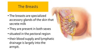
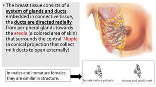
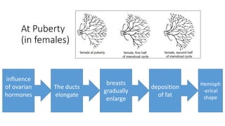
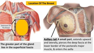
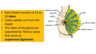
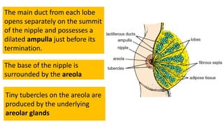
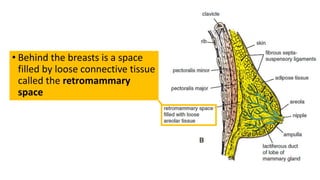
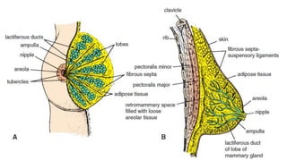
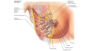
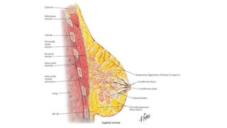
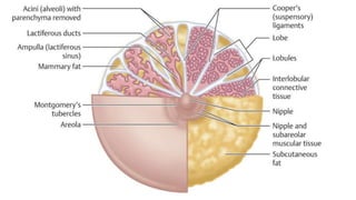
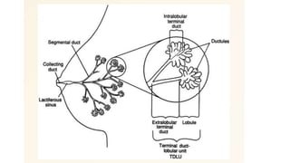
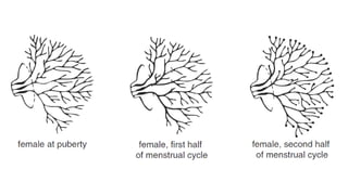
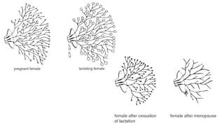
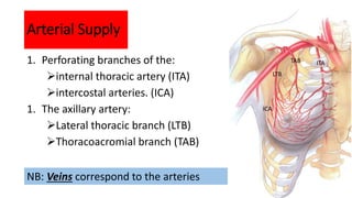
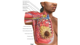
Breast Anatomy
By
Dr.Faris Muhammed
2022
The Breasts
• The breasts are specialized
accessory glands of the skin that
secrete milk
• They are present in both sexes
• situated in the pectoral region
• their blood supply and lymphatic
drainage is largely into the
armpit.
• The breast tissue consists of a
system of glands and ducts,
embedded in connective tissue,
the ducts are directed radially
from peripheral glands towards
the areola (a colored area of skin)
that surrounds the central Nipple
(a conical projection that collect
milk ducts to open externally)
In males and immature females,
they are similar in structure.
At Puberty
(in females)
Hemisph
-erical
shape
influence
of ovarian
hormones
The ducts
elongate
breasts
gradually
enlarge
deposition
of fat
2nd Rib
6th rib
midaxillary
line
Parasternal
Axillary tail A small part, extends upward
and laterally, pierces the deep fascia at the
lower border of the pectoralis major
muscle, & enters the axilla
The greater part of the gland
lies in the superficial fascia
Extensions Of
The Breast
Location Of The Breast
• Each breast consists of 15 to
20 lobes
• Lobes radiate out from the
nipple
• The lobes of the gland are
separated by fibrous septa
that serve as:
suspensory ligaments
Tiny tubercles on the areola are
produced by the underlying
areolar glands
The main duct from each lobe
opens separately on the summit
of the nipple and possesses a
dilated ampulla just before its
termination.
The base of the nipple is
surrounded by the areola
• Behind the breasts is a space
filled by loose connective tissue
called the retromammary
space
Modified Total Mastectomy
Long
Thoracic
Nerve
Arterial Supply
1. Perforating branches of the:
internal thoracic artery (ITA)
intercostal arteries. (ICA)
1. The axillary artery:
Lateral thoracic branch (LTB)
Thoracoacromial branch (TAB)
ITA
LTB
TAB
ICA
NB: Veins correspond to the arteries
Lymph Drainage of The Breast
• The lymph drainage of the mammary gland is of great clinical
importance because of the frequent development of cancer in the
gland and the subsequent dissemination of the malignant cells along
the lymph vessels to the lymph nodes.
1. The lateral quadrants of the breast
drain into the anterior axillary or pectoral group of nodes (situated
just posterior to the lower border of the pectoralis major muscle)
2. The medial quadrants:
drain by means of vessels that pierce the intercostal spaces and enter
the internal thoracic group of nodes (situated within the thoracic
cavity along the course of the internal thoracic artery)
3. A few lymph vessels
a. follow the posterior intercostal arteries and drain posteriorly
into the posterior intercostal nodes (situated along the course
of the posterior intercostal arteries);
b. some vessels communicate with the lymph vessels of the
opposite breast and with those of the anterior abdominal wall
Breast anatomy
Breast anatomy
Breast anatomy
Breast anatomy

Breast anatomy

  • 1.
  • 2.
    The Breasts • Thebreasts are specialized accessory glands of the skin that secrete milk • They are present in both sexes • situated in the pectoral region • their blood supply and lymphatic drainage is largely into the armpit.
  • 3.
    • The breasttissue consists of a system of glands and ducts, embedded in connective tissue, the ducts are directed radially from peripheral glands towards the areola (a colored area of skin) that surrounds the central Nipple (a conical projection that collect milk ducts to open externally) In males and immature females, they are similar in structure.
  • 4.
    At Puberty (in females) Hemisph -erical shape influence ofovarian hormones The ducts elongate breasts gradually enlarge deposition of fat
  • 5.
    2nd Rib 6th rib midaxillary line Parasternal Axillarytail A small part, extends upward and laterally, pierces the deep fascia at the lower border of the pectoralis major muscle, & enters the axilla The greater part of the gland lies in the superficial fascia Extensions Of The Breast Location Of The Breast
  • 6.
    • Each breastconsists of 15 to 20 lobes • Lobes radiate out from the nipple • The lobes of the gland are separated by fibrous septa that serve as: suspensory ligaments
  • 7.
    Tiny tubercles onthe areola are produced by the underlying areolar glands The main duct from each lobe opens separately on the summit of the nipple and possesses a dilated ampulla just before its termination. The base of the nipple is surrounded by the areola
  • 8.
    • Behind thebreasts is a space filled by loose connective tissue called the retromammary space
  • 16.
  • 17.
    Arterial Supply 1. Perforatingbranches of the: internal thoracic artery (ITA) intercostal arteries. (ICA) 1. The axillary artery: Lateral thoracic branch (LTB) Thoracoacromial branch (TAB) ITA LTB TAB ICA NB: Veins correspond to the arteries
  • 18.
    Lymph Drainage ofThe Breast • The lymph drainage of the mammary gland is of great clinical importance because of the frequent development of cancer in the gland and the subsequent dissemination of the malignant cells along the lymph vessels to the lymph nodes.
  • 19.
    1. The lateralquadrants of the breast drain into the anterior axillary or pectoral group of nodes (situated just posterior to the lower border of the pectoralis major muscle) 2. The medial quadrants: drain by means of vessels that pierce the intercostal spaces and enter the internal thoracic group of nodes (situated within the thoracic cavity along the course of the internal thoracic artery)
  • 20.
    3. A fewlymph vessels a. follow the posterior intercostal arteries and drain posteriorly into the posterior intercostal nodes (situated along the course of the posterior intercostal arteries); b. some vessels communicate with the lymph vessels of the opposite breast and with those of the anterior abdominal wall