Renal Replacement Therapy: modes and evidenceMohd Saif Khan
Renal replacement therapy is a supportive care often required in critically ill patients who develop acute renal failure and its complications. Complexity arises when such patients become hemodynamically unstable and pose special challenge to critical care clinicians in ICU to carefully choose dialytic modality to tackle volume and solute overload. This presentation is about short description of modalities of RRT and current evidence regarding initiation, dose and type of modality.
A detailed discussion on a very much in demand topic. Covered all aspects of the procedure which are important for an Emergency, Medical and Intensive Care physician should know. Nurses can also benefit from the presentation as we have tried to keep it as simple and straight forward as possible.
Establishing and maintaining normal extracellular volume (ECV) is required to achieve normotension. The achievement of an optimal fluid status, as expressed by "dry weight" (DW), should allow for controlling blood pressure (BP) in the large majority of HD patients
Case report: A 49-year-old previously healthy man was admitted to the ICU after cardiac arrest following a short history with headache, blurred speech and reduced consciousness. After cardiopulmonary resuscitation perfusion rhythm was regained, but the patient didn`t regain consciousness. The arterial blood gas analysis at the ICU revealed a severe metabolic acidosis with pH at 6.86 and lactate levels of 16 mmol/L. The white blood cells count was also markedly increased (312 * 109/L), and blood smear showed immature cells indicating acute leukemia. The severe metabolic acidosis, at first thought to be due to systemic hypoperfusion, did not improve in spite of fluid and vasopressor resuscitation. A CT scan of the head performed the next day, revealed massive cerebellar haemorrhage, edema in both hemispheres and signs of anoxic brain damage. Immunophenotyping of peripheral blood was consistent with Acute Myeloid Leukemia (AML).
Hypokalemia, Hypoxic Ischemic Encephalopathy, Nosocomial Pneumonia and Urinar...Jack Frost
Hypokalemia, Hypoxic Ischemic Encephalopathy, Nosocomial Pneumonia and Urinary Tract Infection.This presentation contains real names of persons involve of this particular study. This names should not be copied or rewritten. Used the data of this study as basis only. All rights reserved 2009.
Movement disorders: A complication of chronic Hyperglycemia? A case reportApollo Hospitals
The association of chorea with a specific lesion on brain imaging is described as an atypical manifestation of chronic hyperglycemia. This is a rare syndrome, affecting more elderly, presenting a diabetes poorly controlled. There is hyperglycemia without ketosis and moderate hyperosmolarity.
IgA nephropathy is a condition characterized by deposition of IgA immunoglobulins in glomeruli. This condition is fairly common in Western countries. The scope of the disease is wide and case by case. Cases of IgA nephropathy are rare. Our case report is of a young man who developed rapid onset IgA nephropathy leading to end stage renal disease ESRD . This case report describes a 26 years age young man who presented and eventually presented with microscopic hematuria and severe proteinuria. Hemodialysis for his burned out IgA nephropathy. Dr. Thenmozhi. P | Yuvaraj. B "IgA Nephropathy (Burger's Disease): Case Report" Published in International Journal of Trend in Scientific Research and Development (ijtsrd), ISSN: 2456-6470, Volume-7 | Issue-1 , February 2023, URL: https://www.ijtsrd.com/papers/ijtsrd52706.pdf Paper URL: https://www.ijtsrd.com/medicine/other/52706/iga-nephropathy-burgers-disease-case-report/dr-thenmozhi-p
Hypokalemia, Hypoxic Ischemic Encephalopathy, Nosocomial Pneumonia and Urinar...Jack Frost
Hypokalemia, Hypoxic Ischemic Encephalopathy, Nosocomial Pneumonia and Urinary Tract Infection. This presentation contains real names of persons involve of this particular study. This names should not be copied or rewritten. Used the data of this study as basis only. All rights reserved 2009.
Diabetic ketoacidosis induced cerebral infarct - A missing link in pathogenes...Apollo Hospitals
Diabetic ketoacidosis (DKA) is a known complication of acute pancreatitis (AP). We report a case of DKA precipitated by AP. This patient developed watershed infarct in brain during her course of disease which was possibly attributed to hypercoagulability, stasis and endothelial injury triggered by ketosis.
Recomendações da OMS sobre cuidados maternos e neonatais para uma experiência pós-natal positiva.
Em consonância com os ODS – Objetivos do Desenvolvimento Sustentável e a Estratégia Global para a Saúde das Mulheres, Crianças e Adolescentes, e aplicando uma abordagem baseada nos direitos humanos, os esforços de cuidados pós-natais devem expandir-se para além da cobertura e da simples sobrevivência, de modo a incluir cuidados de qualidade.
Estas diretrizes visam melhorar a qualidade dos cuidados pós-natais essenciais e de rotina prestados às mulheres e aos recém-nascidos, com o objetivo final de melhorar a saúde e o bem-estar materno e neonatal.
Uma “experiência pós-natal positiva” é um resultado importante para todas as mulheres que dão à luz e para os seus recém-nascidos, estabelecendo as bases para a melhoria da saúde e do bem-estar a curto e longo prazo. Uma experiência pós-natal positiva é definida como aquela em que as mulheres, pessoas que gestam, os recém-nascidos, os casais, os pais, os cuidadores e as famílias recebem informação consistente, garantia e apoio de profissionais de saúde motivados; e onde um sistema de saúde flexível e com recursos reconheça as necessidades das mulheres e dos bebês e respeite o seu contexto cultural.
Estas diretrizes consolidadas apresentam algumas recomendações novas e já bem fundamentadas sobre cuidados pós-natais de rotina para mulheres e neonatos que recebem cuidados no pós-parto em unidades de saúde ou na comunidade, independentemente dos recursos disponíveis.
É fornecido um conjunto abrangente de recomendações para cuidados durante o período puerperal, com ênfase nos cuidados essenciais que todas as mulheres e recém-nascidos devem receber, e com a devida atenção à qualidade dos cuidados; isto é, a entrega e a experiência do cuidado recebido. Estas diretrizes atualizam e ampliam as recomendações da OMS de 2014 sobre cuidados pós-natais da mãe e do recém-nascido e complementam as atuais diretrizes da OMS sobre a gestão de complicações pós-natais.
O estabelecimento da amamentação e o manejo das principais intercorrências é contemplada.
Recomendamos muito.
Vamos discutir essas recomendações no nosso curso de pós-graduação em Aleitamento no Instituto Ciclos.
Esta publicação só está disponível em inglês até o momento.
Prof. Marcus Renato de Carvalho
www.agostodourado.com
ARTIFICIAL INTELLIGENCE IN HEALTHCARE.pdfAnujkumaranit
Artificial intelligence (AI) refers to the simulation of human intelligence processes by machines, especially computer systems. It encompasses tasks such as learning, reasoning, problem-solving, perception, and language understanding. AI technologies are revolutionizing various fields, from healthcare to finance, by enabling machines to perform tasks that typically require human intelligence.
Explore natural remedies for syphilis treatment in Singapore. Discover alternative therapies, herbal remedies, and lifestyle changes that may complement conventional treatments. Learn about holistic approaches to managing syphilis symptoms and supporting overall health.
These lecture slides, by Dr Sidra Arshad, offer a quick overview of physiological basis of a normal electrocardiogram.
Learning objectives:
1. Define an electrocardiogram (ECG) and electrocardiography
2. Describe how dipoles generated by the heart produce the waveforms of the ECG
3. Describe the components of a normal electrocardiogram of a typical bipolar leads (limb II)
4. Differentiate between intervals and segments
5. Enlist some common indications for obtaining an ECG
Study Resources:
1. Chapter 11, Guyton and Hall Textbook of Medical Physiology, 14th edition
2. Chapter 9, Human Physiology - From Cells to Systems, Lauralee Sherwood, 9th edition
3. Chapter 29, Ganong’s Review of Medical Physiology, 26th edition
4. Electrocardiogram, StatPearls - https://www.ncbi.nlm.nih.gov/books/NBK549803/
5. ECG in Medical Practice by ABM Abdullah, 4th edition
6. ECG Basics, http://www.nataliescasebook.com/tag/e-c-g-basics
Title: Sense of Taste
Presenter: Dr. Faiza, Assistant Professor of Physiology
Qualifications:
MBBS (Best Graduate, AIMC Lahore)
FCPS Physiology
ICMT, CHPE, DHPE (STMU)
MPH (GC University, Faisalabad)
MBA (Virtual University of Pakistan)
Learning Objectives:
Describe the structure and function of taste buds.
Describe the relationship between the taste threshold and taste index of common substances.
Explain the chemical basis and signal transduction of taste perception for each type of primary taste sensation.
Recognize different abnormalities of taste perception and their causes.
Key Topics:
Significance of Taste Sensation:
Differentiation between pleasant and harmful food
Influence on behavior
Selection of food based on metabolic needs
Receptors of Taste:
Taste buds on the tongue
Influence of sense of smell, texture of food, and pain stimulation (e.g., by pepper)
Primary and Secondary Taste Sensations:
Primary taste sensations: Sweet, Sour, Salty, Bitter, Umami
Chemical basis and signal transduction mechanisms for each taste
Taste Threshold and Index:
Taste threshold values for Sweet (sucrose), Salty (NaCl), Sour (HCl), and Bitter (Quinine)
Taste index relationship: Inversely proportional to taste threshold
Taste Blindness:
Inability to taste certain substances, particularly thiourea compounds
Example: Phenylthiocarbamide
Structure and Function of Taste Buds:
Composition: Epithelial cells, Sustentacular/Supporting cells, Taste cells, Basal cells
Features: Taste pores, Taste hairs/microvilli, and Taste nerve fibers
Location of Taste Buds:
Found in papillae of the tongue (Fungiform, Circumvallate, Foliate)
Also present on the palate, tonsillar pillars, epiglottis, and proximal esophagus
Mechanism of Taste Stimulation:
Interaction of taste substances with receptors on microvilli
Signal transduction pathways for Umami, Sweet, Bitter, Sour, and Salty tastes
Taste Sensitivity and Adaptation:
Decrease in sensitivity with age
Rapid adaptation of taste sensation
Role of Saliva in Taste:
Dissolution of tastants to reach receptors
Washing away the stimulus
Taste Preferences and Aversions:
Mechanisms behind taste preference and aversion
Influence of receptors and neural pathways
Impact of Sensory Nerve Damage:
Degeneration of taste buds if the sensory nerve fiber is cut
Abnormalities of Taste Detection:
Conditions: Ageusia, Hypogeusia, Dysgeusia (parageusia)
Causes: Nerve damage, neurological disorders, infections, poor oral hygiene, adverse drug effects, deficiencies, aging, tobacco use, altered neurotransmitter levels
Neurotransmitters and Taste Threshold:
Effects of serotonin (5-HT) and norepinephrine (NE) on taste sensitivity
Supertasters:
25% of the population with heightened sensitivity to taste, especially bitterness
Increased number of fungiform papillae
Pulmonary Thromboembolism - etilogy, types, medical- Surgical and nursing man...VarunMahajani
Disruption of blood supply to lung alveoli due to blockage of one or more pulmonary blood vessels is called as Pulmonary thromboembolism. In this presentation we will discuss its causes, types and its management in depth.
Couples presenting to the infertility clinic- Do they really have infertility...Sujoy Dasgupta
Dr Sujoy Dasgupta presented the study on "Couples presenting to the infertility clinic- Do they really have infertility? – The unexplored stories of non-consummation" in the 13th Congress of the Asia Pacific Initiative on Reproduction (ASPIRE 2024) at Manila on 24 May, 2024.
Title: Sense of Smell
Presenter: Dr. Faiza, Assistant Professor of Physiology
Qualifications:
MBBS (Best Graduate, AIMC Lahore)
FCPS Physiology
ICMT, CHPE, DHPE (STMU)
MPH (GC University, Faisalabad)
MBA (Virtual University of Pakistan)
Learning Objectives:
Describe the primary categories of smells and the concept of odor blindness.
Explain the structure and location of the olfactory membrane and mucosa, including the types and roles of cells involved in olfaction.
Describe the pathway and mechanisms of olfactory signal transmission from the olfactory receptors to the brain.
Illustrate the biochemical cascade triggered by odorant binding to olfactory receptors, including the role of G-proteins and second messengers in generating an action potential.
Identify different types of olfactory disorders such as anosmia, hyposmia, hyperosmia, and dysosmia, including their potential causes.
Key Topics:
Olfactory Genes:
3% of the human genome accounts for olfactory genes.
400 genes for odorant receptors.
Olfactory Membrane:
Located in the superior part of the nasal cavity.
Medially: Folds downward along the superior septum.
Laterally: Folds over the superior turbinate and upper surface of the middle turbinate.
Total surface area: 5-10 square centimeters.
Olfactory Mucosa:
Olfactory Cells: Bipolar nerve cells derived from the CNS (100 million), with 4-25 olfactory cilia per cell.
Sustentacular Cells: Produce mucus and maintain ionic and molecular environment.
Basal Cells: Replace worn-out olfactory cells with an average lifespan of 1-2 months.
Bowman’s Gland: Secretes mucus.
Stimulation of Olfactory Cells:
Odorant dissolves in mucus and attaches to receptors on olfactory cilia.
Involves a cascade effect through G-proteins and second messengers, leading to depolarization and action potential generation in the olfactory nerve.
Quality of a Good Odorant:
Small (3-20 Carbon atoms), volatile, water-soluble, and lipid-soluble.
Facilitated by odorant-binding proteins in mucus.
Membrane Potential and Action Potential:
Resting membrane potential: -55mV.
Action potential frequency in the olfactory nerve increases with odorant strength.
Adaptation Towards the Sense of Smell:
Rapid adaptation within the first second, with further slow adaptation.
Psychological adaptation greater than receptor adaptation, involving feedback inhibition from the central nervous system.
Primary Sensations of Smell:
Camphoraceous, Musky, Floral, Pepperminty, Ethereal, Pungent, Putrid.
Odor Detection Threshold:
Examples: Hydrogen sulfide (0.0005 ppm), Methyl-mercaptan (0.002 ppm).
Some toxic substances are odorless at lethal concentrations.
Characteristics of Smell:
Odor blindness for single substances due to lack of appropriate receptor protein.
Behavioral and emotional influences of smell.
Transmission of Olfactory Signals:
From olfactory cells to glomeruli in the olfactory bulb, involving lateral inhibition.
Primitive, less old, and new olfactory systems with different path
NVBDCP.pptx Nation vector borne disease control programSapna Thakur
NVBDCP was launched in 2003-2004 . Vector-Borne Disease: Disease that results from an infection transmitted to humans and other animals by blood-feeding arthropods, such as mosquitoes, ticks, and fleas. Examples of vector-borne diseases include Dengue fever, West Nile Virus, Lyme disease, and malaria.
Knee anatomy and clinical tests 2024.pdfvimalpl1234
This includes all relevant anatomy and clinical tests compiled from standard textbooks, Campbell,netter etc..It is comprehensive and best suited for orthopaedicians and orthopaedic residents.
TEST BANK for Operations Management, 14th Edition by William J. Stevenson, Ve...kevinkariuki227
TEST BANK for Operations Management, 14th Edition by William J. Stevenson, Verified Chapters 1 - 19, Complete Newest Version.pdf
TEST BANK for Operations Management, 14th Edition by William J. Stevenson, Verified Chapters 1 - 19, Complete Newest Version.pdf
Prix Galien International 2024 Forum ProgramLevi Shapiro
June 20, 2024, Prix Galien International and Jerusalem Ethics Forum in ROME. Detailed agenda including panels:
- ADVANCES IN CARDIOLOGY: A NEW PARADIGM IS COMING
- WOMEN’S HEALTH: FERTILITY PRESERVATION
- WHAT’S NEW IN THE TREATMENT OF INFECTIOUS,
ONCOLOGICAL AND INFLAMMATORY SKIN DISEASES?
- ARTIFICIAL INTELLIGENCE AND ETHICS
- GENE THERAPY
- BEYOND BORDERS: GLOBAL INITIATIVES FOR DEMOCRATIZING LIFE SCIENCE TECHNOLOGIES AND PROMOTING ACCESS TO HEALTHCARE
- ETHICAL CHALLENGES IN LIFE SCIENCES
- Prix Galien International Awards Ceremony
Triangles of Neck and Clinical Correlation by Dr. RIG.pptx
Dialysis disequilibrium-syndrome
1. Med J Malaysia Vol 71 No 2 April 2016 91
SUMMARY
Dialysis disequilibrium syndrome (DDS) is a neurological
disorder with varying severity that is postulated to be
associated with cerebral oedema. We described a case of
DDS resulting in irreversible brain injury and death following
acute haemodialysis. A 13-year-old male with no past
medical history and weighing 30kg, presented to hospital
with severe urosepsis complicated by acute kidney injury
(Creatinine 1422mmol/L; Urea 74.2mmol/L, Potassium
6.3mmol/L, Sodium 137mmol/L) and severe metabolic
acidosis (pH 6.99, HC03 1.7mmol/L). Chest radiograph was
normal. Elective intubation was done for respiratory
distress. Acute haemodialysis performed due to refractory
metabolic acidosis. Following haemodialysis, he became
hypotensive which required inotropes. His Riker's score was
low with absence of brainstem reflexes after withholding
sedation. CT Brain showed generalised cerebral oedema
consistent with global hypoxic changes involving the
brainstem. The symptoms of DDS are caused by water
movement into the brain causing cerebral oedema. Two
theories have been proposed: reverse osmotic shift induced
by urea removal and a fall in cerebral intracellular pH.
Prevention is the key to the management of DDS. It is
important to identify high risk patients and haemodialysis
with reduced dialysis efficacy and gradual urea reduction is
recommended. Patients who are vulnerable to DDS should
be monitored closely. Low efficiency haemodialysis is
recommended. Acute peritoneal dialysis might be an
alternative option, but further studies are needed.
KEY WORDS:
acute kidney injury; haemodialysis; dialysis disequilibrium
syndrome; cerebral oedema
INTRODUCTION
Dialysis disequilibrium syndrome (DDS) is defined as a
clinical syndrome of neurologic deterioration that seen in
patients who undergo haemodialysis (HD). It is more likely to
occur in patients during or immediately after their first
treatment. We report a case of DDS induced cerebral oedema
that resulted in irreversible brain injury and death following
HD. Further, we reviewed the relevant literature of the
association of DDS and HD.
CASE REPORT
A 13-year-old male with no past medical illness, presented
with progressive dyspnoea and vomiting which preceded by
fever and urinary tract infection symptoms for three weeks.
On examination, he was alert but severely dehydrated and
tachypnea. His temperature was 35.5°C, blood pressure
121/83mmHg; pulse rate 118bpm, with SpO2 100%. Systemic
examinations were unremarkable.
Blood investigations showed acidemia and hypocapnia with
pH 6.994, pCO2 10mmHg, HCO3 1.5mmol/L. Renal profile
showed Creatinine 1422mmol/L, Urea 74.2mmol/L,
Potassium 6.3mmol/L and Sodium 137mmol/L with
increased anion gap of 28.8mmol/L and serum Osmolarity
was 356.1mOsm/L. He had a raised white cell count of
22.7(x10³/ µL). Urine microscopic examination showed
pyuria. Hence, patient was treated as severe urosepsis.
Unfortunately, he was not responding to aggressive
resuscitation. His arterial blood gas showed refractory severe
metabolic acidosis with pH of 6.897, pCO2 36.2mmHg, HCO3
7.0mmol/L. Chest radiograph was unremarkable. Patient
was electively intubated and admitted to Intensive Care Unit.
Haemodialysis was performed with Qb of 200ml/min, Qd
500ml/min without ultrafiltration for two hours via femoral
catheter in ICU. Low Flux Dialyser (Fresenius F8HPS
polysuphone, surface area of 1.8 m², Kuf as 18ml/hr x
mmHg) and RenaHD-3A dialysate were used. Prior to
dialysis, patient was normotensive and good Riker’s score of -
1. However, patient became hypotensive which required
inotropic supports and dropped in Riker’s score to -3
drastically immediately after dialysis. Neurological
assessment showed bilateral non-reactive and dilated pupils.
His brainstem reflexes were absent. Repeated investigations
revealed the pH 7.354, pCO2 22.4mmHg and HCO3
12.4mmol/L; Creatinine 451mmol/L, Urea 23.2mmol/L,
Potassium 2.4mmol/L, Sodium 143mmol/L with high urea
reduction rate of 54.95% and serum Osmolarity of
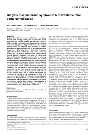
313.9mOsm/L. CT brain showed generalised cerebral oedema
with obliterated basal cisterns, loss of grey-white
differentiation and global hypoxic changes included
brainstem (Figure 1). Diagnosis of brain death was declared
and family members were informed. His condition
deteriorated further and succumbed. No autopsy was done
due to family members’ refusal.
DISCUSSION
Dialysis Disequilibrium Syndrome (DDS) is more likely to occur
in patients during or immediately after their first treatment,
however there were also two case reports of DDS happened
after few haemodialysis sessions for more than one week.1
So
far, we have not found relevant literature in Malaysia.
Dialysis disequilibrium syndrome: A preventable fatal
acute complication
Mah Doo Yee, MRCP1
, Yia Hua Jern, MRCP2
, Cheong Wai Seng, MRCP2
1
Department of Medicine, Sultanah Nora Ismail Hospital, Batu Pahat, Malaysia, 2
Department of Medicine, Sultanah Fatimah
Specialist Hospital, Muar, Malaysia
CASE REPORT
This article was accepted: 3 February 2016
Corresponding Author: Mah Doo Yee, Medical Specialist, Hospital Sultanah Nora Ismail, Department of Internal Medicine, Jalan Korma, Batu Pahat,
83000 Johor
Email: mahdy426@yahoo.com.sg
2. Case Report
92 Med J Malaysia Vol 71 No 2 April 2016
The predisposing factors for DDS in this case are small body
size (paediatric population), severe uraemia, severe
metabolic acidosis, sepsis (widespread immune activation
which may alter blood-brain barrier permeability and
predispose to cerebral oedema)2
and first time dialysis user.
The presenting symptoms vary from mild form: nausea and
headache to severe form: coma and rarely death, as seen in
this patient. However, several mechanisms causing cerebral
oedema have been debated for years. The first mechanism is
‘Reverse urea effect’, which was suggested by Kennedy et al.3
in 1962. The syndrome was attributed to the delayed exit of
urea from the brain post rapid dialysis-induced decline in
blood urea level, thus creating an osmotic gradient that
favoured the shift of water into the brain from the blood
which generates the cerebral oedema as what had happened
in this patient. The second mechanism is “Cerebrospinal fluid
acidosis” theory.4
During haemodialysis, existing systemic
metabolic acidosis is promptly corrected, but the
corresponding CSF pH level remains low. Hence, plasma CO2
diffuses rapidly to the CSF, increasing its pCO2 or to the
production of an unidentified organic acid by the brain
during dialysis. The CSF and brain acidosis somehow brings
about oedema of the brain. However, studies lending support
to this particular theory are few. In this patient, the pCO2 was
reduced instead of raised, from 36.2 mmHg to 22.4 mmHg.
Lastly is the “Idiogenic osmoles” theory. There are organic
osmoles produced by the brain to counteract various hyper-
osmolal states so that brain shrinkage does not occur.
However, there is no increase in idiogenic osmoles has been
observed during experimental hemodialysis.5
In the nutshell,
the “reverse urea effect” theory appears the most promising.
Prevention is the core management for DDS traditionally and
despite the absence of evidence-based guidelines, the
conventional aim is a gradual clearance of urea. However, in
a district hospital which without nephrologist and limited
resources, the low flux conventional haemodialysis is the
only option for this patient. In a paediatric patient with
advanced uraemia, an initial urea clearance of 3 ml/min per
kg, which is calculated from the specifications of the chosen
dialyser and the blood flow attainable is suggested to avoid
disequilibrium. The acute peritoneal dialysis might not be a
good modality in reverting this life threatening severe
metabolic acidosis with high catabolic state.
In hospital with nephrology service, the simplest way to do
this is to perform hemofiltration on the patient instead of
haemodialysis. This method of treatment relies on the
convective removal of solute from the patient in place of
diffusive removal. Thus, the osmolalities of the body fluid
compartments will not change as rapidly as they do during
standard haemodialysis. The other way is to use of a smaller,
less efficient dialyser or lower the targeted blood flow rates by
use of sustained low-efficiency dialysis (SLED) but prolonged
the treatment period, or initiation of continuous renal
replacement therapy (CRRT) with more gradual clearance of
urea.
In conclusion, patients who are vulnerable to DDS should be
monitored closely with low efficiency renal replacement
therapy and nephrology consultation. Acute peritoneal
dialysis might be an alternative option, but further studies
are needed.
REFERENCES
1. Shaikh N, Louron A, Hassens Y. Fatal dialysis disequilibrium syndrome. J
Emerg Shock 2010; 3(3): 300.
2. Green R, Scott LK, Minagar A, Conrad S. Sepsis associated encephalopathy
(SAE): a review. Front Biosci 2004; 9: 1637-41.
3. Sahani MM, Daoud TM, Sam R, Andrews J, Cheng YL, Carl M Kjellstrand
CM el al. Dialysis Disequilibrium Syndrome Revisited. Hemodial Int 2001;
5: 92-6.
4. Arieff Al. Dialysis disequilibrium syndrome: Current concepts on
pathogenesis and prevention. Kidney Int 1994; 45(3): 629-35.
5. Silver SM. Cerebral edema after rapid dialysis is not caused by an increase
in brain organic osmolytes. J Am Soc Nephrol 1995; 6(6): 1600-6.
Fig. 1: Generalised cerebral oedema with obliterated basal cisterns, loss of grey-white differentiation and global hypoxic changes
included brainstem.