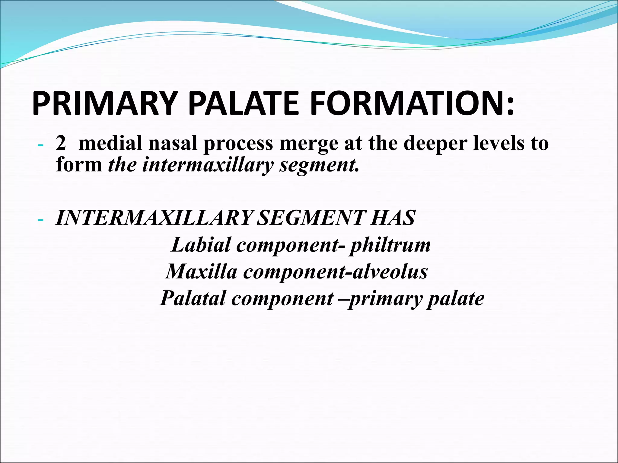
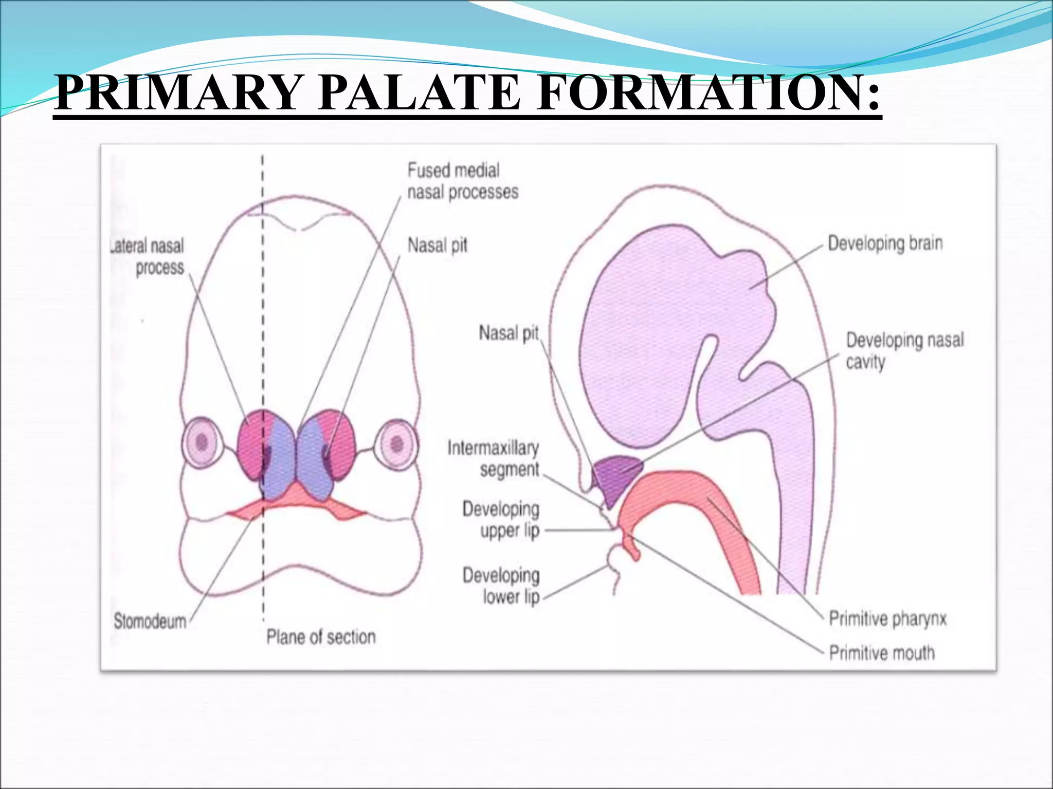
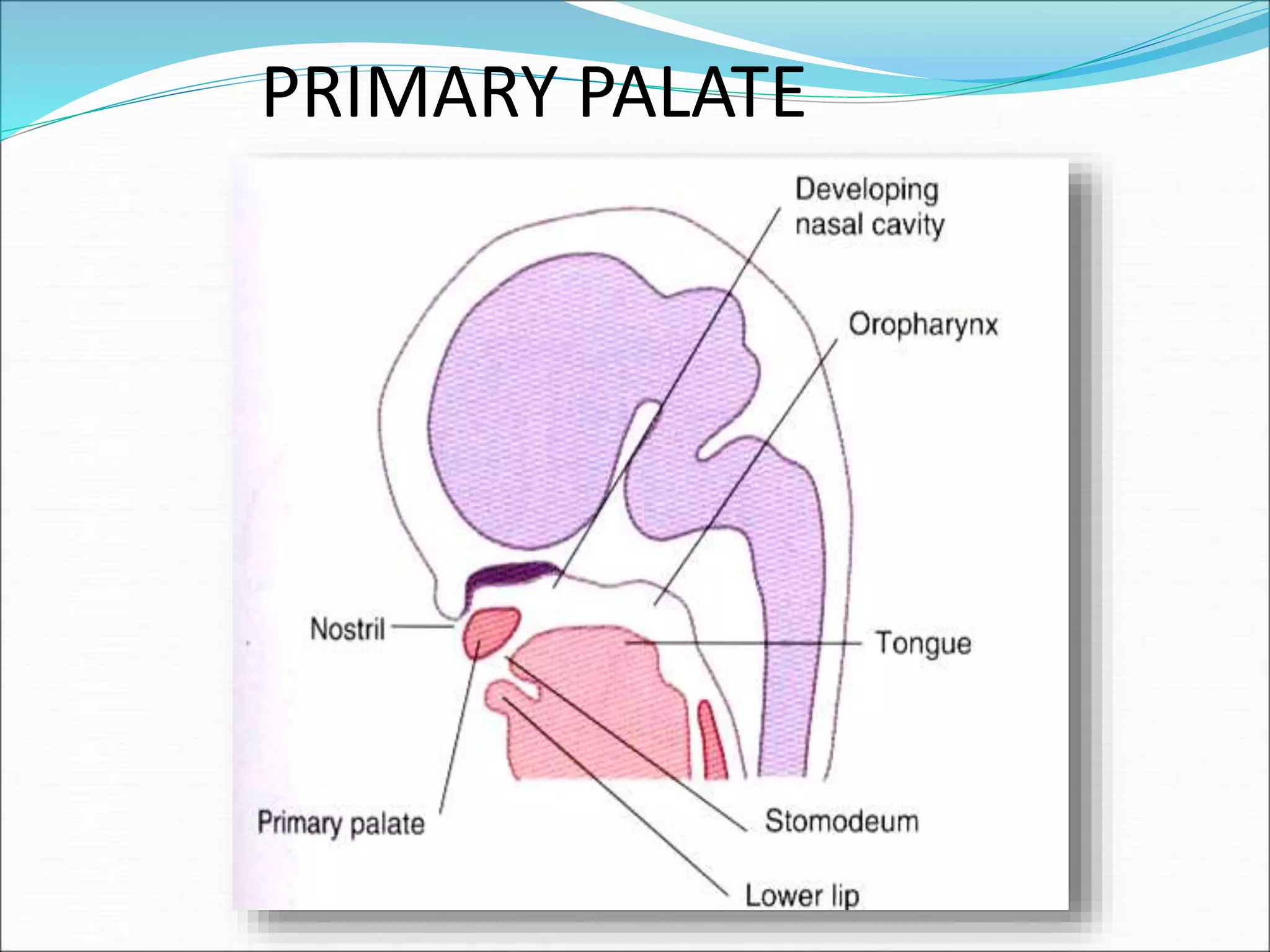
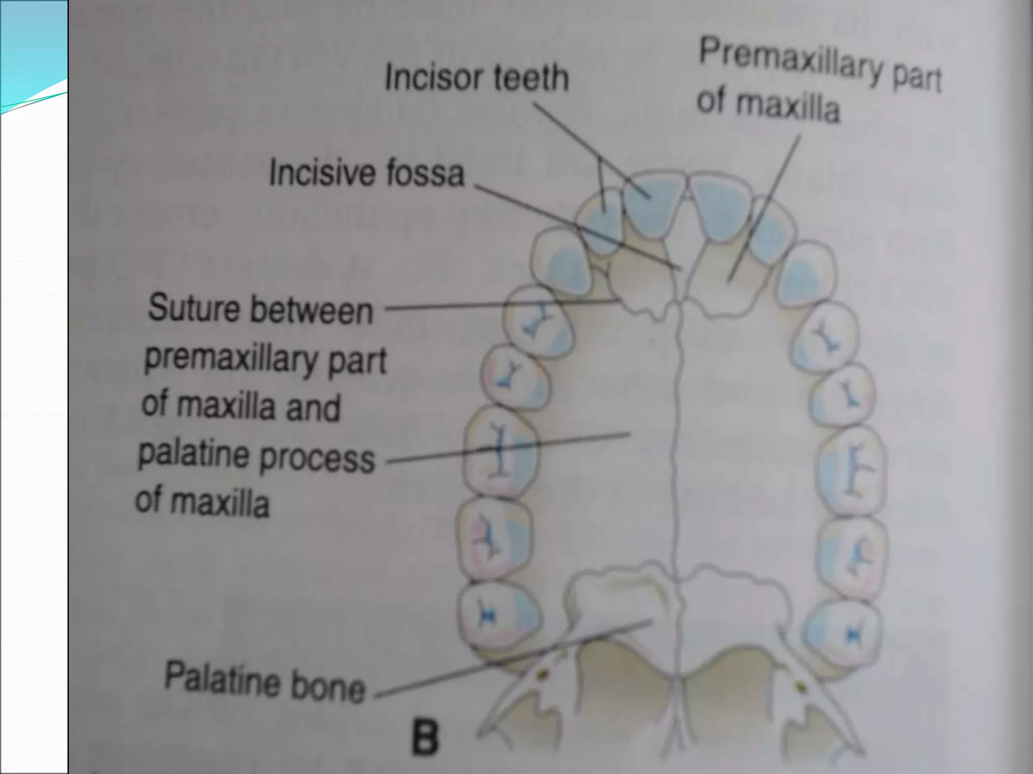
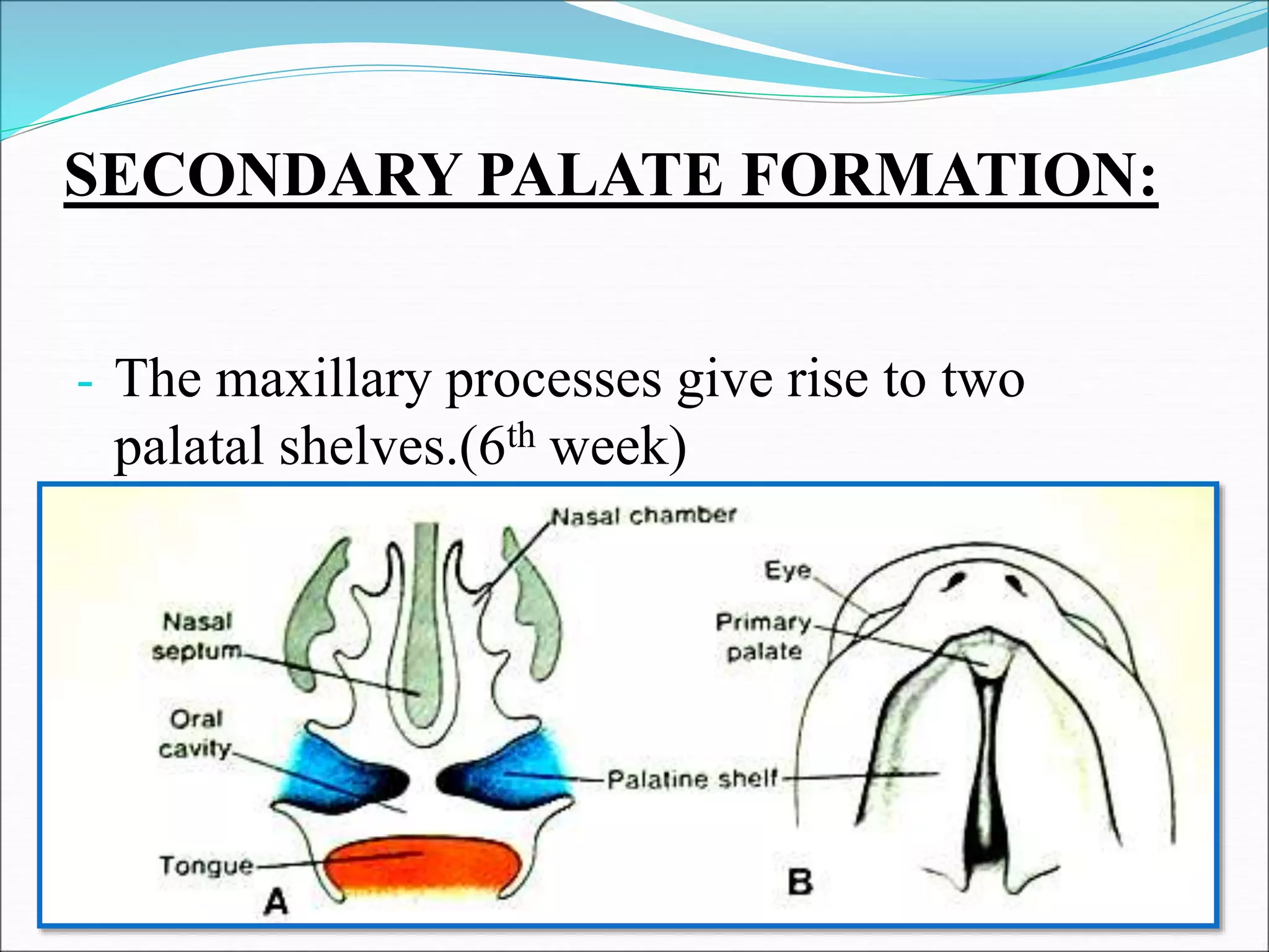
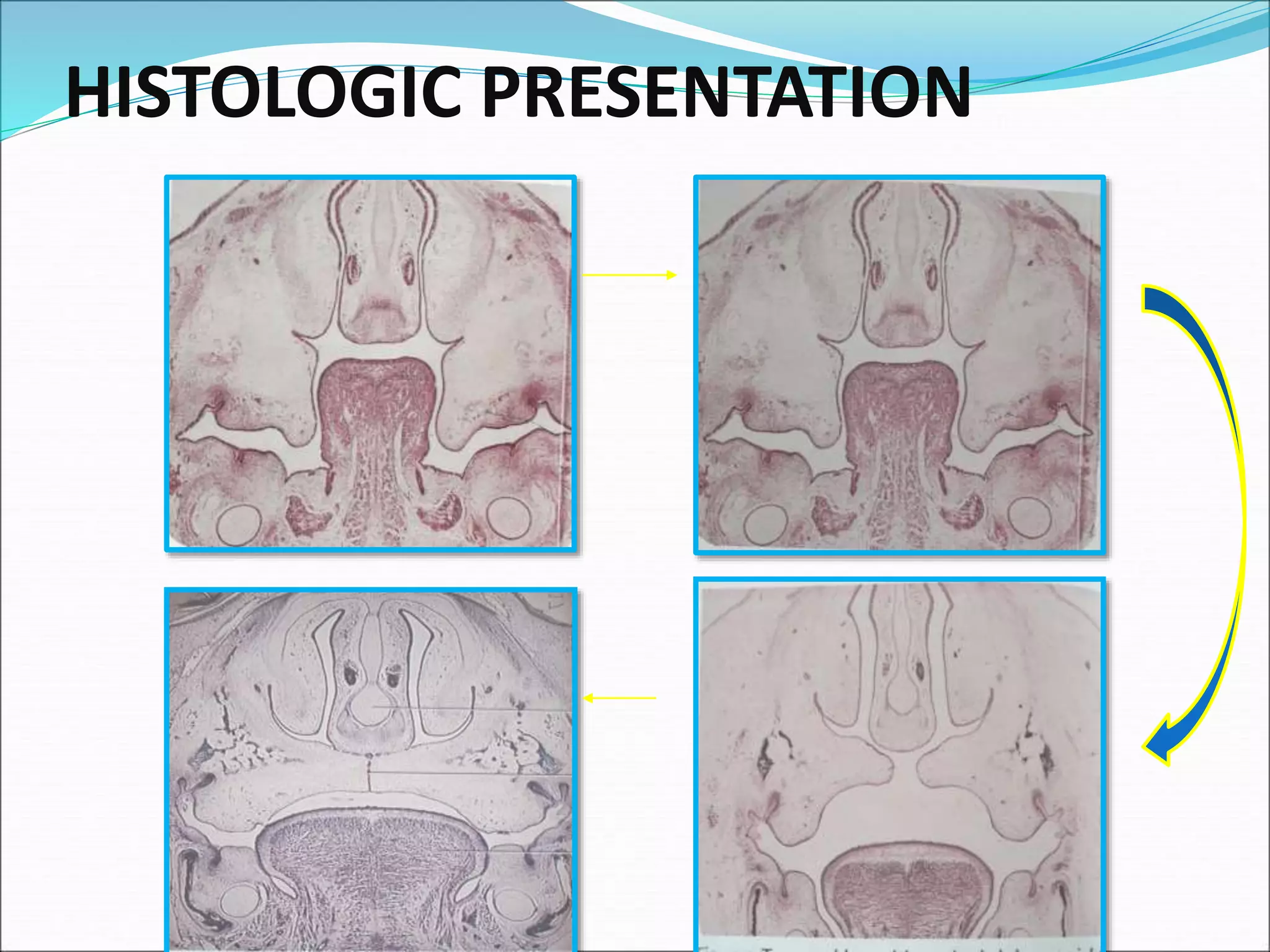
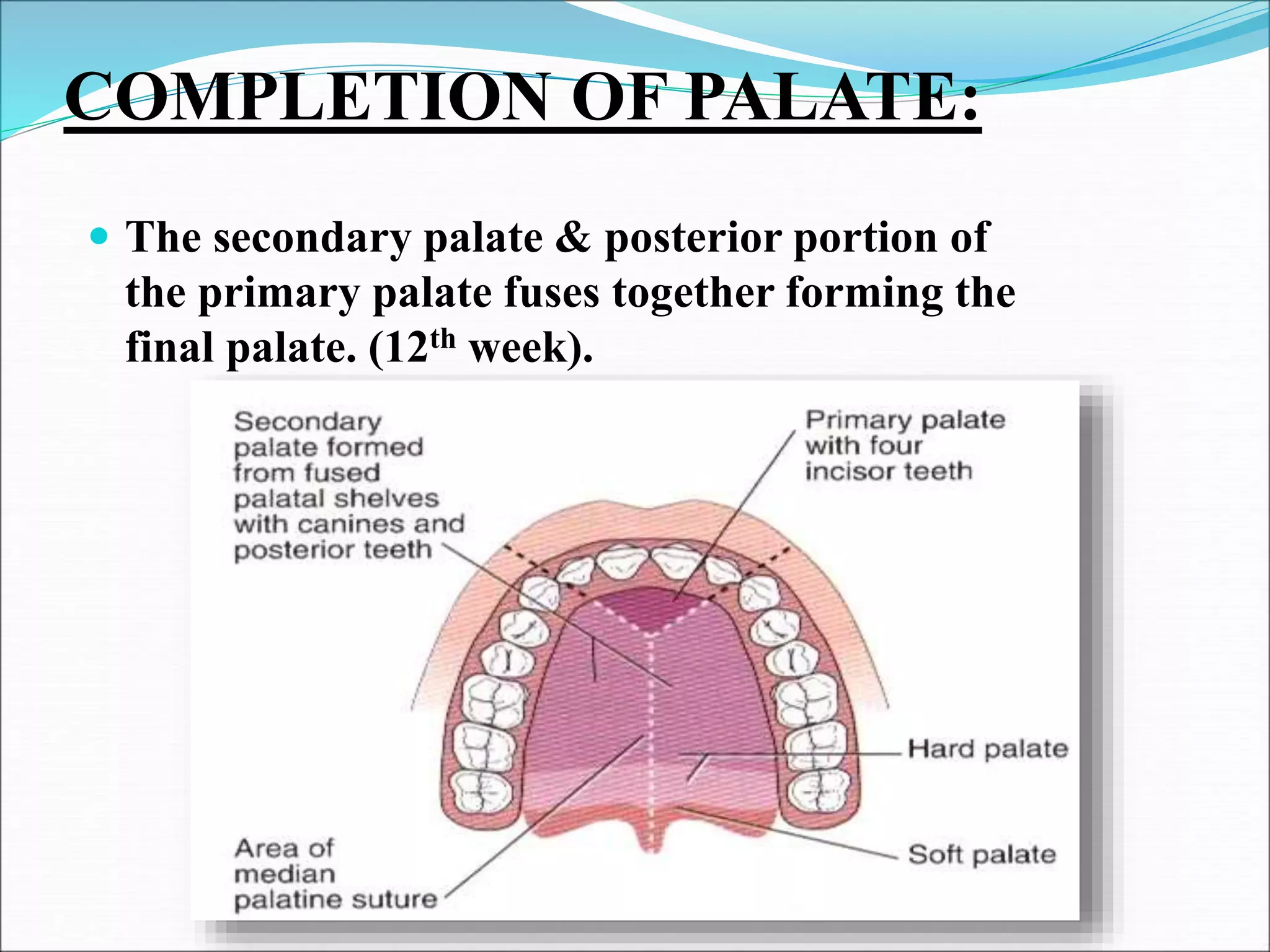
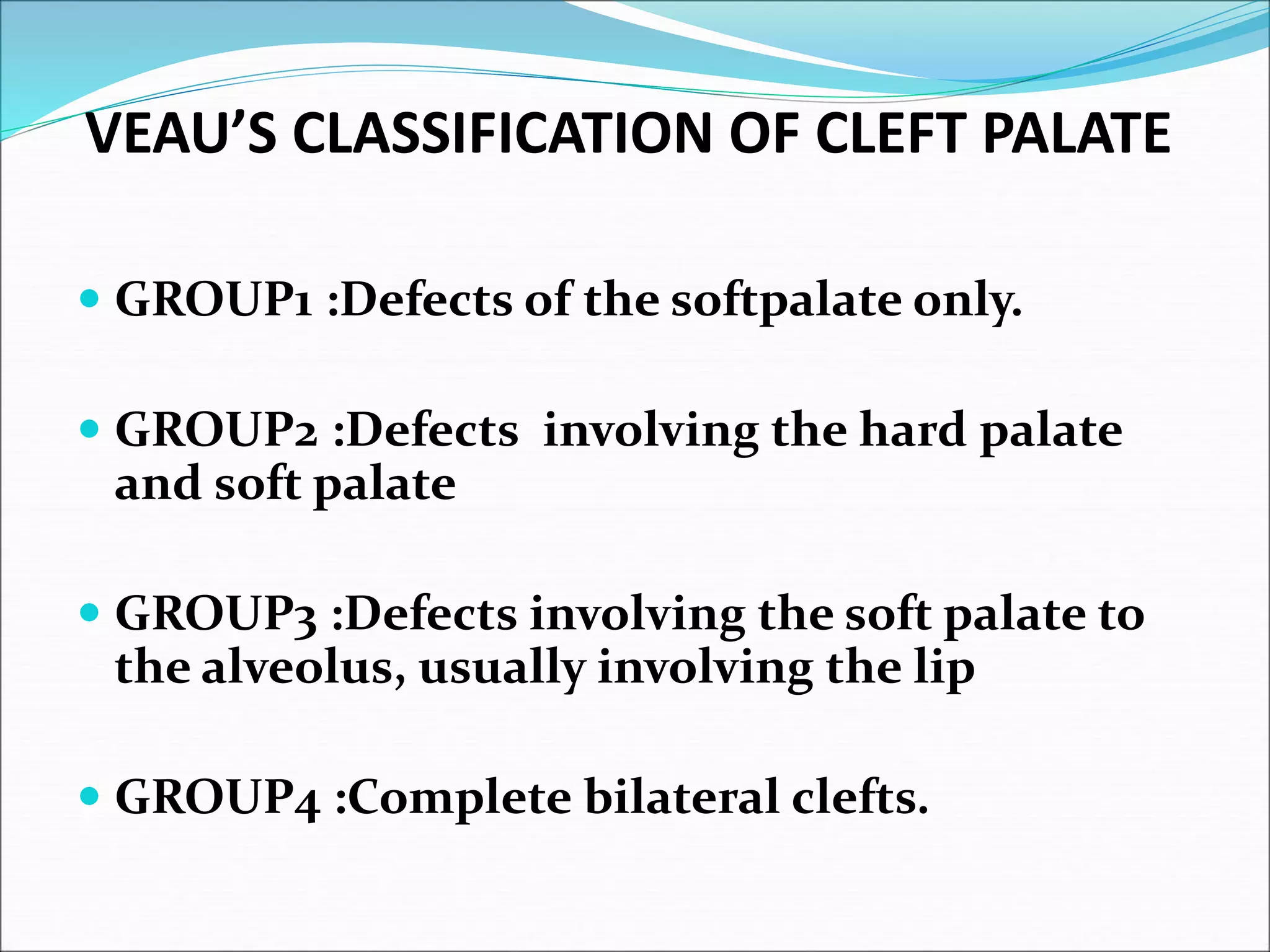
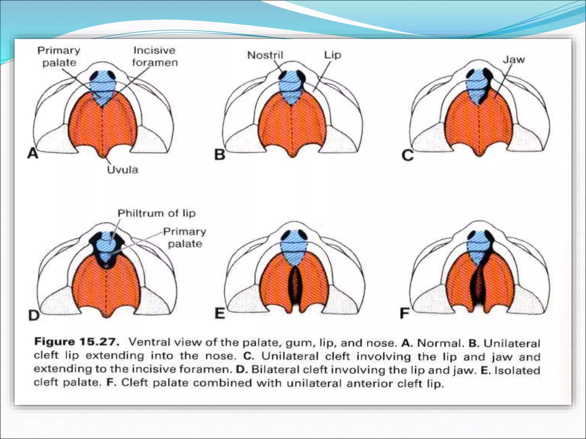
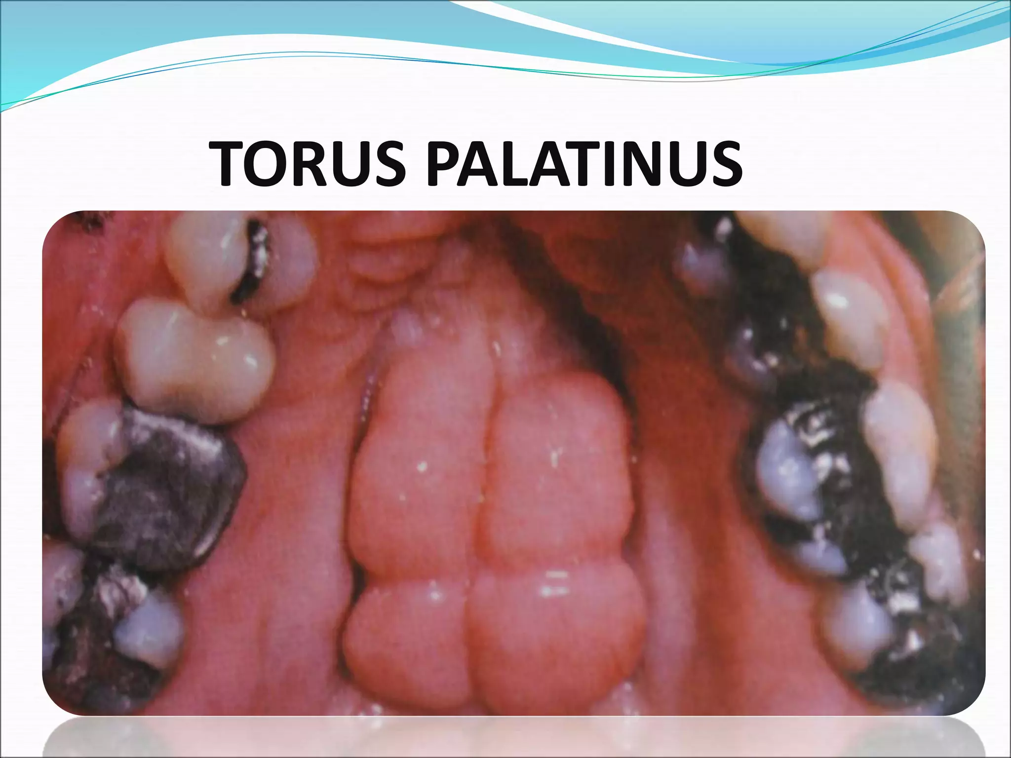
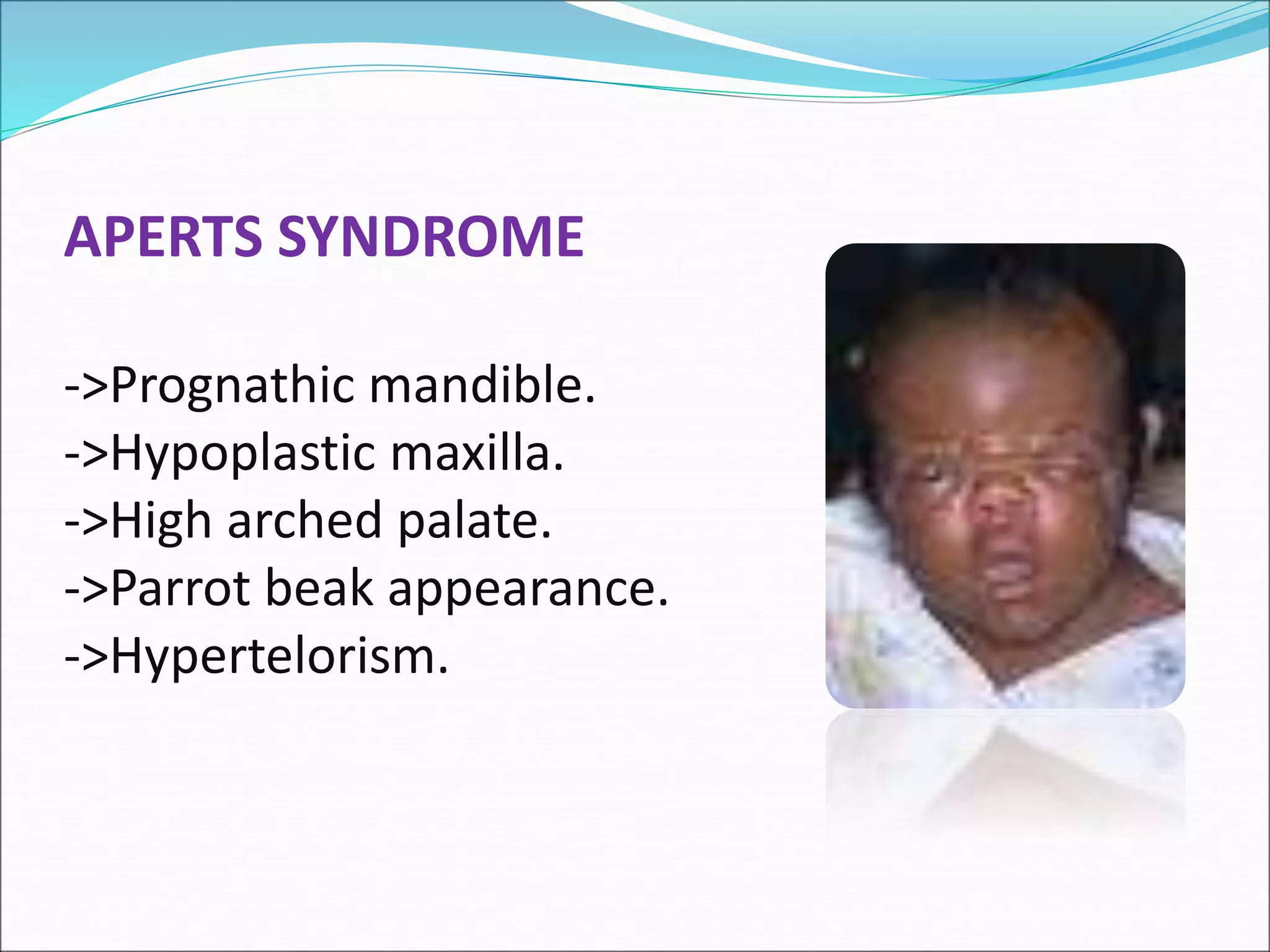
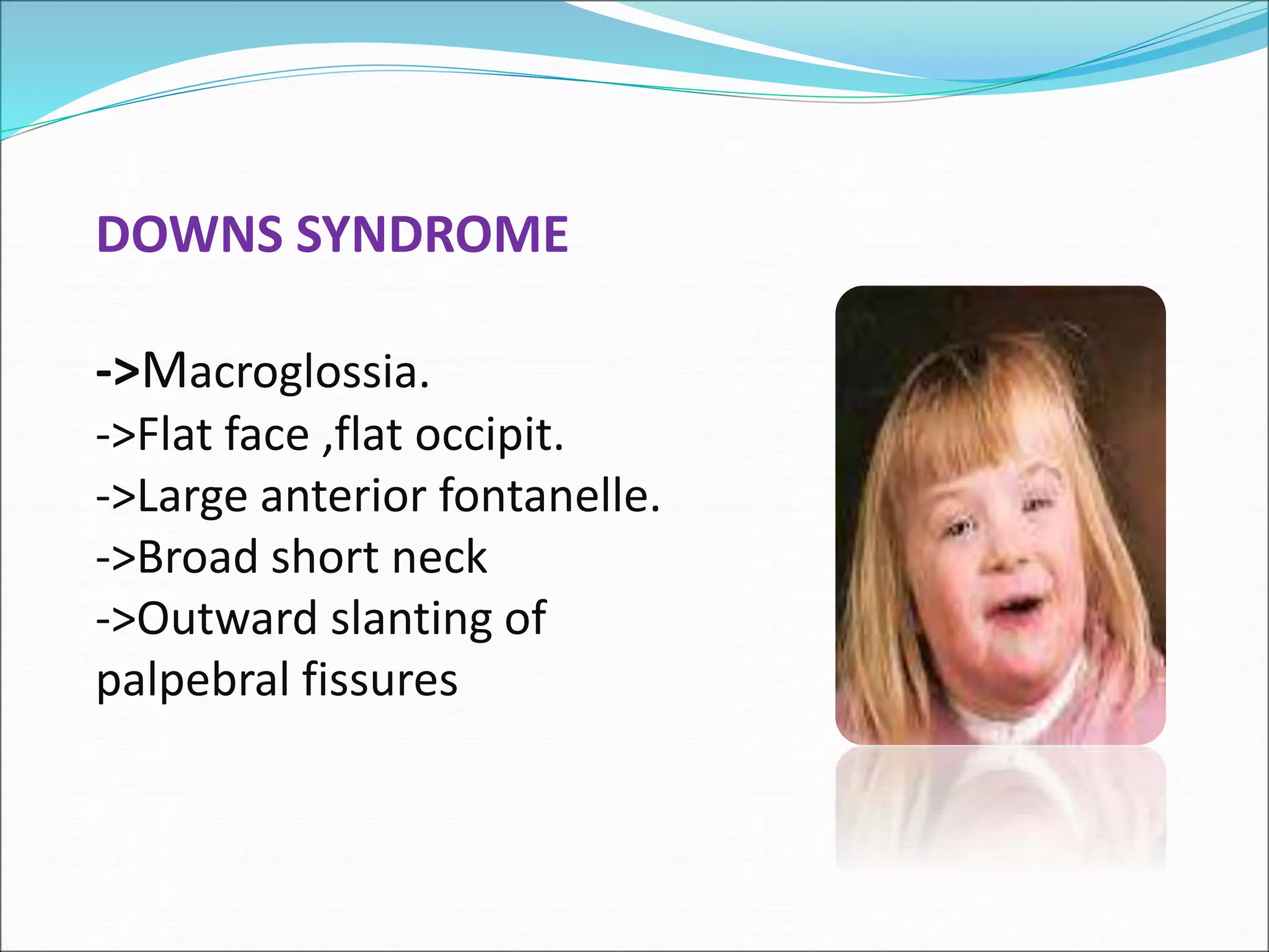
DEVELOPMENT OF
PALATE
DR.VARDENDRA.M
DEPT. OF ORAL PATHOLOGY
CONTENTS:
 INTRODUCTION
 DEVELOPMENT OF PALATE
 PALATAL SHELVES ELEVATION
 FUSION OF PALATAL SHELVES
 APPLIED ASPECTS
 CONCLUSION
 REFERENCES
DEVELOPMENT OF PALATE
INTRODUCTION
• The three elements that make up the palate are : (6th -
9th wk)
i) Two lateral maxillary palatal shelves
ii) Primary palate of the frontonasal prominence
PRIMARY PALATE FORMATION:
- 2 medial nasal process merge at the deeper levels to
form the intermaxillary segment.
- INTERMAXILLARY SEGMENT HAS
Labial component- philtrum
Maxilla component-alveolus
Palatal component –primary palate
PRIMARY PALATE FORMATION:
PRIMARY PALATE
SECONDARY PALATE FORMATION:
- The maxillary processes give rise to two
palatal shelves.(6th week)
6th wk
12th wk
Fusion of
palatal
shelves
8-9th wk
(Elevation of
Palatal shelves)
7th wk
(Tongue)
HISTOLOGIC PRESENTATION
SCHEMATIC PRESENTATION
Palatal shelf elevation
Extrinsic force concept-- jaw
tongue
head position
intrinsic force concept-- hydration of ground substance
mesenchymal cell activity
collagen
Fusion of Palatal Shelves
The site of junction between the 3 palatal
components is marked by the incisive
papilla overlying the incisive canal
SOFT PALATE:
 Posterior 1/4th of secondary palate– soft palate.
Soft palate—bilaminar fold of mucous membrane.
It contains-palatine aponeurisis
-five pairs of muscles
levator veli palatini
tensor veli palatini
musculus uvulae
palatopharyngeus
palatoglossus
-nerves and vessels
-palatine glands
COMPLETION OF PALATE:
 The secondary palate & posterior portion of
the primary palate fuses together forming the
final palate. (12th week).
CLINICAL CONSIDERATIONS:
- Cleft lip & cleft palates are common defects.
- Those anterior to incisive foramen –
lateral cleft lip,
cleft upper jaw &
cleft between primary & secondary palates.
- Clefts posterior to the incisive foramen
include cleft palate & cleft uvula.
- Third category - combination of clefts lying
anterior & posterior to the incisive foramen
INCOMPLETE CLEFT LIP
BILATERAL CLEFT LIP
CLEFT LIP &PALATE AND JAW
OBLIQUE FACIAL CLEFT:
MEDIAN CLEFT
SUBMUCOSAL CLEFT:
CLEFT UVULA
CYST FORMATION
CLASSIFICATION OF CLEFTS
 VARIOUS TYPES:
1.Davis & Ritchie classification.
2.Veaus classification.
3.Kernahan &stark symbolic classification.
4.International confederation of plastic and
reconstructive surgery classification.
VEAU’S CLASSIFICATION OF CLEFT PALATE
 GROUP1 :Defects of the softpalate only.
 GROUP2 :Defects involving the hard palate
and soft palate
 GROUP3 :Defects involving the soft palate to
the alveolus, usually involving the lip
 GROUP4 :Complete bilateral clefts.
ETIOLOGY:
1.HERIDITORY FACTORS
2.ENVIRONMENTAL FACTORS:
 Infections– measles
 radiation—x-rays
 hormones—cortisone
 nutritional deficiencies—B-complex, vit-A,
folate metabolism,
Drugs : Ethyl alcohol
- diphenylhydantoin
Trimethadione
Retinoids
Aminopterin & methotrexate
Pr ednisolone
TRANSITION OF
BLOOD SUPPLY
 Causes of cleft
lip/ palate
Deficient jaw
Growth – tongue
Occupies superior
position
Failure of
mesenchymal
cell consolidation
Lack of degeneration
of medial epithelial
seam
Failure of
elevation
Delayed shelf
elevation
Defective palatal
shelf growth
EXOSTOSIS-TORUS PALATINUS
TORUS PALATINUS
CLEFT PALATE- SYNDROMES
GOLDENHAR SYNDROME
-> Ocular, auricular,
vertebral abnormalities.
PIERRE ROBIN’S SYNDROME
->Cleft palate.
->Glossoptosis.
->Micrognathia.
MEDIAN CLEFT FACE SYNDROME
->Hypertelorism.
->Median cleft of premaxilla,
palate
ORO FACIAL DIGITAL SYNDROME
->Cleft tongue.
->Cleft of mandibular
alveolar process.
APERTS SYNDROME
->Prognathic mandible.
->Hypoplastic maxilla.
->High arched palate.
->Parrot beak appearance.
->Hypertelorism.
TREACHER-COLLINS SYNDROME
->Hypoplasia of mandible
and malar bones.
->Macrostomia.
->Malformation of external ear.
->High arched palate.
->Bird or fish appearance.
CROUZON SYNDROME
->Prognathic mandible.
->Hypoplastic maxilla.
->High arched palate.
->Parrot beak appearance.
->Hypertelorism.
MARFANS SYNDROME
->Long extremities
->Hyperextensibility of joints
->Spidery fingers
->Bifid uvula
->Cvs complications
DOWNS SYNDROME
->Macroglossia.
->Flat face ,flat occipit.
->Large anterior fontanelle.
->Broad short neck
->Outward slanting of
palpebral fissures
CONCLUSION
REFERENCES
 T.w.sadler , ed8,langman’s medical embryology, pg
no.s 272-278
 Marybathbalogh, ed2, illustrated, dental embryology,
histology&anatomy.
 James avery,ed3,oral histology and embryology,pg
no.s 45-49,51-61.
 Tencate’s,ed7,oral histology, development, structure,
and function pg no.s 39-56.
 Berkovitz, ed3,oral anatomy histology and
embryology,pg no.s- 8-12,269-283.
 Shafer’s,ed6,textbook of oral pathology, pg no.s- 22-25.
 Neville ,damm,allen,bouquot,ed3,oral and maxillofacial
pathology,pg no.s-1-5,21-22.
 orban’s,ed12,textbook of oral histology,embryology pg
no.s- 9&10.
 Moore persaud,ed7,the development of human,pg no.s-
230-235
 Profit,ed4,contemporary orthodontics,pg no.s- 44-47.
THANK YOU..

DEVELOPMENT OF PALATE.ppt