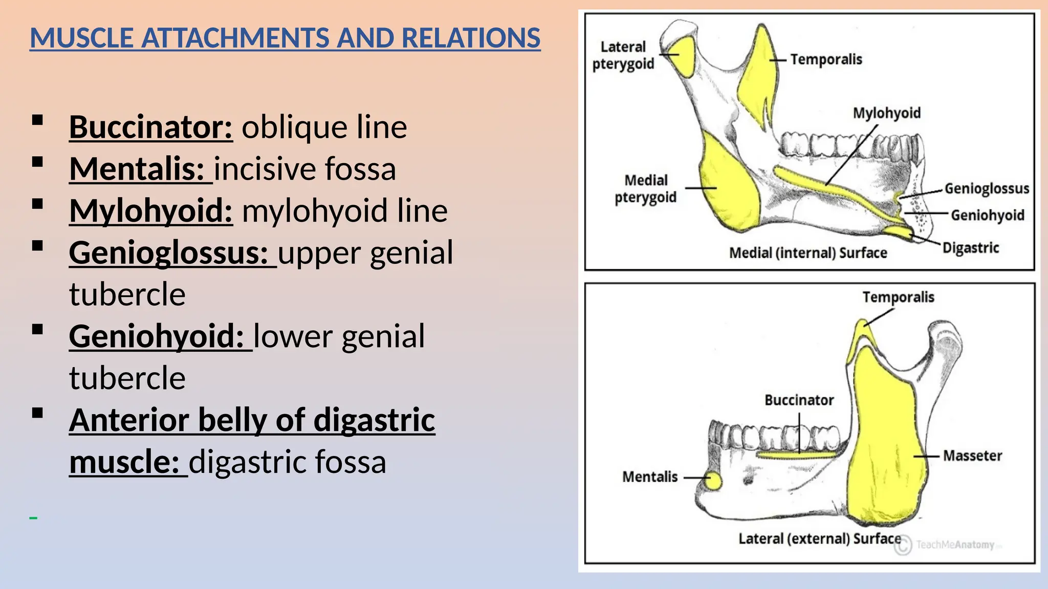
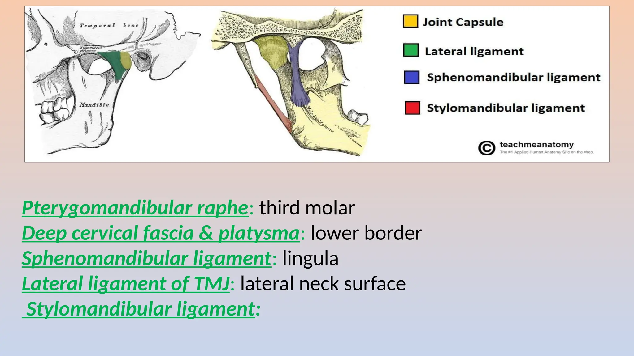
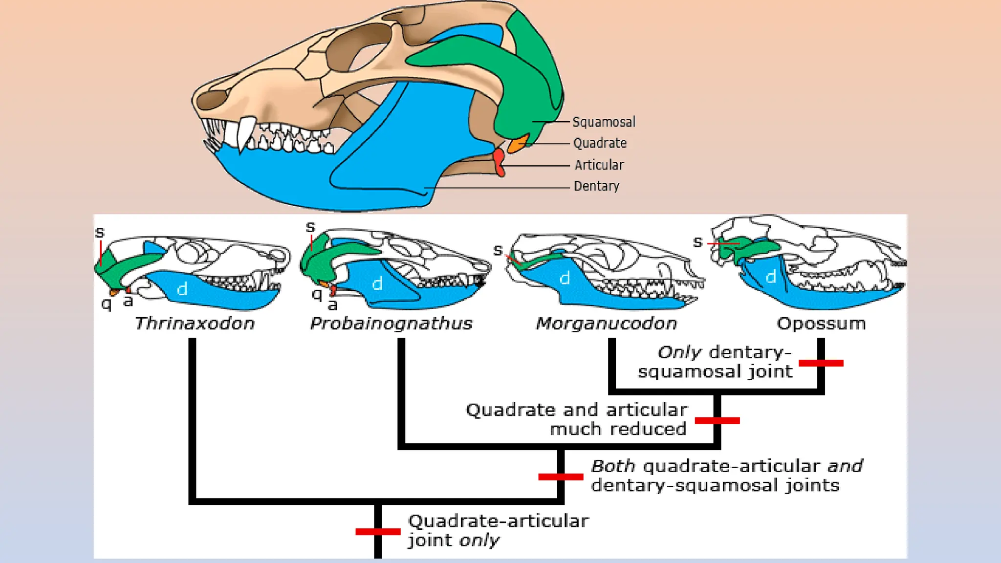
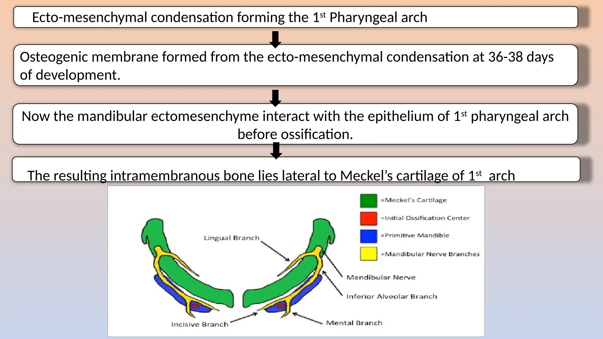
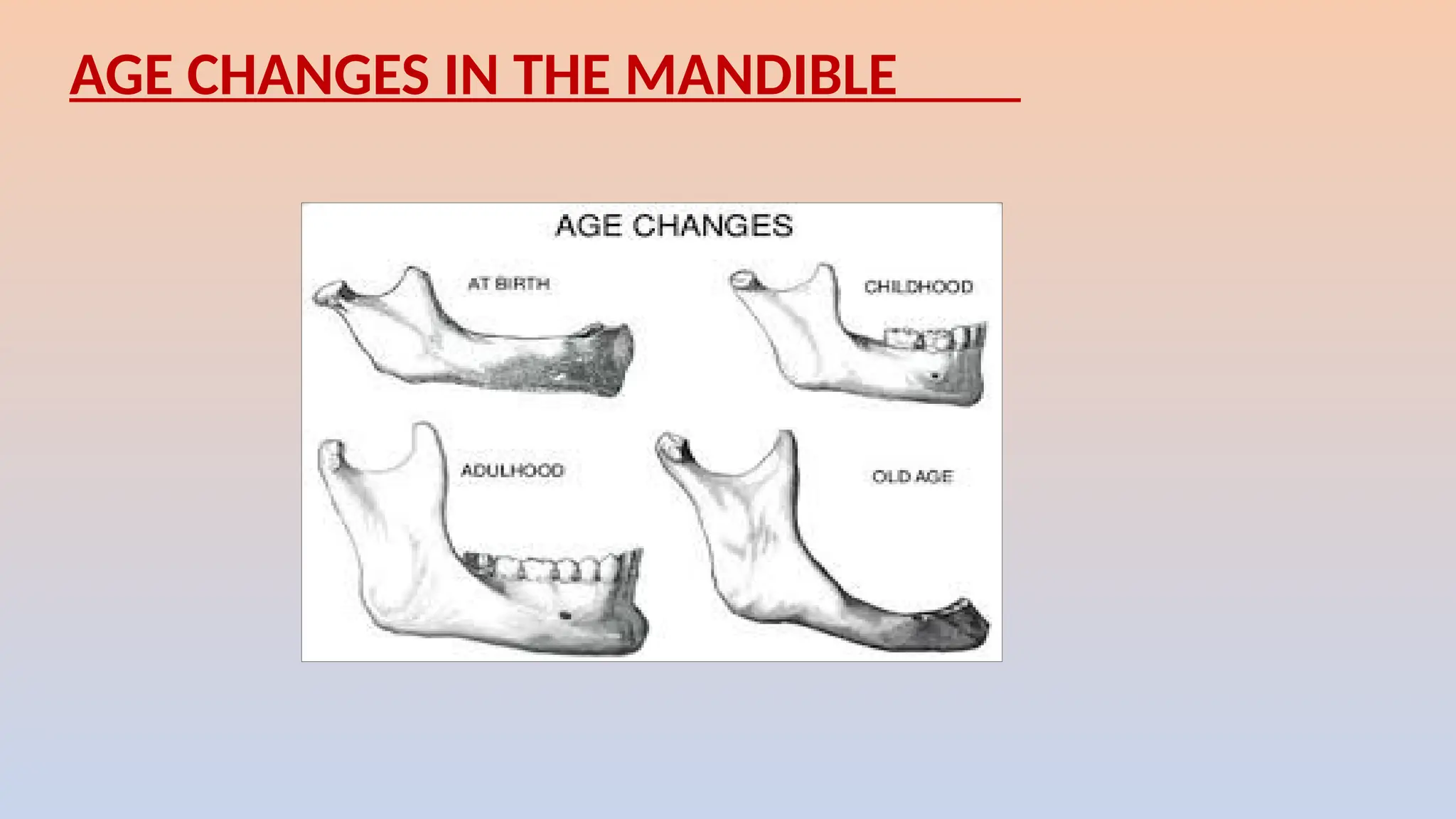
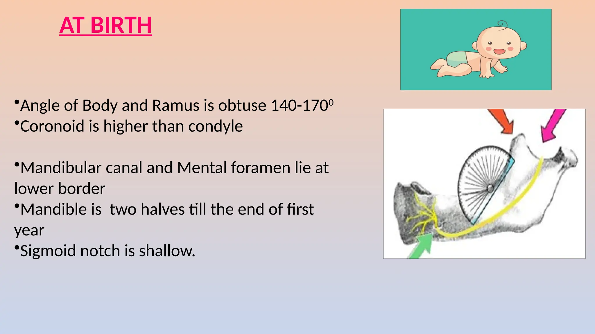
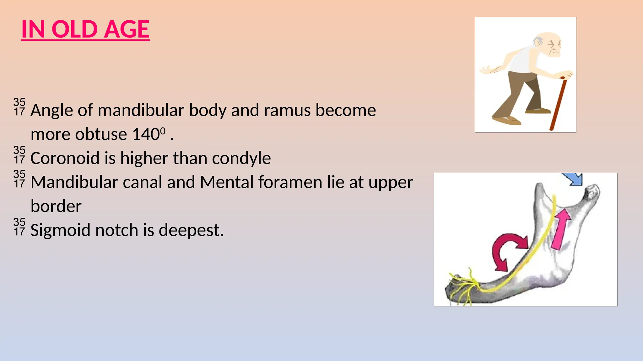
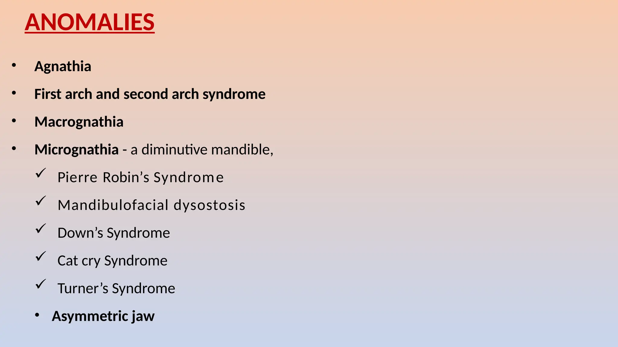
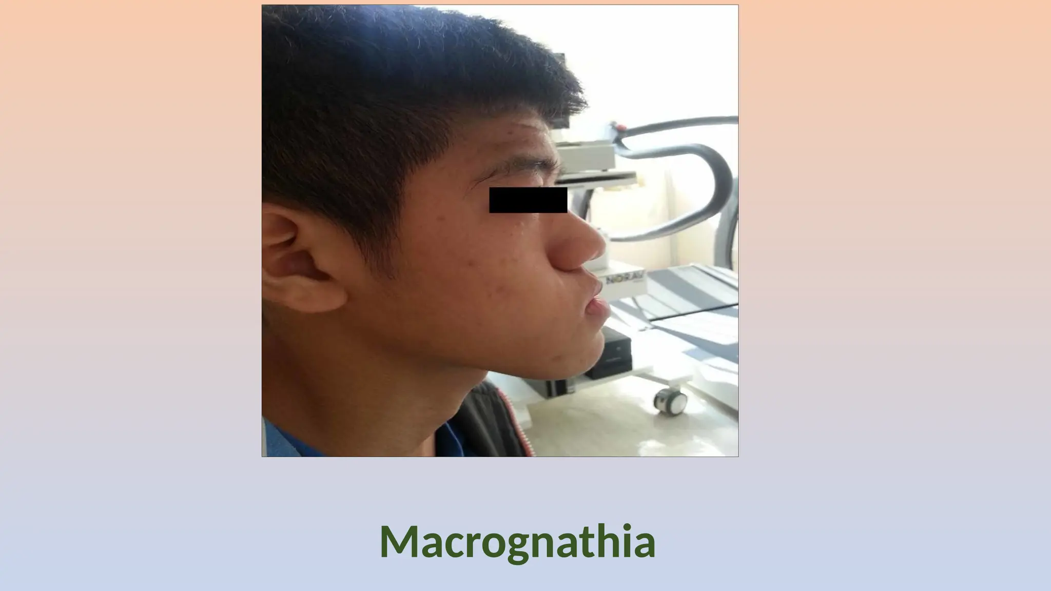
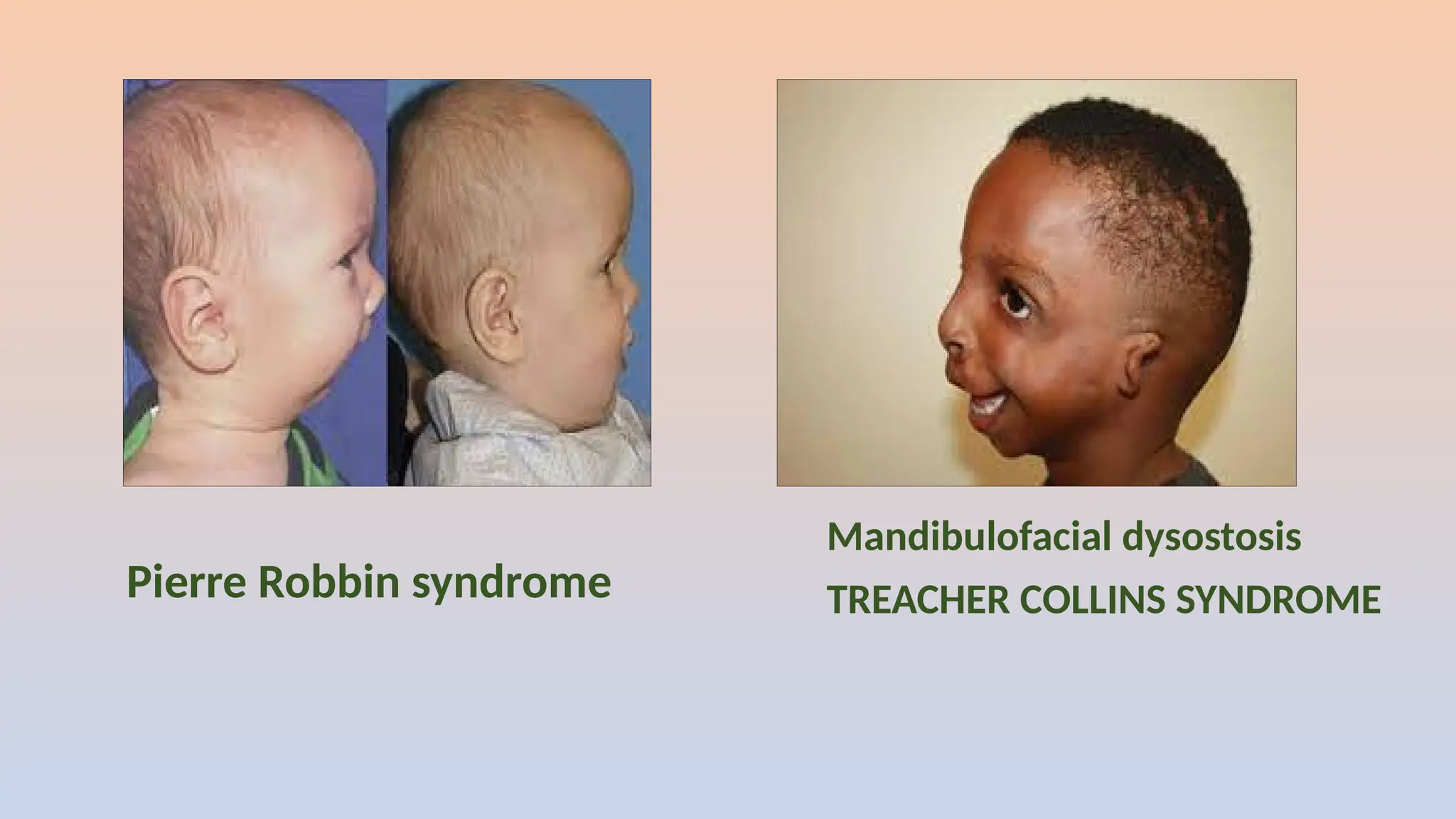
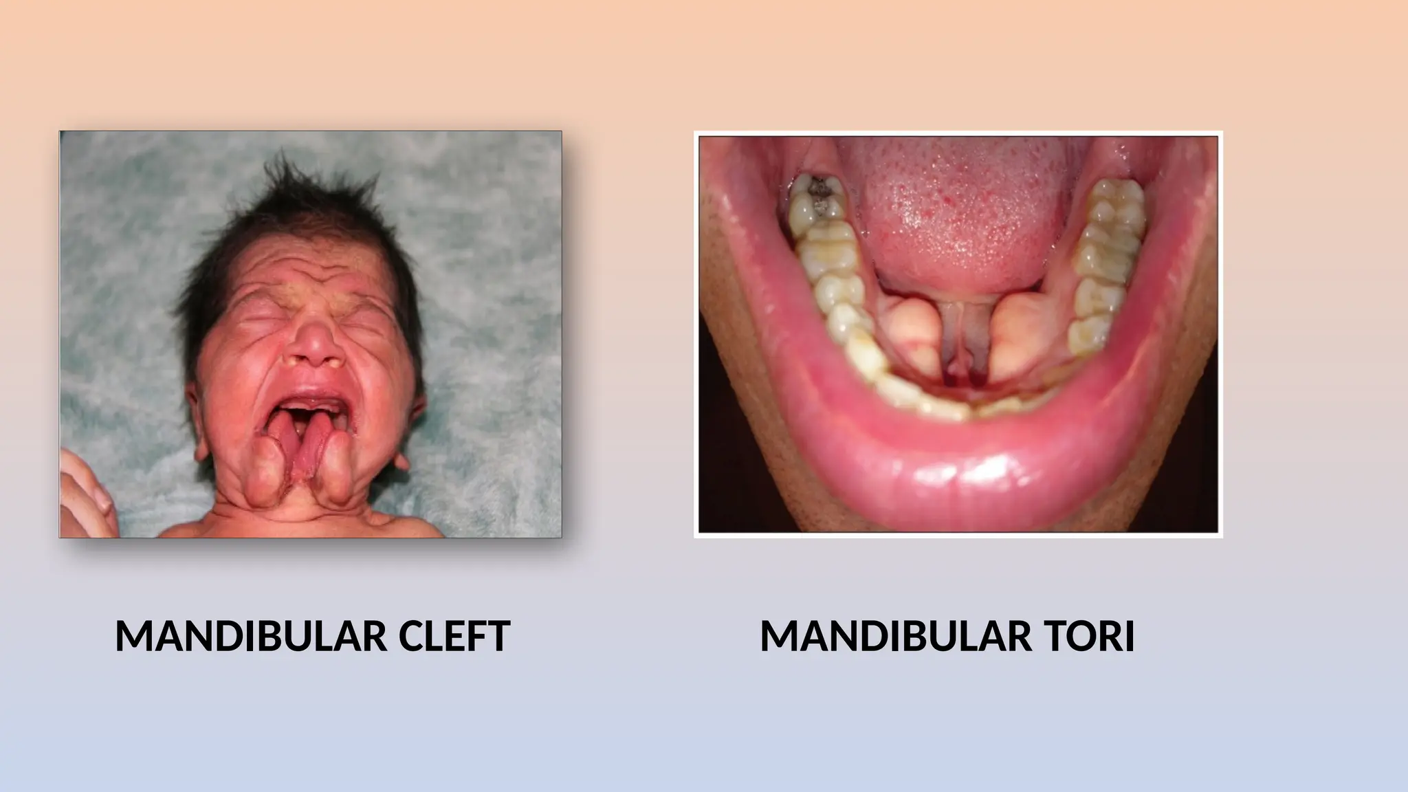
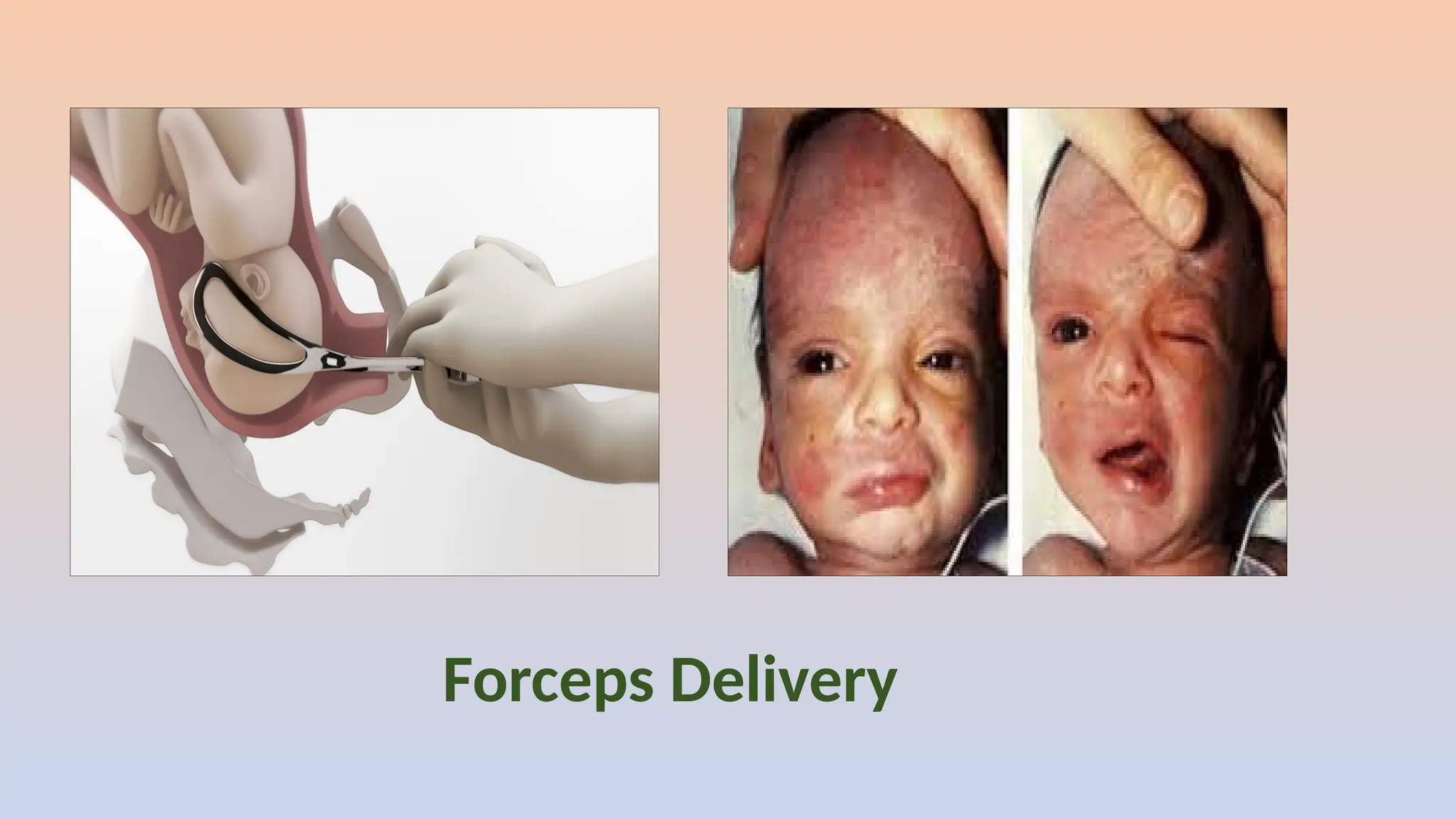
PRE NATAL & POST NATAL GROWTH AND
DEVELOPMENT OF MANDIBLE
DR. TEJASWINI REDDY. B
I MDS
CONTENTS
1.Introduction Of Mandible
2.Anatomy Of Mandible.
3.Evolution Of Mandible.
4.Prenatal Growth & Development Of Mandible.
5.Postnatal Growth & Development Of Mandible.
6.Age Changes.
7.Anomalies .
8.References
INTRODUCTION OF MANDIBLE
 Cranium + Mandible = Skull
 The largest and the strongest bone in the human skull
 It plays an important role in speech, deglutition,
mastication and respiration.
 Derived from
Greek word mandere = to chew / masticate
Latin word mandibula = lower jaw
 It is the only movable joint of skull.
ANATOMY OF MANDIBLE
BODY
Outer
• Symphysis menti – faint ridge
• Mental protuberance-
inferolateral angles- mental
tubercles
• Mental foramen
• Oblique ridge-ant border of
ramus
• Incissive fossa
Inner
• Mylohyoid line-third molar
tooth
• Below mylohyoid line
• Above mylohyoid line
• Mylohyoid groove-extends
on to the body
Upper- sockets
Lower- base
RAMUS
Superior
• Curved downwards-
mandibular notch
• Coronoid process- triangular
upward projection -antero
superior
• Condylar process- postero
superior -head-fibrocartilage-
articulation-TMJ.
• Inferior-base – angle of
mandible
• Lateral-flat -oblique ridge
• Medial-mandibular
foramen -above the
occlusal level
BLOOD SUPPLY AND NERVE SUPPLY
Inferior alveolar nerve and
vessels
Mental nerve and vessels
Mylohyoid nerve and vessels –
mylohyoid groove
Masseteric nerve and vessels-
mandibular notch
Lingual nerve- infront of
mylohyoid groove
Auriculotemporal nerve
and superficial
temporal artery-neck
Facial artery - Antero
inferior angle of
masseter
MUSCLE ATTACHMENTS AND RELATIONS
 Buccinator: oblique line
 Mentalis: incisive fossa
 Mylohyoid: mylohyoid line
 Genioglossus: upper genial
tubercle
 Geniohyoid: lower genial
tubercle
 Anterior belly of digastric
muscle: digastric fossa
 Masseter: except
postero superior ramus
 Temporalis: coronoid
process
 Medial pterygoid:
medial side of ramus
 Lateral pterygoid:
pterygoid fovea-
anterior aspect of neck
Pterygomandibular raphe: third molar
Deep cervical fascia & platysma: lower border
Sphenomandibular ligament: lingula
Lateral ligament of TMJ: lateral neck surface
Stylomandibular ligament:
EVOLUTION OF MANDIBLE
The articular bone is part of the lower jaw
of most vertebrates and is connected to the
angular lower jaw bones .
It originates from the embryonic mandibular
cartilage.
The most caudal portion of the mandibular
cartilage ossifies to form the articular bone.
The upper jaw articulates at the
quadrate bone.
The articular, angular, and
prearticular bones have fused to
form the compound bone.
The mandible is suspended from the
quadrate bone and articulates at this
compound bone.
the articular bone evolves to
form the malleus,. and is
used to determine the fossil
transition to mammals.
After the loss of the quadrate-articular joint, the squamosal and dentary bones form the
new jaw joint in mammals.
PRENATAL GROWTH & DEVELOPMENT OF
MANDIBLE
The Corpus and
the Ramus
In the neural folds ,the mid and hind brain regions forms embryonic neural crest cells.
These cells migrate ventrally to form the mandibular facial prominences and differentiate
into bones and connective tissues.
First structure to develop in this region is the mandibular division of trigeminal nerve
Mandibular nerve induces osteogenesis by producing neurotrophic factors
Now the mandibular ectomesenchyme interact with the epithelium of 1st
pharyngeal arch
before ossification.
Ecto-mesenchymal condensation forming the 1st
Pharyngeal arch
Osteogenic membrane formed from the ecto-mesenchymal condensation at 36-38 days
of development.
The resulting intramembranous bone lies lateral to Meckel’s cartilage of 1st
arch
A Single ossification centre for each half arises at 6th
week of post
conception .
From the ossification centre, ossification spreads upwards to form a
trough for the developing teeth
This spread of intramembranous ossification dorsally and ventrally forms the Body
and Ramus. ossification stops at a point dorsally, that later becomes the mandibular
Lingula
This intial woven bone formed along Meckel’s cartilage is soon replaced by
Lamellar bone. this early remodelling than other bones is responsible for
early intense sucking and swallowing ,which stress the mandible
What happens to Meckel’s cartilage?
• Ventrally - meets its fellow of the opposite
side.
• Dorsally-diverges into tympanic cavity -
ends ossifies -auditory ossicles MALLEUS
AND INCUS.
• Some part of it transforms into
1. Spheno mandibular ligament.
2. Spinous process of sphenoid.
3. Anterior ligament of malleus.
• Meckel’s cartilage disappears by the 24th
week.
Secondary accessory cartilages appear between 10th
and 14th
weeks to form the head of the condyle, coronoid process , and
mental protuberance.
CORONOID PROCESS
20
cartilage of coronoid process
develops within the temporalis
muscle
This 20
cartilage incorporates into
expanding intramembranous
bone of the ramus and disappears
before birth.
MENTAL PROTRUBERANCE
In the mental region, on either side of the symphysis,
two smaller cartilages appear.
They ossify in the 7th
month of intrauterine life
They ossify in the 7th
month of intrauterine life
The ossified cartilages forms mental ossicles in the fibrous tissue
of symphysis
These ossicles incorporates into intramembranous bone when symphysis menti
coverts from syndesmosis into synostosis during 1st
postnatal year.
CONDYLAR PROCESS
Condylar cartilage appears during the 10th
week of post conception as a cone
shaped structure in the ramus region ,which acts as a primordium for future condye
Condylar cartilage - 10th
week of post conception - a cone shaped structure in the
ramus region - acts as a primordium for future condyle.
Condylar cartilage appears during the 10th
week of post conception as a cone
shaped structure in the ramus region ,which acts as a primordium for future cone
By 14th
week, endochondral bone appears in the condyle region.
le
Condylar cartilage appears during the 10th
week of post conception as a cone
shaped structure in the ramus region ,which acts as a primordium for future condye
Cartilage cells differentiate and the condylar head increases by interstitial and
appositional growth.
By 14th
week, endochondral bone appears in the condyle region.
This condylar cartilage serves as important centre for growth of mandible .
Condylar cartilage appears during the 10th
week of post conception as a cone
shaped structure in the ramus region ,which acts as a primordium for future condyl
By the middle of Fetal life the cone shaped cartilage is replaced with bone, but the
upper end persists into adulthood, acting as both growth and articular cartilage.
Change in the mandibular position and form, are related to the direction and
amount of condylar growth.
Differential Growth..
During Fetal Life: 8 weeks - mandible > maxilla
11 weeks - mandible = maxilla
13 – 20 weeks maxilla > mandible
At Birth: Retrognathic
Early postnatal life : Orthognathic
POST NATAL GROWTH AND DEVELOPMENT OF
MANDIBLE
Out of all the facial bones the mandible undergoes
the most growth postnatally and evidences the
greatest variation in morphology.
Post natal growth is discussed in two stages:
1.Mandibular growth in the first year of life.
2.Mandibular growth after the first year of life.
Mandibular growth in the first year of life:
At birth,
Two short mandibular rami.
Minimal condylar development.
Little articular eminence in the glenoid fossa .
Thin fibrocartilage and connective tissue line at the symphysis
separating two halves. Between 4 months to 1 year ,the symphyseal
cartilage is replaced by bone.
During 1st
year the condyle ,ramus ,lateral border and alveolar
border shows appositional growth.
Growth sites in mandible
1) Limited growth takes place at the
symphysis menti until fusion occurs.
2) At the condylar cartilages
3) The posterior border of ramus
4) Alveolar ridges
Mandibular growth after first year of life:
Though mandible appears as a single
bone in the adult, it is developmentally
and functionally divisible into several
skeletal subunits.
The basal bone of the body forms one
unit, to which are attached the alveolar,
coronoid, angular, and condylar
processes and the chin.
• According to the functional matrix theory, bone
growth within the craniofacial skeleton is influenced
primarily by functional needs and neurotrophic
factors influences and is mediated by soft tissue.
• The functioning of the related tongue and perioral
muscles and the expansion of the oral and
pharyngeal cavities provide stimuli for mandibular
growth to reach its full potential.
 The growth pattern of each of these skeletal
subunits is influenced by a functional matrix that
acts upon the bone:
the teeth act as a functional matrix for the
alveolar unit
the action of the temporalis muscle influences
the coronoid process
the masseter and medial pterygoid muscles act
upon the angle and ramus
the lateral pterygoid has some influence on the
condylar process.
REMODELLING OF THE RAMUS
 The Ramal angle slightly uprights- adolescence
in late adulthood- acute.
 Till the uprighting of Ramus
deposition -posterior ramal border
but after uprighting- selective
 Inferior - resorptive
 Superior-depository.
Remodelling of Corpus
 Hunter concept-
 body of mandible-depository-lateral surface
 resorptive- inferior aspect of the medial surface.
depository- superior aspect -medial surface just below the teeth
Medial surface of the ramus- remodeling is the form of ‘L’,
depository - extending from the superior half of medial surface of
corpus to the anterior half of medial surface of the ramus (below
coronoid)
 The resorptive area follows depository area, from inferior half of
medial surface of corpus to posterior half of medial surface of
ramus below the condyle.
 This eccentric remodeling - configuration of adult mandible.
Mandibles of a neonate (top), a 4-year-old
child (middle), and an adult (below)
combination process of periosteal
resorption and endosteal deposition
Growth at the condylar cartilage is pressure adapted. Superior surface of
condyle is depository.
 Only the cap of condyle undergoes endochondral ossification, the rest of the
condyle and the neck of condyle grows by intramembranous ossification.
Growth At The Condylar Cartilage
Enlow's 'V' Principle of Growth.
The condyle grows like an expanding V.
deposition- inner aspect of V
resorption -outer
Due to this, the bone moves in the direction
towards the wide end of 'V.’
 Simultaneously deposition at the ends of
the two arms of the V -widening.
Remodeling Of Coronoid Process
 The coronoid process has a twisted form
 periosteal deposition -lingual surface
resorption- buccal surface
 The basal part - periosteal deposits on the buccal side
resorption - lingual surface.
Chin
 Heavy periosteal growth - lingual surface of the chin
 Dense lamellar bone fusing and overlapping - labial
surface of the chin.
 anterior surface-resorptive .
 deposition in the endosteal surface.
 lingual periosteum is of the symphysis - depository.
AGE CHANGES IN THE MANDIBLE
AT BIRTH
•Angle of Body and Ramus is obtuse 140-1700
•Coronoid is higher than condyle
•Mandibular canal and Mental foramen lie at
lower border
•Mandible is two halves till the end of first
year
•Sigmoid notch is shallow.
IN ADULTS
 Angle of mandibular body and ramus is less obtuse 110-1200
.
 Condyle is higher than coronoid.
 Mandibular canal and Mental foramen lie in the middle due to
increase in the vertical height by the presence of teeth.
 Sigmoid notch is deepest
IN OLD AGE
 Angle of mandibular body and ramus become
more obtuse 1400
.
 Coronoid is higher than condyle
 Mandibular canal and Mental foramen lie at upper
border
 Sigmoid notch is deepest.
• Agnathia
• First arch and second arch syndrome
• Macrognathia
• Micrognathia - a diminutive mandible,
 Pierre Robin’s Syndrome
 Mandibulofacial dysostosis
 Down’s Syndrome
 Cat cry Syndrome
 Turner’s Syndrome
• Asymmetric jaw
ANOMALIES
Agnathia
First arch and second arch syndrome
Macrognathia
Micrognathia
Pierre Robbin syndrome
Mandibulofacial dysostosis
TREACHER COLLINS SYNDROME
HALLERMANN-STREIFF SYNDROME
( OCULOMANDIBULODYSCEPHALY )
Cri-du-chat syndrome
DOWN’S SYNDROME TURNER SYNDROME
MANDIBULAR CLEFT MANDIBULAR TORI
Asymmetric Jaw
Forceps Delivery
REFERENCES
1. Contemporary Orthodontics, William Proffit, Henry Fields, Brent Larson, David Sarver, 6th Edition, 2018
2. Craniofacial Embryogenetics And Development Craniofacial Embryogenetics and Development. Sperber GH,
Sperber SM, Guttmann GD. Shelton, CT: People's Medical Publishing House-USA. 2010
3. Textbook of Craniofacial Growth - Sridhar Premkumar.
4. The V Principle – Donald H Enlow – AJO – Jan 1984
5. Hildyard LT, Moore WJ, Corbett ME. Logarithmic growth of the hominoid mandible. The Anatomical Record.
1976 Nov;186(3):405-11.
6. Human Embryology – I.B. Singh, 11th Edition, 2017
7. Oka, K., Oka, S., Sasaki, T., Ito, Y., Bringas, P. Jr., Nonaka, K. and Chai, Y: regulating chondrogenesis and
osteogenesis during mandibular development. Dev. Biol. 303:391-404, 2007.
8. Mandibular development and its Age changes, Journal of Pharmaceutical Sciences and Research, Vol. 6
(11),2014,360-362.
9. Mandibular Growth Anomalies -Hugo L. Obwegeser
10.B. D. Chaurasia's Human Anatomy-Third edition
THANK YOU

Mandible.pptx characterstics properties definition

  • 2.
    PRE NATAL &POST NATAL GROWTH AND DEVELOPMENT OF MANDIBLE DR. TEJASWINI REDDY. B I MDS
  • 3.
    CONTENTS 1.Introduction Of Mandible 2.AnatomyOf Mandible. 3.Evolution Of Mandible. 4.Prenatal Growth & Development Of Mandible. 5.Postnatal Growth & Development Of Mandible. 6.Age Changes. 7.Anomalies . 8.References
  • 4.
    INTRODUCTION OF MANDIBLE Cranium + Mandible = Skull  The largest and the strongest bone in the human skull  It plays an important role in speech, deglutition, mastication and respiration.  Derived from Greek word mandere = to chew / masticate Latin word mandibula = lower jaw  It is the only movable joint of skull.
  • 5.
  • 6.
    BODY Outer • Symphysis menti– faint ridge • Mental protuberance- inferolateral angles- mental tubercles • Mental foramen • Oblique ridge-ant border of ramus • Incissive fossa
  • 7.
    Inner • Mylohyoid line-thirdmolar tooth • Below mylohyoid line • Above mylohyoid line • Mylohyoid groove-extends on to the body Upper- sockets Lower- base
  • 8.
    RAMUS Superior • Curved downwards- mandibularnotch • Coronoid process- triangular upward projection -antero superior • Condylar process- postero superior -head-fibrocartilage- articulation-TMJ.
  • 9.
    • Inferior-base –angle of mandible • Lateral-flat -oblique ridge • Medial-mandibular foramen -above the occlusal level
  • 10.
    BLOOD SUPPLY ANDNERVE SUPPLY Inferior alveolar nerve and vessels Mental nerve and vessels Mylohyoid nerve and vessels – mylohyoid groove Masseteric nerve and vessels- mandibular notch
  • 11.
    Lingual nerve- infrontof mylohyoid groove Auriculotemporal nerve and superficial temporal artery-neck Facial artery - Antero inferior angle of masseter
  • 12.
    MUSCLE ATTACHMENTS ANDRELATIONS  Buccinator: oblique line  Mentalis: incisive fossa  Mylohyoid: mylohyoid line  Genioglossus: upper genial tubercle  Geniohyoid: lower genial tubercle  Anterior belly of digastric muscle: digastric fossa
  • 13.
     Masseter: except posterosuperior ramus  Temporalis: coronoid process  Medial pterygoid: medial side of ramus  Lateral pterygoid: pterygoid fovea- anterior aspect of neck
  • 14.
    Pterygomandibular raphe: thirdmolar Deep cervical fascia & platysma: lower border Sphenomandibular ligament: lingula Lateral ligament of TMJ: lateral neck surface Stylomandibular ligament:
  • 15.
    EVOLUTION OF MANDIBLE Thearticular bone is part of the lower jaw of most vertebrates and is connected to the angular lower jaw bones . It originates from the embryonic mandibular cartilage. The most caudal portion of the mandibular cartilage ossifies to form the articular bone.
  • 16.
    The upper jawarticulates at the quadrate bone. The articular, angular, and prearticular bones have fused to form the compound bone. The mandible is suspended from the quadrate bone and articulates at this compound bone.
  • 17.
    the articular boneevolves to form the malleus,. and is used to determine the fossil transition to mammals.
  • 18.
    After the lossof the quadrate-articular joint, the squamosal and dentary bones form the new jaw joint in mammals.
  • 20.
    PRENATAL GROWTH &DEVELOPMENT OF MANDIBLE The Corpus and the Ramus
  • 21.
    In the neuralfolds ,the mid and hind brain regions forms embryonic neural crest cells. These cells migrate ventrally to form the mandibular facial prominences and differentiate into bones and connective tissues. First structure to develop in this region is the mandibular division of trigeminal nerve Mandibular nerve induces osteogenesis by producing neurotrophic factors
  • 22.
    Now the mandibularectomesenchyme interact with the epithelium of 1st pharyngeal arch before ossification. Ecto-mesenchymal condensation forming the 1st Pharyngeal arch Osteogenic membrane formed from the ecto-mesenchymal condensation at 36-38 days of development. The resulting intramembranous bone lies lateral to Meckel’s cartilage of 1st arch
  • 23.
    A Single ossificationcentre for each half arises at 6th week of post conception . From the ossification centre, ossification spreads upwards to form a trough for the developing teeth This spread of intramembranous ossification dorsally and ventrally forms the Body and Ramus. ossification stops at a point dorsally, that later becomes the mandibular Lingula This intial woven bone formed along Meckel’s cartilage is soon replaced by Lamellar bone. this early remodelling than other bones is responsible for early intense sucking and swallowing ,which stress the mandible
  • 24.
    What happens toMeckel’s cartilage? • Ventrally - meets its fellow of the opposite side. • Dorsally-diverges into tympanic cavity - ends ossifies -auditory ossicles MALLEUS AND INCUS. • Some part of it transforms into 1. Spheno mandibular ligament. 2. Spinous process of sphenoid. 3. Anterior ligament of malleus. • Meckel’s cartilage disappears by the 24th week.
  • 25.
    Secondary accessory cartilagesappear between 10th and 14th weeks to form the head of the condyle, coronoid process , and mental protuberance.
  • 26.
    CORONOID PROCESS 20 cartilage ofcoronoid process develops within the temporalis muscle This 20 cartilage incorporates into expanding intramembranous bone of the ramus and disappears before birth.
  • 27.
    MENTAL PROTRUBERANCE In themental region, on either side of the symphysis, two smaller cartilages appear. They ossify in the 7th month of intrauterine life They ossify in the 7th month of intrauterine life The ossified cartilages forms mental ossicles in the fibrous tissue of symphysis These ossicles incorporates into intramembranous bone when symphysis menti coverts from syndesmosis into synostosis during 1st postnatal year.
  • 28.
    CONDYLAR PROCESS Condylar cartilageappears during the 10th week of post conception as a cone shaped structure in the ramus region ,which acts as a primordium for future condye Condylar cartilage - 10th week of post conception - a cone shaped structure in the ramus region - acts as a primordium for future condyle. Condylar cartilage appears during the 10th week of post conception as a cone shaped structure in the ramus region ,which acts as a primordium for future cone By 14th week, endochondral bone appears in the condyle region. le Condylar cartilage appears during the 10th week of post conception as a cone shaped structure in the ramus region ,which acts as a primordium for future condye Cartilage cells differentiate and the condylar head increases by interstitial and appositional growth. By 14th week, endochondral bone appears in the condyle region. This condylar cartilage serves as important centre for growth of mandible . Condylar cartilage appears during the 10th week of post conception as a cone shaped structure in the ramus region ,which acts as a primordium for future condyl By the middle of Fetal life the cone shaped cartilage is replaced with bone, but the upper end persists into adulthood, acting as both growth and articular cartilage. Change in the mandibular position and form, are related to the direction and amount of condylar growth.
  • 29.
    Differential Growth.. During FetalLife: 8 weeks - mandible > maxilla 11 weeks - mandible = maxilla 13 – 20 weeks maxilla > mandible At Birth: Retrognathic Early postnatal life : Orthognathic
  • 30.
    POST NATAL GROWTHAND DEVELOPMENT OF MANDIBLE Out of all the facial bones the mandible undergoes the most growth postnatally and evidences the greatest variation in morphology. Post natal growth is discussed in two stages: 1.Mandibular growth in the first year of life. 2.Mandibular growth after the first year of life.
  • 31.
    Mandibular growth inthe first year of life: At birth, Two short mandibular rami. Minimal condylar development. Little articular eminence in the glenoid fossa . Thin fibrocartilage and connective tissue line at the symphysis separating two halves. Between 4 months to 1 year ,the symphyseal cartilage is replaced by bone. During 1st year the condyle ,ramus ,lateral border and alveolar border shows appositional growth.
  • 32.
    Growth sites inmandible 1) Limited growth takes place at the symphysis menti until fusion occurs. 2) At the condylar cartilages 3) The posterior border of ramus 4) Alveolar ridges
  • 33.
    Mandibular growth afterfirst year of life: Though mandible appears as a single bone in the adult, it is developmentally and functionally divisible into several skeletal subunits. The basal bone of the body forms one unit, to which are attached the alveolar, coronoid, angular, and condylar processes and the chin.
  • 34.
    • According tothe functional matrix theory, bone growth within the craniofacial skeleton is influenced primarily by functional needs and neurotrophic factors influences and is mediated by soft tissue. • The functioning of the related tongue and perioral muscles and the expansion of the oral and pharyngeal cavities provide stimuli for mandibular growth to reach its full potential.
  • 35.
     The growthpattern of each of these skeletal subunits is influenced by a functional matrix that acts upon the bone: the teeth act as a functional matrix for the alveolar unit the action of the temporalis muscle influences the coronoid process the masseter and medial pterygoid muscles act upon the angle and ramus the lateral pterygoid has some influence on the condylar process.
  • 36.
    REMODELLING OF THERAMUS  The Ramal angle slightly uprights- adolescence in late adulthood- acute.  Till the uprighting of Ramus deposition -posterior ramal border but after uprighting- selective  Inferior - resorptive  Superior-depository.
  • 37.
    Remodelling of Corpus Hunter concept-  body of mandible-depository-lateral surface  resorptive- inferior aspect of the medial surface. depository- superior aspect -medial surface just below the teeth Medial surface of the ramus- remodeling is the form of ‘L’, depository - extending from the superior half of medial surface of corpus to the anterior half of medial surface of the ramus (below coronoid)  The resorptive area follows depository area, from inferior half of medial surface of corpus to posterior half of medial surface of ramus below the condyle.  This eccentric remodeling - configuration of adult mandible.
  • 38.
    Mandibles of aneonate (top), a 4-year-old child (middle), and an adult (below) combination process of periosteal resorption and endosteal deposition Growth at the condylar cartilage is pressure adapted. Superior surface of condyle is depository.  Only the cap of condyle undergoes endochondral ossification, the rest of the condyle and the neck of condyle grows by intramembranous ossification. Growth At The Condylar Cartilage
  • 39.
    Enlow's 'V' Principleof Growth. The condyle grows like an expanding V. deposition- inner aspect of V resorption -outer Due to this, the bone moves in the direction towards the wide end of 'V.’  Simultaneously deposition at the ends of the two arms of the V -widening.
  • 40.
    Remodeling Of CoronoidProcess  The coronoid process has a twisted form  periosteal deposition -lingual surface resorption- buccal surface  The basal part - periosteal deposits on the buccal side resorption - lingual surface.
  • 41.
    Chin  Heavy periostealgrowth - lingual surface of the chin  Dense lamellar bone fusing and overlapping - labial surface of the chin.  anterior surface-resorptive .  deposition in the endosteal surface.  lingual periosteum is of the symphysis - depository.
  • 42.
    AGE CHANGES INTHE MANDIBLE
  • 43.
    AT BIRTH •Angle ofBody and Ramus is obtuse 140-1700 •Coronoid is higher than condyle •Mandibular canal and Mental foramen lie at lower border •Mandible is two halves till the end of first year •Sigmoid notch is shallow.
  • 44.
    IN ADULTS  Angleof mandibular body and ramus is less obtuse 110-1200 .  Condyle is higher than coronoid.  Mandibular canal and Mental foramen lie in the middle due to increase in the vertical height by the presence of teeth.  Sigmoid notch is deepest
  • 45.
    IN OLD AGE Angle of mandibular body and ramus become more obtuse 1400 .  Coronoid is higher than condyle  Mandibular canal and Mental foramen lie at upper border  Sigmoid notch is deepest.
  • 46.
    • Agnathia • Firstarch and second arch syndrome • Macrognathia • Micrognathia - a diminutive mandible,  Pierre Robin’s Syndrome  Mandibulofacial dysostosis  Down’s Syndrome  Cat cry Syndrome  Turner’s Syndrome • Asymmetric jaw ANOMALIES
  • 47.
  • 48.
    First arch andsecond arch syndrome
  • 49.
  • 50.
  • 51.
    Pierre Robbin syndrome Mandibulofacialdysostosis TREACHER COLLINS SYNDROME
  • 52.
  • 53.
  • 54.
  • 55.
  • 56.
  • 57.
    REFERENCES 1. Contemporary Orthodontics,William Proffit, Henry Fields, Brent Larson, David Sarver, 6th Edition, 2018 2. Craniofacial Embryogenetics And Development Craniofacial Embryogenetics and Development. Sperber GH, Sperber SM, Guttmann GD. Shelton, CT: People's Medical Publishing House-USA. 2010 3. Textbook of Craniofacial Growth - Sridhar Premkumar. 4. The V Principle – Donald H Enlow – AJO – Jan 1984 5. Hildyard LT, Moore WJ, Corbett ME. Logarithmic growth of the hominoid mandible. The Anatomical Record. 1976 Nov;186(3):405-11. 6. Human Embryology – I.B. Singh, 11th Edition, 2017 7. Oka, K., Oka, S., Sasaki, T., Ito, Y., Bringas, P. Jr., Nonaka, K. and Chai, Y: regulating chondrogenesis and osteogenesis during mandibular development. Dev. Biol. 303:391-404, 2007. 8. Mandibular development and its Age changes, Journal of Pharmaceutical Sciences and Research, Vol. 6 (11),2014,360-362. 9. Mandibular Growth Anomalies -Hugo L. Obwegeser 10.B. D. Chaurasia's Human Anatomy-Third edition
  • 58.

Editor's Notes

  • #6 Line at wich Med tri projectg area. ILA –form MT - Sharp conti of Depression just below inci tee
  • #7 1.Prom obliq ridge rns dow n fwds from belw 4. Below the postend of mylohyoid line.
  • #8 crow’sbeak,flatnd- Knuckle like-upwd pro-
  • #10 Brch of post div of mand n -Mand frmen 3. Brch of IAN-lie in 4. Brch of ant div - Pas thru
  • #11 1.Post div- 2.Post div-STA term brch f ECA 3.Brch of ECA -Palpable
  • #12 2 IF gives origin
  • #13 1 whol lat surf rmus 2 inserted into Below n bhnd the Mhygrov Insrt into-fromneck
  • #14 1,attach imme behind 3 2.Attch to whole
  • #20 4weeks after conception
  • #23 Mec Car lacks phosphatase-wich is found in ossifying carti
  • #36 2.Ant inf –resor Inorder to inc Ht,len,wid
  • #37 H-res ant, dep post, ram shifd 2 mostpost and cor lengthened
  • #45 Los of teeth causes resorption of bone and eventually loss of height
  • #47 Abst or hypo Derived fm the 1st Par as conseq of atropy in dev f mand
  • #48 It manifests as comb tiss def and hypo of face ,max n mand aarch,ext ear,mid ear,hyoid etc
  • #49 Cond of abnory largejaw,many fac eg; horm –acrome-growth hor, genetical in- Habsburg jaw,cong dis-cherub bilat jaws,
  • #50 Cond of ab underdev small jaw
  • #51 1 micro,glossoptosis,clefpal 2.micgn,micotia,coloboma notch in lowereyelids
  • #52 1,piecofchrom5miss,micgn,micceph,hyptelo 2,hypoplasmand,micropthalmia,hypotrichosis
  • #53 1,shotneck,ears,flatndface,protrutongue 2,dimimand,shortneckwithwebbedapp,crowdi,bruxi,
  • #54 Medclef f lipnmand,Rare,fail f mesod pene n merg f mandproces Bonyoutgrowths on lingsurf,bilat,sym, cause;envifac brux,vitdef,ca richsupple.occurs late adolo,canine pm reg
  • #55 Vert-condylarlevel Hor-growth rotat
  • #56 It is suden traumatic insult due2forcp Ankylosis,bruxi,postcrsbi,