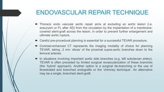
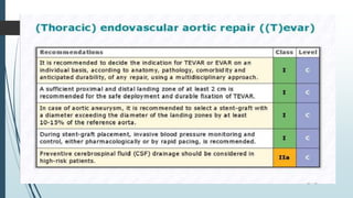
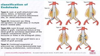
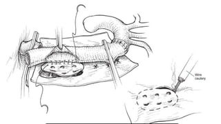
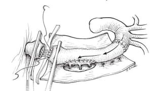
SlideShare a Scribd company logo
THORACOABDOMINAL
AORTIC ANEURYSM
management
Dr. Dhaval A. Bhimani
ANATOMY
Crawford
classification
1. Type 24 %
2. Type 26 %
3. Type 26 %
4. Type 24 %
ANATOMIC PRINCIPLES
ī‚´ Arises distal to the origin of Left SCA.
ī‚´ TAA span the diaphragmatic hiatus at the level of Tl2
and introduce important considerations for pleural entry,
peritoneal access, and diaphragmatic conservation for
aortic repair.
ī‚´ In addition, phrenic nerve localization and preservation
are imperative within the pericardium as it crosses the
left atrium and terminates distally on the abdominal
surface of the diaphragm.
ī‚´ The critical vascular zone of the spinal cord from T4 to Ll vertebral levels is
characterized by the least prominent blood supply and is the zone at which
interference with the circulation is most likely to result in paraplegia.
ī‚´ The artery of Adamkiewicz is the largest anterior medullary feeder for the
supply of the lumbar cord and arises from the lower intercostal or lumbar
artery on left in 65% to 80% of cases between T6 and 14 levels.
ī‚´ The lower intercostal and lumbar artery supplying the artery of
Adamkiewicz should be preserved or reconstructed to maintain blood
supply to the lumbar spinal cord to minimize the risk of spinal cord injury.
ī‚´ Perfusion through medullary arteries maintains spinal cord blood supply
proximal to the aortic cross-clamp and determines postoperative
neurologic function.
ī‚´ Visceral branches are located on the ventral aorta as the celiac
axis, superior mesenteric, inferior mesenteric, and paired or
multiple renal arteries.
ī‚´ The anterior spinal artery is a principal component of the extensive
intraspinal and paraspinous collateral blood supply to the anterior
spinal cord, with 75% of all segmental arteries providing direct
anterior spinal artery-supplying branches.
ī‚´ This extensive collateral network imparts a responsibility to define
dominant intercostal and lumbar arteries for each individual patient
to guide operative planning for reconstruction and preservation of
blood supply to the spinal cord.
IMAGING, option available ??
ī‚´ Determined by following factors
1. patient-related factors
â€ĸ hemodynamic stability
â€ĸ renal function
â€ĸ contrast allergy
2. institutional capabilities ( technologic capability, expertise).
CT-induced contrast nephropathy and magnetic resonance (MR) associated gadolinium
nephrogenic systemic fibrosis are principal considerations for patients with borderline
kidney function (serum creatinine > 1.8 to 2.0 mg/dl).
Chest X- Ray
ī‚´ specificity for acute aortic pathology of
86% in prospective study of patients
undergoing evaluation for acute thoracic
aortic disease.
ī‚´ However, it is insufficient to definitely
exclude thoracic aortic aneurysm in high-
risk patients and lacks anatomic detail
necessary for directed treatment.
CT angiography
(CTA)
ī‚´ CTA is accepted as the primary
diagnostic imaging modality for the
thoracic aorta with a demonstrated
accuracy of92% for all-inclusive
abnormalities of the thoracic aorta and
has an established efficacy in the
prediction of the need for hypothermic
circulatory arrest during surgical repair.
ī‚´ Image acquisition should include
ascending aorta, aortic arch,thoracic
branch vessels and femoral vessels to
determine aneurysm extension and to
guide potential endovascular access.
MR angiography (MRA)
ī‚´ provides similar advantages to CTA without the limitations of radiation exposure or
iodinated contrast.
ī‚´ Phase-contrast techniques and two-dimensional time-of-flight modalities have
increased the application of MRA in the setting of thoracic aortic aneurysm and
dissection, with beneficial applications in the determination of flow dynamics within
the false channel.
ī‚´ First-line adoption of this imaging modality in the setting of thoracic aortic
aneurysm remains limited by institutional capabilities and the time required for
acquisition.
ī‚´ In addition to providing insight regarding aortic size and anatomic aneurysm
characteristics, both CTA and MRA guide the selection of safe sites for arterial
cannulation and cross clamp application.
Recommendation
Treatment
1. Conservative, medical
2. Endovascular repair
3. Open surgical repair
4. Hybrid approch
Principle of medical therapy
ī‚´ The main aim of medical therapy in this condition is to reduce shear stress on the
diseased segment of the aorta by reducing blood pressure and cardiac
contractility.
ī‚´ Cessation of smoking is important, as studies have shown that self-reported
current smoking induced a signiīŦcantly faster expansion (by approximately 0.4
mm/year).
ī‚´ Moderate physical activity probably prevents the progression of aortic
atherosclerosis but data are sparse.
ī‚´ To prevent blood pressure spikes, competitive sports should be avoided in patients
with an enlarged aorta.
ī‚´ It is mechanistically logical that medical therapy to reduce dP/dt and to control
blood pressure would be beneficial for the treatment of all patients with thoracic
aortic aneurysms.
ī‚´ In a randomized study of adults with Marfan syndrome, Shores et al found that
treatment with propranolol (versus no b-blocker therapy) over 10 years resulted in
a significantly slower rate of aortic dilatation, fewer aortic events, and lower
mortality.
ī‚´ There is some early experimental evidence to suggest that oxidative stress may
play a role in the pathogenesis of atherosclerotic thoracic aortic aneurysms and
that perhaps statin therapy and angiotensin II receptor blocker therapy may
potentially have a protective effect.
ī‚´ Once b- blocker therapy is maximized (or in the event that b-blockers are
contraindicated or not tolerated), any persistent hypertension should be treated
with other antihypertensive agents to bring the blood pressure down to a low-
normal range, eg, a systolic pressure of 105 to 120 mm Hg
ī‚´ Patients should be informed of the typical symptoms of acute aortic dissection.
Moreover, patients should be instructed that should they ever experience the
abrupt onset of significant chest, neck, back, or abdominal pain, they should
present immediately to an emergency department for evaluation.
ī‚´ In addition, patients should be instructed to inform the emergency physician of the
existence of a thoracic aortic aneurysm and explain explicitly that their physician
recommends an urgent CT scan of the chest (or transesophageal
echocardiography or MRI if a CT is contraindicated) to rule out acute aortic
dissection or rupture.
ENDOVASCULAR REPAIR TECHNIQUE
ī‚´ Thoracic endo vascular aortic repair aims at excluding an aortic lesion (i.e.
aneurysm or FL after AD) from the circulation by the implantation of a membrane-
covered stent-graft across the lesion, in order to prevent further enlargement and
ultimate aortic rupture.
ī‚´ Careful pre-procedural planning is essential for a successful TEVAR procedure.
ī‚´ Contrast-enhanced CT represents the imaging modality of choice for planning
TEVAR, taking ,3 mm ‘slices’ of the proximal supra-aortic branches down to the
femoral arteries.
ī‚´ In situations involving important aortic side branches (e.g. left subclavian artery),
TEVAR is often preceded by limited surgical revascularization of these branches
(the ‘hybrid’ approach). Another option is a surgical de-branching or the use of
fenestrated and branched endografts or the ‘chimney technique’. An alternative
may be a single, branched stent-graft.
ī‚´ In high-risk patients, preventive cerebrospinal īŦ‚uid(CSF)drainage can be
beneīŦcial, as it has proven efīŦcacy in spinal cord protection during open
thoraco-abdominal aneurysm surgery.
ī‚´ Reversal of paraplegia can be achieved by the immediate initiation of CSF
drainage and pharmacological elevation of blood pressure to >90 mm Hg
mean arterial pressure.
ī‚´ Hypotensive episodes during the procedure should be avoided.
ī‚´ Follow up Ct scan to be done after 1 week to rule out endoleak.
ī‚´ Type I and Type III
ī‚´Endoleaks are regarded as treatment failures and warrant
further treatment to prevent the continuing risk of rupture.
ī‚´Type II
ī‚´Endoleaks are normally managed conservatively by a ‘wait-and-
watch’ strategy to detect aneurysmal expansion, except for
supra-aortic arteries.
ī‚´Types IV and V
ī‚´Endoleaks are indirect and have a benign course. Treatment is
required in cases of aneurysm expansion.
Surgical management
ī‚´ Anaesthesia
ī‚´ Monitoring
ī‚´ Position
ī‚´ Operative technique
ī‚´ Extracorporeal circulation
ī‚´ Spinal cord protection strategy
Anaesthesia and monitoring
ī‚´ General anaesthesia
ī‚´ Double lumen endotracheal tube.
ī‚´ Monitoring :-
ī‚´ Large-bore peripheral access, central venous pressure monitoring, continuous
arterial pressure assessment, pulse oximetry, and foley catheter are imperative to
guide intraoperative resuscitation and support.
ī‚´ Transesophageal echocardiography, continuous electrocardiographic monitoring,
and a pulmonary artery catheter provide additional adjuncts for intraoperative
cardiac evaluation and are routine in our institution.
ī‚´ Femoral artery access is needed with cardiopulmonary bypass to maintain
balanced pressures.
ī‚´ Temperature monitoring is achieved at two access sites to estimate
cerebral (blood, esophageal, tympanic membrane, nasopharynx) and
visceral (bladder, rectal) temperatures.
ī‚´ Cerebrospinal fluid (CSF) drainage is the principal modality for spinal cord
protection , due to the established benefit and low-risk of this protective
strategy.
ī‚´ Motor-evoked potential (MEP) and somato sensory evoked potential
(SSEP) monitoring may be applied in addition to CSF drainage for spinal
cord protection and monitoring.
Incision and Exposure: Thoracic
Aneurysms
ī‚´ An extended posterolateral thoracotomy is performed to expose the entire
length of the thoracic aorta.
ī‚´ The latissimus dorsi and a minimal portion of the serratus anterior muscles
are divided.
ī‚´ Aneurysms of the upper or middle thoracic aorta are accessed through a
single intercostal space, which may be facilitated by division of one of the
ribs posteriorly.
ī‚´ Two separate sites of entry through the fourth interspace and the seventh
or eighth interspace for more extensive descending thoracoabdominal
aneurysms can be obtained and recommended.
ī‚´ The fourth interspace is critical to proximal clamp application.
ī‚´ The thoracic duct and adjacent lymph vessels should be avoided. Intraoperative
lymph leaks should be immediately repaired.
ī‚´ The identification of the esophagus may be augmented by the direct palpation of
the nasogastric tube or transesophageal echocardiography scope to avoid
inadvertent injury.
ī‚´ FOR thoracoabdominal aneurysm-
ī‚´ Incision is started posterolaterally over the ribs of the seventh, eighth, or ninth
interspace dependent on the proximal extent of the aneurysm.
ī‚´ The incision is then advanced across the ninth interspace at the costal margin to
curve inferiorly to run parallel and left-lateral to the midline and rectus sheath.
ī‚´ The abdominal muscles are divided and the peritoneum is preserved. Special care
is needed at the lateral edge of the rectus abdominis muscle where the peritoneum
and transversalis fascia are closely apposed to the abdominal wall.
ī‚´ The diaphragm may be taken down with a
curved incision along the costal margin with
care to preserve a 3-cm rim along the
posterior aspect of the rib.
ī‚´ The tendinous center of the diaphragm is
then conserved when anatomically feasible
to improve postoperative respiratory recovery
and weaning time.
ī‚´ This limited phrenotomy technique allows
passage of the graft through the natural
hiatus of the diaphragm.
ī‚´ The incision is continued down to the crura,
and the left crus may be divided to expose
the aorta beneath.
ī‚´ A radial diaphragmatic incision may also be
utilized.
ī‚´ Progressive retraction of the peritoneum and its contents will facilitate exposure of
the retroperitoneum and a self-retaining retractor is necessary.
ī‚´ The aorta is located medial to the iliopsoas muscle.
ī‚´ Mobilization of the left kidney from the bed of the psoas muscle may provide
additional exposure.
ī‚´ Importantly, this step is deferred in patients with a retroaortic left renal vein.
Anterior visceral branches are identified and sharply dissected with attention to
mobilization should reimplantation or bypass be required.
ī‚´ Dissection is performed proximal and distal to the aneurysmal segment of aorta
and each site is tapped in preparation for cross-clamping.
Hemodynamic Support during Aortic
Cross-Clamping
ī‚´ Proximal aortic cross-clamp application induces a significant
increase in cardiac afterload.
ī‚´ Sudden afterload reduction following clamp release is
associated with an acute relative hypovolemia and systemic
hypotension.
Strategy:-
ī‚´ Approach to cross-clamp application and distal aortic perfusion involves active
distal aortic perfusion with femoral-to-femoral bypass for all but type IV and V
aortic aneurysms, for which we can minimize supraceliac clamp times with no
active distal perfusion.
ī‚´ maintenance of distal aortic blood pressure is imperative and is achieved through
the titration of pharmacologic agents and pump flow rates to maintain optimal
perfusion.
ī‚´ Infusion of nitroglycerine, trimethaphan, or sodium nitroprusside prior to the
application of the aortic cross-clamp may be performed to lower systolic blood
pressure to 70 to 80 mmHg.
ī‚´ Sodium bicarbonate, calcium, and rapid volume infusion may be initiated prior to
unclamping to prevent acute hypotension.
ī‚´ Vasopressor agents may also be utilized to augment these resuscitative measures
to maintain blood pressure upon clamp release.
ī‚´ Progressive clamp application and release over a period of 2 to 4 minutes may
blunt the imposed physiologic insult and hemodynamic response.
ī‚´ In assessment of end-organ ischemia, cross-clamp time is measured from the first
click of clamp application until its complete release.
Extracorporeal Circulation
ī‚´ Extracorporeal circulation support provides after load reduction and
continuous end-organ perfusion during the aortic cross-clamp period.
ī‚´ Techniques for the maintenance of extracorporeal circulation include
īƒ˜ passive aorto-aortic shunt,
īƒ˜ left atriofemoral bypass, and
īƒ˜ femorofemoral cardiopulmonary bypass.
Spinal Cord Protection
ī‚´ Central to methods of cord protection is an understanding of the axial network and
the supplying segmental, subclavian, and hypogastric arteries and the protective
effect of permissive hypothermia on neural tissue.
ī‚´ This collateral network may increase flow through alternative routes when another
is reduced; however, this network may also result in steal that will result in
decreased nutrient flow to the cord.
ī‚´ Steal may occur secondary to the absence of visceral and iliac artery perfusion,
during the cross-clamp period, or as a result of pharmacologically induced
arteriovenous shunting when bleeding intercostals into an excluded aortic
segment.
ī‚´ liberal use of multi head
perfusion cannulae and
pediatric coronary sinus
catheters for the maintenance
of perfusion to the visceral and
subclavian aortic branches and
dominant intercostal arteries,
respectively.
ī‚´ The detection of potentially reversible delayed paraplegia may be achieved by
MEP and SSEP monitoring with immediate assessment of function postoperatively.
ī‚´ The adoption of the following provocative techniques for reduction of spinal cord
ischemic-induced injury has resulted in a significant decrease in the incidence of
paralysis following open repair that is dependent on the extent of repair.
ī‚´ 15% for type I,
ī‚´ 30% for type II,
ī‚´ 7% for type III, and
ī‚´ 4% for type Iv.
Predictors of delayed neurologic deficit
ī‚´ emergent operative status,
ī‚´ prolonged aortic cross-clamp time,
ī‚´ level of cross-clamp, hypogastric artery exclusion,
ī‚´ aortic rupture,
ī‚´ preoperative renal dysfunction,
ī‚´ prior abdominal aortic aneurysm repair,
ī‚´ acute dissection, and
ī‚´ extent II involvement.
Strategies
īļ Distal aortic perfusion.
īļ Lower body pressure monitoring is imperative to maintain appropriate cord
perfusion which may necessitate flow rates as high as 3 to 3.5 L/min.
īļ Intrathecal vasodilators and topical cooling.
īļ Experimental porcine studies have established the protective effect of
intrathecal administration of vasodilators during the aortic cross-clamp period.
This technique dilates spinal arteries and prevents spasm and may be
combined with local cooling of the cord with 4°C saline to extend the potential
aortic cross-clamp time.
īļ Topical cooling of the spinal cord is associated with increases in CSF pressure;
however, this technique has proven to be a protective adjunct in the reduction
of postoperative paralysis and paraplegia.
īļ Cerebrospinal fluid drainage.
īļ In randomized prospective study, the incidence of paraplegia or
paraparesis was reduced with CSF drainage from 13.0% to 2.6%, P =
0.03.
īļ CSF pressure is maintained < 10 mmHg with mean perfusion
pressures of 85 to 90 mmHg to enhance spinal cord perfusion.
īļ Lumbar drains are generally removed on postoperative day 2 in
patients without neurologic deficit.
īļ Preference in high-risk patients is to leave the lumbar drain inplace for
72 hours, while in select low risk patients one can consider removal at
24 hours.
īļ Reattachment of intercostal and lumbar arteries.
īļ acceptable rates of paralysis and paraplegia following sacrifice without
reimplantation of as many as 15 intercostal and lumbar arteries during
thoracoabdominal aneurysm reconstruction.
īļ Prompt ligation of nonimplanted intercostal arteries is recommended to
avoid steal from the cord circulation.
īļ While multimodality approaches have implemented complete intercostal
reimplantation.
īļ Hypothermia.
īļ Moderate (29 to 32°C) to profound (20%) hypothermia is associated with
improved outcomes following thoracoabdominal and descending aortic
operations.
īļ The proposed mechanism of hypothermia-induced protection involves the
reduction of excitatory neurotransmitter release, decreased free oxygen radical
production, decreased postischemic edema, and stabilized central nervous
system blood flow.
īļ Pharmacologic agents.
īļ Mannitol (0.25 to 1.0 g/kg), high-dose barbiturates, methylprednisolone (30
mg/kg), calcium-channel blockers, adenosine 2A agonist, naloxone, and local
anesthetic agents have a demonstrated efficacy in experimental models of
spinal cord ischemia and multimodality clinical protocols.
īļ The proposed mechanism of spinal cord protection involves decreased spinal
cord edema and improved free oxygen radical scavenging
īļ Minimizing aortic cross-clamp times.
RECOMMENDATION
TECHNIQUE FOR AORTIC ANASTOMOSES
ī‚´ TUBE GRAFT PLACEMENT & REIMPLANTATION OF INTERCOSTAL ARTERIES
REIMPLANTATION OF VISERAL
ARTERIES
HYBRID APPROCH
ī‚´ Hybrid techniques that incorporate open and endovascular repair
techniques provide a promising approach to treatment in patients with
complex thoracic aortic aneurysms with or without concomitant dissection.
ī‚´ In patients with descending thoracic aortic aneurysms with proximal
extension into the aortic arch, we can successfully performed open arch
debranching and revascularization of branch vessels, followed by TEVAR
of the arch and descending thoracic aorta.
ī‚´ Similaraly, TAAA extending and involving viseral vessel, hybrid approch
can be tried.
SPECIAL FEATURES OF
POSTOPERATIVE CARE
ī‚´ Particular attention is paid to maintaining adequate ventricular preload by
volume infusions.
ī‚´ Amount of chest drainage is closely monitored.
ī‚´ close attention to pulmonary subsystem management is mandatory in
thoracotomy involving cases.
ī‚´ Prompt initiation of cerebrospinal
ī‚´ fluid drainage may, in some instances, reverse the neurologic deficit, and if
a drain is not already in place, it should be inserted when signs of spinal
cord ischemia develop postoperatively.
ī‚´ Avoiding prolonged periods of hypotension is also essential to
ensure optimal spinal cord perfusion.
FOLLOW-UP RECOMMENDATIONS
ī‚´ The current standard for postoperative endovascular and open
descending thoracic and thoracoabdominal aortic aneurysm
repair involves
ī‚´ physical examination,
ī‚´ Repeat imaging with CTA chest and chest radiography at 1, 6, and 12 months.
ī‚´ The initial interval to postoperative evaluation may be modified as determined by
the immediate in-hospital postoperative course.
Literature rerview
ī‚´What to look for
1. In hospital mortality
2. Morbidity
3. Type of different strategy developed over years.
LITERATURE
THANK YOU

More Related Content

What's hot

Aorto pulmonary window may 2021
Aorto   pulmonary window  may 2021Aorto   pulmonary window  may 2021
Aorto pulmonary window may 2021
rajasthan govt
 
Femoral Site Complications
Femoral Site ComplicationsFemoral Site Complications
Femoral Site Complications
Cathy Lewis
 
Vsd device closure
Vsd device closureVsd device closure
Vsd device closure
Pavan Rasalkar
 
Bypass graft intervention2
Bypass graft intervention2Bypass graft intervention2
Bypass graft intervention2
Dr Virbhan Balai
 
TAVR/TAVI/TRANCATHETER SAORTIC VALVE REPLECEMENT
TAVR/TAVI/TRANCATHETER SAORTIC VALVE REPLECEMENTTAVR/TAVI/TRANCATHETER SAORTIC VALVE REPLECEMENT
TAVR/TAVI/TRANCATHETER SAORTIC VALVE REPLECEMENT
Dr Virbhan Balai
 
Vascular closure devices
Vascular closure devicesVascular closure devices
Vascular closure devices
Dr Siva subramaniyan
 
Revisiting bentall procedure
Revisiting bentall procedureRevisiting bentall procedure
Revisiting bentall procedure
Dicky A Wartono
 
Indications and timing of intervention in congenital heart disease
Indications and timing of intervention in congenital heart diseaseIndications and timing of intervention in congenital heart disease
Indications and timing of intervention in congenital heart disease
Ramachandra Barik
 
Management of Small Aortic Root
Management of Small Aortic RootManagement of Small Aortic Root
Management of Small Aortic Root
DR.NABAJYOTI HAZARIKA
 
PVBD
PVBDPVBD
Intracoronary optical coherence tomography
Intracoronary optical coherence tomographyIntracoronary optical coherence tomography
Intracoronary optical coherence tomography
Ramachandra Barik
 
Vascular Closure Devices
Vascular Closure DevicesVascular Closure Devices
Vascular Closure Devices
Asad Moosa
 
Seminar congenital cardiac disorders (pda,TA and AP Window)
Seminar congenital cardiac disorders (pda,TA and AP Window)Seminar congenital cardiac disorders (pda,TA and AP Window)
Seminar congenital cardiac disorders (pda,TA and AP Window)Uma Binoy
 
AORTIC ARCH SURGERY
AORTIC ARCH SURGERYAORTIC ARCH SURGERY
AORTIC ARCH SURGERYDicky A Wartono
 
Groin management 2013
Groin management 2013Groin management 2013
Groin management 2013
Cardiac Cath Lab Information
 
CORONARY BALLOONS PRACTICAL ASPECTS.pptx
CORONARY BALLOONS PRACTICAL ASPECTS.pptxCORONARY BALLOONS PRACTICAL ASPECTS.pptx
CORONARY BALLOONS PRACTICAL ASPECTS.pptx
RohitWalse2
 
FFR(fractional flow reserve)
FFR(fractional flow reserve)FFR(fractional flow reserve)
FFR(fractional flow reserve)
DIPAK PATADE
 
Asd ppt
Asd pptAsd ppt
Step by Step Rotational Athrectomy
Step by Step Rotational AthrectomyStep by Step Rotational Athrectomy
Step by Step Rotational Athrectomy
Dr Virbhan Balai
 

What's hot (20)

Aorto pulmonary window may 2021
Aorto   pulmonary window  may 2021Aorto   pulmonary window  may 2021
Aorto pulmonary window may 2021
 
Tevar
TevarTevar
Tevar
 
Femoral Site Complications
Femoral Site ComplicationsFemoral Site Complications
Femoral Site Complications
 
Vsd device closure
Vsd device closureVsd device closure
Vsd device closure
 
Bypass graft intervention2
Bypass graft intervention2Bypass graft intervention2
Bypass graft intervention2
 
TAVR/TAVI/TRANCATHETER SAORTIC VALVE REPLECEMENT
TAVR/TAVI/TRANCATHETER SAORTIC VALVE REPLECEMENTTAVR/TAVI/TRANCATHETER SAORTIC VALVE REPLECEMENT
TAVR/TAVI/TRANCATHETER SAORTIC VALVE REPLECEMENT
 
Vascular closure devices
Vascular closure devicesVascular closure devices
Vascular closure devices
 
Revisiting bentall procedure
Revisiting bentall procedureRevisiting bentall procedure
Revisiting bentall procedure
 
Indications and timing of intervention in congenital heart disease
Indications and timing of intervention in congenital heart diseaseIndications and timing of intervention in congenital heart disease
Indications and timing of intervention in congenital heart disease
 
Management of Small Aortic Root
Management of Small Aortic RootManagement of Small Aortic Root
Management of Small Aortic Root
 
PVBD
PVBDPVBD
PVBD
 
Intracoronary optical coherence tomography
Intracoronary optical coherence tomographyIntracoronary optical coherence tomography
Intracoronary optical coherence tomography
 
Vascular Closure Devices
Vascular Closure DevicesVascular Closure Devices
Vascular Closure Devices
 
Seminar congenital cardiac disorders (pda,TA and AP Window)
Seminar congenital cardiac disorders (pda,TA and AP Window)Seminar congenital cardiac disorders (pda,TA and AP Window)
Seminar congenital cardiac disorders (pda,TA and AP Window)
 
AORTIC ARCH SURGERY
AORTIC ARCH SURGERYAORTIC ARCH SURGERY
AORTIC ARCH SURGERY
 
Groin management 2013
Groin management 2013Groin management 2013
Groin management 2013
 
CORONARY BALLOONS PRACTICAL ASPECTS.pptx
CORONARY BALLOONS PRACTICAL ASPECTS.pptxCORONARY BALLOONS PRACTICAL ASPECTS.pptx
CORONARY BALLOONS PRACTICAL ASPECTS.pptx
 
FFR(fractional flow reserve)
FFR(fractional flow reserve)FFR(fractional flow reserve)
FFR(fractional flow reserve)
 
Asd ppt
Asd pptAsd ppt
Asd ppt
 
Step by Step Rotational Athrectomy
Step by Step Rotational AthrectomyStep by Step Rotational Athrectomy
Step by Step Rotational Athrectomy
 

Similar to Thoraco abdominal aortic aneurysm(TAAA) management

09 hashem et al
09 hashem et al09 hashem et al
09 hashem et al
Jahangir Alam
 
Mediastinal injury (PNEUMOMEdIASTINUM)- Bhadra.pptx
Mediastinal injury (PNEUMOMEdIASTINUM)- Bhadra.pptxMediastinal injury (PNEUMOMEdIASTINUM)- Bhadra.pptx
Mediastinal injury (PNEUMOMEdIASTINUM)- Bhadra.pptx
VigneshSNair3
 
External hemorrhage
External hemorrhage External hemorrhage
External hemorrhage
BEDEER ELSHERBINY
 
Blunt Thoracic Aortic Injury BTAI
Blunt Thoracic Aortic Injury BTAIBlunt Thoracic Aortic Injury BTAI
Blunt Thoracic Aortic Injury BTAI
KHALID ALRAJHI
 
SURGICAL MANAGEMENT of thoracic abdominal aortic aneurysms.pptx
SURGICAL MANAGEMENT of thoracic abdominal aortic aneurysms.pptxSURGICAL MANAGEMENT of thoracic abdominal aortic aneurysms.pptx
SURGICAL MANAGEMENT of thoracic abdominal aortic aneurysms.pptx
Jaydeep Malakar
 
Aortic Diseases Ain Shams Post graduate CTS Course
Aortic Diseases Ain Shams Post graduate CTS CourseAortic Diseases Ain Shams Post graduate CTS Course
Aortic Diseases Ain Shams Post graduate CTS Course
Mohammed Nabil Abd al jawad
 
TEVAR.pptx
TEVAR.pptxTEVAR.pptx
TEVAR.pptx
Hossam atef
 
Transcatheter therapy for mitral regurgitation
Transcatheter therapy for mitral regurgitationTranscatheter therapy for mitral regurgitation
Transcatheter therapy for mitral regurgitation
SR,CARDIOLOGY,JIPMER,PUDUCHERRY
 
aneurysm.pptx
aneurysm.pptxaneurysm.pptx
aneurysm.pptx
SubhashreeMahapatro
 
Shock in Trauma.pptx
Shock in Trauma.pptxShock in Trauma.pptx
Shock in Trauma.pptx
prabhatbhati3
 
Aortic Aneurysm: Diagnosis, Management, Exercise Testing, And Training
Aortic Aneurysm: Diagnosis, Management, Exercise Testing, And TrainingAortic Aneurysm: Diagnosis, Management, Exercise Testing, And Training
Aortic Aneurysm: Diagnosis, Management, Exercise Testing, And Training
Javidsultandar
 
Stanford Type A Aortic Dissection: a Complex Disease for Patients and Cardiot...
Stanford Type A Aortic Dissection: a Complex Disease for Patients and Cardiot...Stanford Type A Aortic Dissection: a Complex Disease for Patients and Cardiot...
Stanford Type A Aortic Dissection: a Complex Disease for Patients and Cardiot...
Crimsonpublisherssmoaj
 
mediastinum-1.pptx
mediastinum-1.pptxmediastinum-1.pptx
mediastinum-1.pptx
FlyHeyjude
 
A Complex Case Of Polianeurysmatic Disease
A Complex Case Of Polianeurysmatic DiseaseA Complex Case Of Polianeurysmatic Disease
A Complex Case Of Polianeurysmatic Disease
Salvatore Ronsivalle
 
Thoraco Abdominal Aortic Aneurysm technique for present ok.pptx
Thoraco Abdominal Aortic Aneurysm technique for present  ok.pptxThoraco Abdominal Aortic Aneurysm technique for present  ok.pptx
Thoraco Abdominal Aortic Aneurysm technique for present ok.pptx
Peter Flash
 
Carotid artery disease
Carotid artery diseaseCarotid artery disease
Carotid artery disease
Blerim Ademi
 
Hemodynamic monitoring- Dr Sandeep Gampa
Hemodynamic monitoring- Dr Sandeep GampaHemodynamic monitoring- Dr Sandeep Gampa
Hemodynamic monitoring- Dr Sandeep GampaSiddharth Pandey
 
New microsoft power point presentation
New microsoft power point presentationNew microsoft power point presentation
New microsoft power point presentation
mohammed Assuit)
 
AORTIC DISSECTION and management of aortic dissection
AORTIC DISSECTION and management of aortic dissectionAORTIC DISSECTION and management of aortic dissection
AORTIC DISSECTION and management of aortic dissection
drhanifmohdali
 

Similar to Thoraco abdominal aortic aneurysm(TAAA) management (20)

09 hashem et al
09 hashem et al09 hashem et al
09 hashem et al
 
Mediastinal injury (PNEUMOMEdIASTINUM)- Bhadra.pptx
Mediastinal injury (PNEUMOMEdIASTINUM)- Bhadra.pptxMediastinal injury (PNEUMOMEdIASTINUM)- Bhadra.pptx
Mediastinal injury (PNEUMOMEdIASTINUM)- Bhadra.pptx
 
External hemorrhage
External hemorrhage External hemorrhage
External hemorrhage
 
Blunt Thoracic Aortic Injury BTAI
Blunt Thoracic Aortic Injury BTAIBlunt Thoracic Aortic Injury BTAI
Blunt Thoracic Aortic Injury BTAI
 
SURGICAL MANAGEMENT of thoracic abdominal aortic aneurysms.pptx
SURGICAL MANAGEMENT of thoracic abdominal aortic aneurysms.pptxSURGICAL MANAGEMENT of thoracic abdominal aortic aneurysms.pptx
SURGICAL MANAGEMENT of thoracic abdominal aortic aneurysms.pptx
 
Aortic Diseases Ain Shams Post graduate CTS Course
Aortic Diseases Ain Shams Post graduate CTS CourseAortic Diseases Ain Shams Post graduate CTS Course
Aortic Diseases Ain Shams Post graduate CTS Course
 
TEVAR.pptx
TEVAR.pptxTEVAR.pptx
TEVAR.pptx
 
Transcatheter therapy for mitral regurgitation
Transcatheter therapy for mitral regurgitationTranscatheter therapy for mitral regurgitation
Transcatheter therapy for mitral regurgitation
 
aneurysm.pptx
aneurysm.pptxaneurysm.pptx
aneurysm.pptx
 
Shock in Trauma.pptx
Shock in Trauma.pptxShock in Trauma.pptx
Shock in Trauma.pptx
 
Aortic Aneurysm: Diagnosis, Management, Exercise Testing, And Training
Aortic Aneurysm: Diagnosis, Management, Exercise Testing, And TrainingAortic Aneurysm: Diagnosis, Management, Exercise Testing, And Training
Aortic Aneurysm: Diagnosis, Management, Exercise Testing, And Training
 
Stanford Type A Aortic Dissection: a Complex Disease for Patients and Cardiot...
Stanford Type A Aortic Dissection: a Complex Disease for Patients and Cardiot...Stanford Type A Aortic Dissection: a Complex Disease for Patients and Cardiot...
Stanford Type A Aortic Dissection: a Complex Disease for Patients and Cardiot...
 
JET surgical substrates
JET surgical substratesJET surgical substrates
JET surgical substrates
 
mediastinum-1.pptx
mediastinum-1.pptxmediastinum-1.pptx
mediastinum-1.pptx
 
A Complex Case Of Polianeurysmatic Disease
A Complex Case Of Polianeurysmatic DiseaseA Complex Case Of Polianeurysmatic Disease
A Complex Case Of Polianeurysmatic Disease
 
Thoraco Abdominal Aortic Aneurysm technique for present ok.pptx
Thoraco Abdominal Aortic Aneurysm technique for present  ok.pptxThoraco Abdominal Aortic Aneurysm technique for present  ok.pptx
Thoraco Abdominal Aortic Aneurysm technique for present ok.pptx
 
Carotid artery disease
Carotid artery diseaseCarotid artery disease
Carotid artery disease
 
Hemodynamic monitoring- Dr Sandeep Gampa
Hemodynamic monitoring- Dr Sandeep GampaHemodynamic monitoring- Dr Sandeep Gampa
Hemodynamic monitoring- Dr Sandeep Gampa
 
New microsoft power point presentation
New microsoft power point presentationNew microsoft power point presentation
New microsoft power point presentation
 
AORTIC DISSECTION and management of aortic dissection
AORTIC DISSECTION and management of aortic dissectionAORTIC DISSECTION and management of aortic dissection
AORTIC DISSECTION and management of aortic dissection
 

More from Dhaval Bhimani

Total parentral nutrition in cardiac surgery
Total parentral nutrition in cardiac surgeryTotal parentral nutrition in cardiac surgery
Total parentral nutrition in cardiac surgery
Dhaval Bhimani
 
HOCM(hypertrophic obstructive cardiomyopathy)
HOCM(hypertrophic obstructive cardiomyopathy)HOCM(hypertrophic obstructive cardiomyopathy)
HOCM(hypertrophic obstructive cardiomyopathy)
Dhaval Bhimani
 
Infective Endocarditis- surgical indication & principle of surgery
Infective Endocarditis- surgical indication & principle of surgeryInfective Endocarditis- surgical indication & principle of surgery
Infective Endocarditis- surgical indication & principle of surgery
Dhaval Bhimani
 
Neurocognitive function in on pump vs off pump CABG
Neurocognitive function in on pump vs off pump CABGNeurocognitive function in on pump vs off pump CABG
Neurocognitive function in on pump vs off pump CABG
Dhaval Bhimani
 
pathophysiology of Left to right shunt
pathophysiology of Left to right shuntpathophysiology of Left to right shunt
pathophysiology of Left to right shunt
Dhaval Bhimani
 
Av canal defect
Av canal defectAv canal defect
Av canal defect
Dhaval Bhimani
 

More from Dhaval Bhimani (6)

Total parentral nutrition in cardiac surgery
Total parentral nutrition in cardiac surgeryTotal parentral nutrition in cardiac surgery
Total parentral nutrition in cardiac surgery
 
HOCM(hypertrophic obstructive cardiomyopathy)
HOCM(hypertrophic obstructive cardiomyopathy)HOCM(hypertrophic obstructive cardiomyopathy)
HOCM(hypertrophic obstructive cardiomyopathy)
 
Infective Endocarditis- surgical indication & principle of surgery
Infective Endocarditis- surgical indication & principle of surgeryInfective Endocarditis- surgical indication & principle of surgery
Infective Endocarditis- surgical indication & principle of surgery
 
Neurocognitive function in on pump vs off pump CABG
Neurocognitive function in on pump vs off pump CABGNeurocognitive function in on pump vs off pump CABG
Neurocognitive function in on pump vs off pump CABG
 
pathophysiology of Left to right shunt
pathophysiology of Left to right shuntpathophysiology of Left to right shunt
pathophysiology of Left to right shunt
 
Av canal defect
Av canal defectAv canal defect
Av canal defect
 

Recently uploaded

Dimensions of Healthcare Quality
Dimensions of Healthcare QualityDimensions of Healthcare Quality
Dimensions of Healthcare Quality
Naeemshahzad51
 
POLYCYSTIC OVARIAN SYNDROME (PCOS)......
POLYCYSTIC OVARIAN SYNDROME (PCOS)......POLYCYSTIC OVARIAN SYNDROME (PCOS)......
POLYCYSTIC OVARIAN SYNDROME (PCOS)......
Ameena Kadar
 
💘Ludhiana ℂall Girls 📞]][89011★83002][[ 📱 ❤ESCORTS service in Ludhiana💃đŸ’ĻLudhi...
💘Ludhiana ℂall Girls 📞]][89011★83002][[ 📱 ❤ESCORTS service in Ludhiana💃đŸ’ĻLudhi...💘Ludhiana ℂall Girls 📞]][89011★83002][[ 📱 ❤ESCORTS service in Ludhiana💃đŸ’ĻLudhi...
💘Ludhiana ℂall Girls 📞]][89011★83002][[ 📱 ❤ESCORTS service in Ludhiana💃đŸ’ĻLudhi...
ranishasharma67
 
Artificial Intelligence to Optimize Cardiovascular Therapy
Artificial Intelligence to Optimize Cardiovascular TherapyArtificial Intelligence to Optimize Cardiovascular Therapy
Artificial Intelligence to Optimize Cardiovascular Therapy
Iris Thiele Isip-Tan
 
Demystifying-Gene-Editing-The-Promise-and-Peril-of-CRISPR.pdf
Demystifying-Gene-Editing-The-Promise-and-Peril-of-CRISPR.pdfDemystifying-Gene-Editing-The-Promise-and-Peril-of-CRISPR.pdf
Demystifying-Gene-Editing-The-Promise-and-Peril-of-CRISPR.pdf
SasikiranMarri
 
How many patients does case series should have In comparison to case reports.pdf
How many patients does case series should have In comparison to case reports.pdfHow many patients does case series should have In comparison to case reports.pdf
How many patients does case series should have In comparison to case reports.pdf
pubrica101
 
India Clinical Trials Market: Industry Size and Growth Trends [2030] Analyzed...
India Clinical Trials Market: Industry Size and Growth Trends [2030] Analyzed...India Clinical Trials Market: Industry Size and Growth Trends [2030] Analyzed...
India Clinical Trials Market: Industry Size and Growth Trends [2030] Analyzed...
Kumar Satyam
 
Navigating Healthcare with Telemedicine
Navigating Healthcare with  TelemedicineNavigating Healthcare with  Telemedicine
Navigating Healthcare with Telemedicine
Iris Thiele Isip-Tan
 
ICH Guidelines for Pharmacovigilance.pdf
ICH Guidelines for Pharmacovigilance.pdfICH Guidelines for Pharmacovigilance.pdf
ICH Guidelines for Pharmacovigilance.pdf
NEHA GUPTA
 
Myopia Management & Control Strategies.pptx
Myopia Management & Control Strategies.pptxMyopia Management & Control Strategies.pptx
Myopia Management & Control Strategies.pptx
RitonDeb1
 
CANCER CANCER CANCER CANCER CANCER CANCER
CANCER  CANCER  CANCER  CANCER  CANCER CANCERCANCER  CANCER  CANCER  CANCER  CANCER CANCER
CANCER CANCER CANCER CANCER CANCER CANCER
KRISTELLEGAMBOA2
 
CHAPTER 1 SEMESTER V PREVENTIVE-PEDIATRICS.pdf
CHAPTER 1 SEMESTER V PREVENTIVE-PEDIATRICS.pdfCHAPTER 1 SEMESTER V PREVENTIVE-PEDIATRICS.pdf
CHAPTER 1 SEMESTER V PREVENTIVE-PEDIATRICS.pdf
Sachin Sharma
 
Essential Metrics for Palliative Care Management
Essential Metrics for Palliative Care ManagementEssential Metrics for Palliative Care Management
Essential Metrics for Palliative Care Management
Care Coordinations
 
GLOBAL WARMING BY PRIYA BHOJWANI @..pptx
GLOBAL WARMING BY PRIYA BHOJWANI @..pptxGLOBAL WARMING BY PRIYA BHOJWANI @..pptx
GLOBAL WARMING BY PRIYA BHOJWANI @..pptx
priyabhojwani1200
 
BOWEL ELIMINATION BY ANUSHRI SRIVASTAVA.pptx
BOWEL ELIMINATION BY ANUSHRI SRIVASTAVA.pptxBOWEL ELIMINATION BY ANUSHRI SRIVASTAVA.pptx
BOWEL ELIMINATION BY ANUSHRI SRIVASTAVA.pptx
AnushriSrivastav
 
The Importance of Community Nursing Care.pdf
The Importance of Community Nursing Care.pdfThe Importance of Community Nursing Care.pdf
The Importance of Community Nursing Care.pdf
AD Healthcare
 
Global launch of the Healthy Ageing and Prevention Index 2nd wave – alongside...
Global launch of the Healthy Ageing and Prevention Index 2nd wave – alongside...Global launch of the Healthy Ageing and Prevention Index 2nd wave – alongside...
Global launch of the Healthy Ageing and Prevention Index 2nd wave – alongside...
ILC- UK
 
Antibiotic Stewardship by Anushri Srivastava.pptx
Antibiotic Stewardship by Anushri Srivastava.pptxAntibiotic Stewardship by Anushri Srivastava.pptx
Antibiotic Stewardship by Anushri Srivastava.pptx
AnushriSrivastav
 
A Community health , health for prisoners
A Community health  , health for prisonersA Community health  , health for prisoners
A Community health , health for prisoners
Ahmed Elmi
 
ventilator, child on ventilator, newborn
ventilator, child on ventilator, newbornventilator, child on ventilator, newborn
ventilator, child on ventilator, newborn
Pooja Rani
 

Recently uploaded (20)

Dimensions of Healthcare Quality
Dimensions of Healthcare QualityDimensions of Healthcare Quality
Dimensions of Healthcare Quality
 
POLYCYSTIC OVARIAN SYNDROME (PCOS)......
POLYCYSTIC OVARIAN SYNDROME (PCOS)......POLYCYSTIC OVARIAN SYNDROME (PCOS)......
POLYCYSTIC OVARIAN SYNDROME (PCOS)......
 
💘Ludhiana ℂall Girls 📞]][89011★83002][[ 📱 ❤ESCORTS service in Ludhiana💃đŸ’ĻLudhi...
💘Ludhiana ℂall Girls 📞]][89011★83002][[ 📱 ❤ESCORTS service in Ludhiana💃đŸ’ĻLudhi...💘Ludhiana ℂall Girls 📞]][89011★83002][[ 📱 ❤ESCORTS service in Ludhiana💃đŸ’ĻLudhi...
💘Ludhiana ℂall Girls 📞]][89011★83002][[ 📱 ❤ESCORTS service in Ludhiana💃đŸ’ĻLudhi...
 
Artificial Intelligence to Optimize Cardiovascular Therapy
Artificial Intelligence to Optimize Cardiovascular TherapyArtificial Intelligence to Optimize Cardiovascular Therapy
Artificial Intelligence to Optimize Cardiovascular Therapy
 
Demystifying-Gene-Editing-The-Promise-and-Peril-of-CRISPR.pdf
Demystifying-Gene-Editing-The-Promise-and-Peril-of-CRISPR.pdfDemystifying-Gene-Editing-The-Promise-and-Peril-of-CRISPR.pdf
Demystifying-Gene-Editing-The-Promise-and-Peril-of-CRISPR.pdf
 
How many patients does case series should have In comparison to case reports.pdf
How many patients does case series should have In comparison to case reports.pdfHow many patients does case series should have In comparison to case reports.pdf
How many patients does case series should have In comparison to case reports.pdf
 
India Clinical Trials Market: Industry Size and Growth Trends [2030] Analyzed...
India Clinical Trials Market: Industry Size and Growth Trends [2030] Analyzed...India Clinical Trials Market: Industry Size and Growth Trends [2030] Analyzed...
India Clinical Trials Market: Industry Size and Growth Trends [2030] Analyzed...
 
Navigating Healthcare with Telemedicine
Navigating Healthcare with  TelemedicineNavigating Healthcare with  Telemedicine
Navigating Healthcare with Telemedicine
 
ICH Guidelines for Pharmacovigilance.pdf
ICH Guidelines for Pharmacovigilance.pdfICH Guidelines for Pharmacovigilance.pdf
ICH Guidelines for Pharmacovigilance.pdf
 
Myopia Management & Control Strategies.pptx
Myopia Management & Control Strategies.pptxMyopia Management & Control Strategies.pptx
Myopia Management & Control Strategies.pptx
 
CANCER CANCER CANCER CANCER CANCER CANCER
CANCER  CANCER  CANCER  CANCER  CANCER CANCERCANCER  CANCER  CANCER  CANCER  CANCER CANCER
CANCER CANCER CANCER CANCER CANCER CANCER
 
CHAPTER 1 SEMESTER V PREVENTIVE-PEDIATRICS.pdf
CHAPTER 1 SEMESTER V PREVENTIVE-PEDIATRICS.pdfCHAPTER 1 SEMESTER V PREVENTIVE-PEDIATRICS.pdf
CHAPTER 1 SEMESTER V PREVENTIVE-PEDIATRICS.pdf
 
Essential Metrics for Palliative Care Management
Essential Metrics for Palliative Care ManagementEssential Metrics for Palliative Care Management
Essential Metrics for Palliative Care Management
 
GLOBAL WARMING BY PRIYA BHOJWANI @..pptx
GLOBAL WARMING BY PRIYA BHOJWANI @..pptxGLOBAL WARMING BY PRIYA BHOJWANI @..pptx
GLOBAL WARMING BY PRIYA BHOJWANI @..pptx
 
BOWEL ELIMINATION BY ANUSHRI SRIVASTAVA.pptx
BOWEL ELIMINATION BY ANUSHRI SRIVASTAVA.pptxBOWEL ELIMINATION BY ANUSHRI SRIVASTAVA.pptx
BOWEL ELIMINATION BY ANUSHRI SRIVASTAVA.pptx
 
The Importance of Community Nursing Care.pdf
The Importance of Community Nursing Care.pdfThe Importance of Community Nursing Care.pdf
The Importance of Community Nursing Care.pdf
 
Global launch of the Healthy Ageing and Prevention Index 2nd wave – alongside...
Global launch of the Healthy Ageing and Prevention Index 2nd wave – alongside...Global launch of the Healthy Ageing and Prevention Index 2nd wave – alongside...
Global launch of the Healthy Ageing and Prevention Index 2nd wave – alongside...
 
Antibiotic Stewardship by Anushri Srivastava.pptx
Antibiotic Stewardship by Anushri Srivastava.pptxAntibiotic Stewardship by Anushri Srivastava.pptx
Antibiotic Stewardship by Anushri Srivastava.pptx
 
A Community health , health for prisoners
A Community health  , health for prisonersA Community health  , health for prisoners
A Community health , health for prisoners
 
ventilator, child on ventilator, newborn
ventilator, child on ventilator, newbornventilator, child on ventilator, newborn
ventilator, child on ventilator, newborn
 

Thoraco abdominal aortic aneurysm(TAAA) management

  • 3. Crawford classification 1. Type 24 % 2. Type 26 % 3. Type 26 % 4. Type 24 %
  • 4. ANATOMIC PRINCIPLES ī‚´ Arises distal to the origin of Left SCA. ī‚´ TAA span the diaphragmatic hiatus at the level of Tl2 and introduce important considerations for pleural entry, peritoneal access, and diaphragmatic conservation for aortic repair. ī‚´ In addition, phrenic nerve localization and preservation are imperative within the pericardium as it crosses the left atrium and terminates distally on the abdominal surface of the diaphragm.
  • 5. ī‚´ The critical vascular zone of the spinal cord from T4 to Ll vertebral levels is characterized by the least prominent blood supply and is the zone at which interference with the circulation is most likely to result in paraplegia. ī‚´ The artery of Adamkiewicz is the largest anterior medullary feeder for the supply of the lumbar cord and arises from the lower intercostal or lumbar artery on left in 65% to 80% of cases between T6 and 14 levels. ī‚´ The lower intercostal and lumbar artery supplying the artery of Adamkiewicz should be preserved or reconstructed to maintain blood supply to the lumbar spinal cord to minimize the risk of spinal cord injury. ī‚´ Perfusion through medullary arteries maintains spinal cord blood supply proximal to the aortic cross-clamp and determines postoperative neurologic function.
  • 6. ī‚´ Visceral branches are located on the ventral aorta as the celiac axis, superior mesenteric, inferior mesenteric, and paired or multiple renal arteries. ī‚´ The anterior spinal artery is a principal component of the extensive intraspinal and paraspinous collateral blood supply to the anterior spinal cord, with 75% of all segmental arteries providing direct anterior spinal artery-supplying branches. ī‚´ This extensive collateral network imparts a responsibility to define dominant intercostal and lumbar arteries for each individual patient to guide operative planning for reconstruction and preservation of blood supply to the spinal cord.
  • 7. IMAGING, option available ?? ī‚´ Determined by following factors 1. patient-related factors â€ĸ hemodynamic stability â€ĸ renal function â€ĸ contrast allergy 2. institutional capabilities ( technologic capability, expertise). CT-induced contrast nephropathy and magnetic resonance (MR) associated gadolinium nephrogenic systemic fibrosis are principal considerations for patients with borderline kidney function (serum creatinine > 1.8 to 2.0 mg/dl).
  • 8. Chest X- Ray ī‚´ specificity for acute aortic pathology of 86% in prospective study of patients undergoing evaluation for acute thoracic aortic disease. ī‚´ However, it is insufficient to definitely exclude thoracic aortic aneurysm in high- risk patients and lacks anatomic detail necessary for directed treatment.
  • 9. CT angiography (CTA) ī‚´ CTA is accepted as the primary diagnostic imaging modality for the thoracic aorta with a demonstrated accuracy of92% for all-inclusive abnormalities of the thoracic aorta and has an established efficacy in the prediction of the need for hypothermic circulatory arrest during surgical repair. ī‚´ Image acquisition should include ascending aorta, aortic arch,thoracic branch vessels and femoral vessels to determine aneurysm extension and to guide potential endovascular access.
  • 10.
  • 11. MR angiography (MRA) ī‚´ provides similar advantages to CTA without the limitations of radiation exposure or iodinated contrast. ī‚´ Phase-contrast techniques and two-dimensional time-of-flight modalities have increased the application of MRA in the setting of thoracic aortic aneurysm and dissection, with beneficial applications in the determination of flow dynamics within the false channel. ī‚´ First-line adoption of this imaging modality in the setting of thoracic aortic aneurysm remains limited by institutional capabilities and the time required for acquisition. ī‚´ In addition to providing insight regarding aortic size and anatomic aneurysm characteristics, both CTA and MRA guide the selection of safe sites for arterial cannulation and cross clamp application.
  • 12.
  • 14.
  • 15.
  • 16.
  • 17. Treatment 1. Conservative, medical 2. Endovascular repair 3. Open surgical repair 4. Hybrid approch
  • 18. Principle of medical therapy ī‚´ The main aim of medical therapy in this condition is to reduce shear stress on the diseased segment of the aorta by reducing blood pressure and cardiac contractility. ī‚´ Cessation of smoking is important, as studies have shown that self-reported current smoking induced a signiīŦcantly faster expansion (by approximately 0.4 mm/year). ī‚´ Moderate physical activity probably prevents the progression of aortic atherosclerosis but data are sparse. ī‚´ To prevent blood pressure spikes, competitive sports should be avoided in patients with an enlarged aorta.
  • 19. ī‚´ It is mechanistically logical that medical therapy to reduce dP/dt and to control blood pressure would be beneficial for the treatment of all patients with thoracic aortic aneurysms. ī‚´ In a randomized study of adults with Marfan syndrome, Shores et al found that treatment with propranolol (versus no b-blocker therapy) over 10 years resulted in a significantly slower rate of aortic dilatation, fewer aortic events, and lower mortality. ī‚´ There is some early experimental evidence to suggest that oxidative stress may play a role in the pathogenesis of atherosclerotic thoracic aortic aneurysms and that perhaps statin therapy and angiotensin II receptor blocker therapy may potentially have a protective effect.
  • 20. ī‚´ Once b- blocker therapy is maximized (or in the event that b-blockers are contraindicated or not tolerated), any persistent hypertension should be treated with other antihypertensive agents to bring the blood pressure down to a low- normal range, eg, a systolic pressure of 105 to 120 mm Hg ī‚´ Patients should be informed of the typical symptoms of acute aortic dissection. Moreover, patients should be instructed that should they ever experience the abrupt onset of significant chest, neck, back, or abdominal pain, they should present immediately to an emergency department for evaluation. ī‚´ In addition, patients should be instructed to inform the emergency physician of the existence of a thoracic aortic aneurysm and explain explicitly that their physician recommends an urgent CT scan of the chest (or transesophageal echocardiography or MRI if a CT is contraindicated) to rule out acute aortic dissection or rupture.
  • 21.
  • 22. ENDOVASCULAR REPAIR TECHNIQUE ī‚´ Thoracic endo vascular aortic repair aims at excluding an aortic lesion (i.e. aneurysm or FL after AD) from the circulation by the implantation of a membrane- covered stent-graft across the lesion, in order to prevent further enlargement and ultimate aortic rupture. ī‚´ Careful pre-procedural planning is essential for a successful TEVAR procedure. ī‚´ Contrast-enhanced CT represents the imaging modality of choice for planning TEVAR, taking ,3 mm ‘slices’ of the proximal supra-aortic branches down to the femoral arteries. ī‚´ In situations involving important aortic side branches (e.g. left subclavian artery), TEVAR is often preceded by limited surgical revascularization of these branches (the ‘hybrid’ approach). Another option is a surgical de-branching or the use of fenestrated and branched endografts or the ‘chimney technique’. An alternative may be a single, branched stent-graft.
  • 23.
  • 24. ī‚´ In high-risk patients, preventive cerebrospinal īŦ‚uid(CSF)drainage can be beneīŦcial, as it has proven efīŦcacy in spinal cord protection during open thoraco-abdominal aneurysm surgery. ī‚´ Reversal of paraplegia can be achieved by the immediate initiation of CSF drainage and pharmacological elevation of blood pressure to >90 mm Hg mean arterial pressure. ī‚´ Hypotensive episodes during the procedure should be avoided. ī‚´ Follow up Ct scan to be done after 1 week to rule out endoleak.
  • 25.
  • 26. ī‚´ Type I and Type III ī‚´Endoleaks are regarded as treatment failures and warrant further treatment to prevent the continuing risk of rupture. ī‚´Type II ī‚´Endoleaks are normally managed conservatively by a ‘wait-and- watch’ strategy to detect aneurysmal expansion, except for supra-aortic arteries. ī‚´Types IV and V ī‚´Endoleaks are indirect and have a benign course. Treatment is required in cases of aneurysm expansion.
  • 27. Surgical management ī‚´ Anaesthesia ī‚´ Monitoring ī‚´ Position ī‚´ Operative technique ī‚´ Extracorporeal circulation ī‚´ Spinal cord protection strategy
  • 28. Anaesthesia and monitoring ī‚´ General anaesthesia ī‚´ Double lumen endotracheal tube. ī‚´ Monitoring :- ī‚´ Large-bore peripheral access, central venous pressure monitoring, continuous arterial pressure assessment, pulse oximetry, and foley catheter are imperative to guide intraoperative resuscitation and support. ī‚´ Transesophageal echocardiography, continuous electrocardiographic monitoring, and a pulmonary artery catheter provide additional adjuncts for intraoperative cardiac evaluation and are routine in our institution. ī‚´ Femoral artery access is needed with cardiopulmonary bypass to maintain balanced pressures.
  • 29. ī‚´ Temperature monitoring is achieved at two access sites to estimate cerebral (blood, esophageal, tympanic membrane, nasopharynx) and visceral (bladder, rectal) temperatures. ī‚´ Cerebrospinal fluid (CSF) drainage is the principal modality for spinal cord protection , due to the established benefit and low-risk of this protective strategy. ī‚´ Motor-evoked potential (MEP) and somato sensory evoked potential (SSEP) monitoring may be applied in addition to CSF drainage for spinal cord protection and monitoring.
  • 30. Incision and Exposure: Thoracic Aneurysms ī‚´ An extended posterolateral thoracotomy is performed to expose the entire length of the thoracic aorta. ī‚´ The latissimus dorsi and a minimal portion of the serratus anterior muscles are divided. ī‚´ Aneurysms of the upper or middle thoracic aorta are accessed through a single intercostal space, which may be facilitated by division of one of the ribs posteriorly. ī‚´ Two separate sites of entry through the fourth interspace and the seventh or eighth interspace for more extensive descending thoracoabdominal aneurysms can be obtained and recommended. ī‚´ The fourth interspace is critical to proximal clamp application.
  • 31.
  • 32.
  • 33. ī‚´ The thoracic duct and adjacent lymph vessels should be avoided. Intraoperative lymph leaks should be immediately repaired. ī‚´ The identification of the esophagus may be augmented by the direct palpation of the nasogastric tube or transesophageal echocardiography scope to avoid inadvertent injury. ī‚´ FOR thoracoabdominal aneurysm- ī‚´ Incision is started posterolaterally over the ribs of the seventh, eighth, or ninth interspace dependent on the proximal extent of the aneurysm. ī‚´ The incision is then advanced across the ninth interspace at the costal margin to curve inferiorly to run parallel and left-lateral to the midline and rectus sheath. ī‚´ The abdominal muscles are divided and the peritoneum is preserved. Special care is needed at the lateral edge of the rectus abdominis muscle where the peritoneum and transversalis fascia are closely apposed to the abdominal wall.
  • 34. ī‚´ The diaphragm may be taken down with a curved incision along the costal margin with care to preserve a 3-cm rim along the posterior aspect of the rib. ī‚´ The tendinous center of the diaphragm is then conserved when anatomically feasible to improve postoperative respiratory recovery and weaning time. ī‚´ This limited phrenotomy technique allows passage of the graft through the natural hiatus of the diaphragm. ī‚´ The incision is continued down to the crura, and the left crus may be divided to expose the aorta beneath. ī‚´ A radial diaphragmatic incision may also be utilized.
  • 35. ī‚´ Progressive retraction of the peritoneum and its contents will facilitate exposure of the retroperitoneum and a self-retaining retractor is necessary. ī‚´ The aorta is located medial to the iliopsoas muscle. ī‚´ Mobilization of the left kidney from the bed of the psoas muscle may provide additional exposure. ī‚´ Importantly, this step is deferred in patients with a retroaortic left renal vein. Anterior visceral branches are identified and sharply dissected with attention to mobilization should reimplantation or bypass be required. ī‚´ Dissection is performed proximal and distal to the aneurysmal segment of aorta and each site is tapped in preparation for cross-clamping.
  • 36. Hemodynamic Support during Aortic Cross-Clamping ī‚´ Proximal aortic cross-clamp application induces a significant increase in cardiac afterload. ī‚´ Sudden afterload reduction following clamp release is associated with an acute relative hypovolemia and systemic hypotension.
  • 37. Strategy:- ī‚´ Approach to cross-clamp application and distal aortic perfusion involves active distal aortic perfusion with femoral-to-femoral bypass for all but type IV and V aortic aneurysms, for which we can minimize supraceliac clamp times with no active distal perfusion. ī‚´ maintenance of distal aortic blood pressure is imperative and is achieved through the titration of pharmacologic agents and pump flow rates to maintain optimal perfusion. ī‚´ Infusion of nitroglycerine, trimethaphan, or sodium nitroprusside prior to the application of the aortic cross-clamp may be performed to lower systolic blood pressure to 70 to 80 mmHg.
  • 38. ī‚´ Sodium bicarbonate, calcium, and rapid volume infusion may be initiated prior to unclamping to prevent acute hypotension. ī‚´ Vasopressor agents may also be utilized to augment these resuscitative measures to maintain blood pressure upon clamp release. ī‚´ Progressive clamp application and release over a period of 2 to 4 minutes may blunt the imposed physiologic insult and hemodynamic response. ī‚´ In assessment of end-organ ischemia, cross-clamp time is measured from the first click of clamp application until its complete release.
  • 39. Extracorporeal Circulation ī‚´ Extracorporeal circulation support provides after load reduction and continuous end-organ perfusion during the aortic cross-clamp period. ī‚´ Techniques for the maintenance of extracorporeal circulation include īƒ˜ passive aorto-aortic shunt, īƒ˜ left atriofemoral bypass, and īƒ˜ femorofemoral cardiopulmonary bypass.
  • 40. Spinal Cord Protection ī‚´ Central to methods of cord protection is an understanding of the axial network and the supplying segmental, subclavian, and hypogastric arteries and the protective effect of permissive hypothermia on neural tissue. ī‚´ This collateral network may increase flow through alternative routes when another is reduced; however, this network may also result in steal that will result in decreased nutrient flow to the cord. ī‚´ Steal may occur secondary to the absence of visceral and iliac artery perfusion, during the cross-clamp period, or as a result of pharmacologically induced arteriovenous shunting when bleeding intercostals into an excluded aortic segment.
  • 41. ī‚´ liberal use of multi head perfusion cannulae and pediatric coronary sinus catheters for the maintenance of perfusion to the visceral and subclavian aortic branches and dominant intercostal arteries, respectively.
  • 42. ī‚´ The detection of potentially reversible delayed paraplegia may be achieved by MEP and SSEP monitoring with immediate assessment of function postoperatively. ī‚´ The adoption of the following provocative techniques for reduction of spinal cord ischemic-induced injury has resulted in a significant decrease in the incidence of paralysis following open repair that is dependent on the extent of repair. ī‚´ 15% for type I, ī‚´ 30% for type II, ī‚´ 7% for type III, and ī‚´ 4% for type Iv.
  • 43. Predictors of delayed neurologic deficit ī‚´ emergent operative status, ī‚´ prolonged aortic cross-clamp time, ī‚´ level of cross-clamp, hypogastric artery exclusion, ī‚´ aortic rupture, ī‚´ preoperative renal dysfunction, ī‚´ prior abdominal aortic aneurysm repair, ī‚´ acute dissection, and ī‚´ extent II involvement.
  • 44. Strategies īļ Distal aortic perfusion. īļ Lower body pressure monitoring is imperative to maintain appropriate cord perfusion which may necessitate flow rates as high as 3 to 3.5 L/min. īļ Intrathecal vasodilators and topical cooling. īļ Experimental porcine studies have established the protective effect of intrathecal administration of vasodilators during the aortic cross-clamp period. This technique dilates spinal arteries and prevents spasm and may be combined with local cooling of the cord with 4°C saline to extend the potential aortic cross-clamp time. īļ Topical cooling of the spinal cord is associated with increases in CSF pressure; however, this technique has proven to be a protective adjunct in the reduction of postoperative paralysis and paraplegia.
  • 45. īļ Cerebrospinal fluid drainage. īļ In randomized prospective study, the incidence of paraplegia or paraparesis was reduced with CSF drainage from 13.0% to 2.6%, P = 0.03. īļ CSF pressure is maintained < 10 mmHg with mean perfusion pressures of 85 to 90 mmHg to enhance spinal cord perfusion. īļ Lumbar drains are generally removed on postoperative day 2 in patients without neurologic deficit. īļ Preference in high-risk patients is to leave the lumbar drain inplace for 72 hours, while in select low risk patients one can consider removal at 24 hours.
  • 46. īļ Reattachment of intercostal and lumbar arteries. īļ acceptable rates of paralysis and paraplegia following sacrifice without reimplantation of as many as 15 intercostal and lumbar arteries during thoracoabdominal aneurysm reconstruction. īļ Prompt ligation of nonimplanted intercostal arteries is recommended to avoid steal from the cord circulation. īļ While multimodality approaches have implemented complete intercostal reimplantation.
  • 47.
  • 48.
  • 49. īļ Hypothermia. īļ Moderate (29 to 32°C) to profound (20%) hypothermia is associated with improved outcomes following thoracoabdominal and descending aortic operations. īļ The proposed mechanism of hypothermia-induced protection involves the reduction of excitatory neurotransmitter release, decreased free oxygen radical production, decreased postischemic edema, and stabilized central nervous system blood flow.
  • 50. īļ Pharmacologic agents. īļ Mannitol (0.25 to 1.0 g/kg), high-dose barbiturates, methylprednisolone (30 mg/kg), calcium-channel blockers, adenosine 2A agonist, naloxone, and local anesthetic agents have a demonstrated efficacy in experimental models of spinal cord ischemia and multimodality clinical protocols. īļ The proposed mechanism of spinal cord protection involves decreased spinal cord edema and improved free oxygen radical scavenging īļ Minimizing aortic cross-clamp times.
  • 52.
  • 53. TECHNIQUE FOR AORTIC ANASTOMOSES ī‚´ TUBE GRAFT PLACEMENT & REIMPLANTATION OF INTERCOSTAL ARTERIES
  • 55. HYBRID APPROCH ī‚´ Hybrid techniques that incorporate open and endovascular repair techniques provide a promising approach to treatment in patients with complex thoracic aortic aneurysms with or without concomitant dissection. ī‚´ In patients with descending thoracic aortic aneurysms with proximal extension into the aortic arch, we can successfully performed open arch debranching and revascularization of branch vessels, followed by TEVAR of the arch and descending thoracic aorta. ī‚´ Similaraly, TAAA extending and involving viseral vessel, hybrid approch can be tried.
  • 56.
  • 57.
  • 58. SPECIAL FEATURES OF POSTOPERATIVE CARE ī‚´ Particular attention is paid to maintaining adequate ventricular preload by volume infusions. ī‚´ Amount of chest drainage is closely monitored. ī‚´ close attention to pulmonary subsystem management is mandatory in thoracotomy involving cases. ī‚´ Prompt initiation of cerebrospinal ī‚´ fluid drainage may, in some instances, reverse the neurologic deficit, and if a drain is not already in place, it should be inserted when signs of spinal cord ischemia develop postoperatively. ī‚´ Avoiding prolonged periods of hypotension is also essential to ensure optimal spinal cord perfusion.
  • 59. FOLLOW-UP RECOMMENDATIONS ī‚´ The current standard for postoperative endovascular and open descending thoracic and thoracoabdominal aortic aneurysm repair involves ī‚´ physical examination, ī‚´ Repeat imaging with CTA chest and chest radiography at 1, 6, and 12 months. ī‚´ The initial interval to postoperative evaluation may be modified as determined by the immediate in-hospital postoperative course.
  • 60.
  • 61. Literature rerview ī‚´What to look for 1. In hospital mortality 2. Morbidity 3. Type of different strategy developed over years.
  • 62.
  • 63.
  • 64.